Valgus Stress | Grade I-III | Usually Conservative | Combined ACL
- sMCL is PRIMARY restraint to valgus at 30 degrees
- Most MCL injuries heal conservatively (90%+)
- Combined MCL/ACL: treat ACL, MCL heals with rehab
- Surgery indicated: chronic instability, Grade III with ACL, multiligament
- Test at 30 degrees flexion to isolate MCL
- “Test at 0 degrees: posteromedial corner involvement if unstable
- “Stener lesion equivalent: sMCL displaces over pes anserinus
- “sMCL femoral attachment lies just proximal-posterior to medial epicondyle
- “POL (posterior oblique ligament) is the key dynamic posteromedial stabiliser
90%+ of MCL injuries heal without surgery. Even Grade III isolated MCL usually heals. Functional bracing and early ROM is key.
Valgus stress at 30 degrees isolates the MCL. At 0 degrees, posteromedial corner and cruciates also contribute. Compare sides.
Address ACL surgically, MCL usually heals. Exception: Grade III MCL with valgus laxity at 0 degrees may need MCL surgery at same time.
Surgery for: chronic instability, failed conservative, multiligament injury, Stener equivalent with MCL trapped.
- Laxity
- None
- Endpoint
- Firm
- Treatment
- Functional brace, early ROM
- Laxity
- 1-5mm increased
- Endpoint
- Present
- Treatment
- Hinged brace 4-6 weeks, rehab
- Laxity
- 5-10mm increased
- Endpoint
- Absent
- Treatment
- Hinged brace 6-8 weeks, usually heals
- Laxity
- Significant
- Endpoint
- Absent
- Treatment
- ACL reconstruction, MCL usually heals (may need repair)
VALMCL Injury Mechanism
Hook:VAL-gus stress causes MCL injury!
Overview and Epidemiology
MCL has excellent healing capacity due to extraarticular location and good blood supply. Even Grade III tears usually heal with bracing and rehabilitation. This differentiates it from ACL/PCL.
- Most common knee ligament injury
- Contact sports: football, rugby, hockey
- Skiing (combined ACL/MCL common)
- Males greater than females
- Often combined injuries
- Valgus stress: Most common
- Contact: Blow to lateral knee
- Non-contact: Cutting, pivoting
- External rotation: May also injure
- Combined ACL: Common mechanism
Pathophysiology and Mechanisms
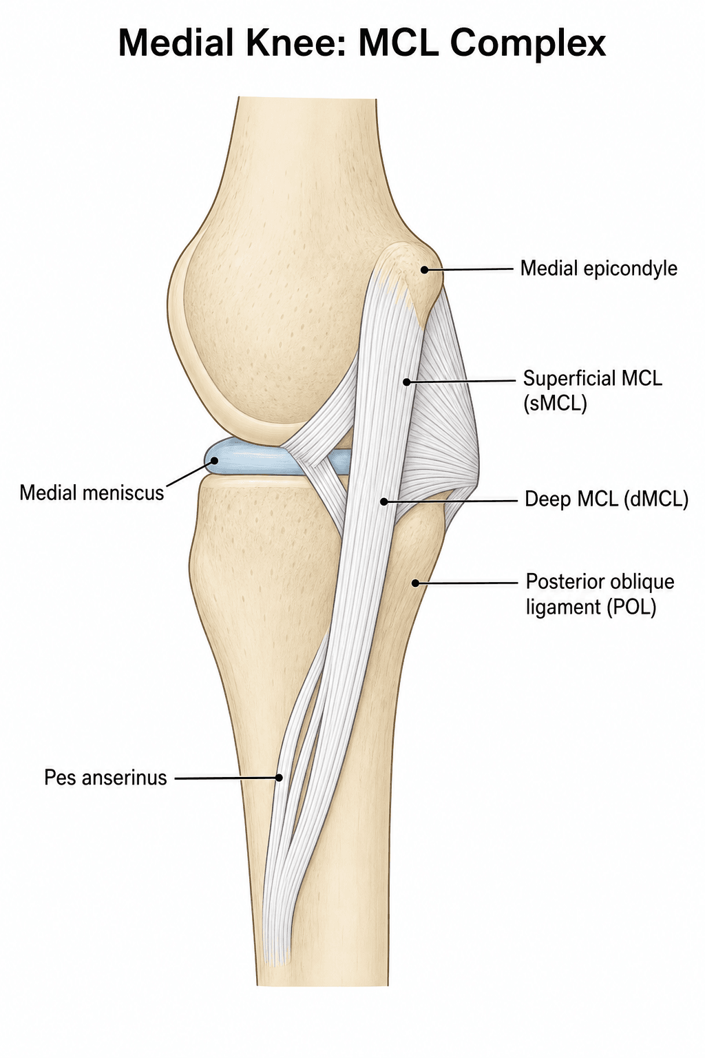
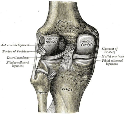
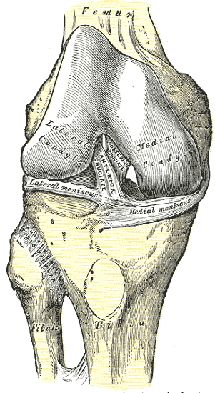
Medial Knee Complex Anatomy
The medial side is best understood as three primary static stabilisers (LaPrade quantitative anatomy): the superficial MCL, the deep MCL and the posterior oblique ligament.
- Primary static restraint to valgus and a key secondary restraint to external/internal tibial rotation
- Femoral attachment lies just proximal and posterior to the medial epicondyle (a small depression, on average a few mm from the epicondyle), NOT several cm proximal to the joint
- Average ligament length around 9 to 10 cm
- Two tibial attachments: a proximal soft-tissue (meniscal) attachment and a distal firm bony attachment roughly 6 cm distal to the joint line, deep to the pes anserinus
- A capsular thickening with meniscofemoral and meniscotibial components
- Firmly anchors the body of the medial meniscus
- Contributes mainly to anteromedial rotatory restraint
- Fan-shaped condensation posterior to the sMCL, blending with the semimembranosus
- Principal restraint to valgus and rotation near full extension and the key structure in posteromedial corner (PMC) injury
Valgus laxity at 0 degrees extension indicates injury to posteromedial corner and possibly cruciates in addition to MCL. This is a more severe injury pattern requiring careful evaluation.
Classification Systems
MCL Injury Grading
- Pathology
- Fiber stretch, intact
- Examination
- Tender, firm endpoint
- Laxity
- 0-5mm, no increase
- Pathology
- Partial tear
- Examination
- Lax with endpoint
- Laxity
- 5-10mm increased
- Pathology
- Complete rupture
- Examination
- Lax without endpoint
- Laxity
- Greater than 10mm increased
Compare to contralateral side - absolute values vary between individuals.
Clinical Assessment
- Mechanism: Valgus blow, contact
- Pain: Medial knee, at time of injury
- Swelling: Often localized medial
- Instability: Giving way with valgus
- Associated injuries: Pop (ACL), locking (meniscus)
- Tenderness: Along MCL course
- Valgus stress 30 degrees: Isolates MCL
- Valgus stress 0 degrees: PMC involved if lax
- ACL tests: Lachman, pivot shift
- Meniscus: McMurray, joint line
Patient supine. At 30 degrees flexion, stabilize thigh, apply valgus force to ankle. Compare opening and endpoint to contralateral side. Repeat at 0 degrees - laxity here indicates posteromedial corner injury.
Key Clinical Pearls
Tenderness along course helps identify injury location - femoral, mid-substance, or tibial.
Always perform Lachman and pivot shift. Combined injuries are common.
Differential Diagnosis of Acute Medial Knee Pain
- Key Distinguishing Feature
- Valgus laxity at 30 degrees, tenderness along MCL course
- Best Test
- Valgus stress at 30 degrees
- Key Distinguishing Feature
- Joint-line tenderness, mechanical locking, effusion
- Best Test
- McMurray, MRI
- Key Distinguishing Feature
- Valgus laxity at 0 degrees plus rotatory instability
- Best Test
- Valgus stress at 0 degrees, MRI
- Key Distinguishing Feature
- Inability to weight bear, bony tenderness
- Best Test
- Radiograph, CT
- Key Distinguishing Feature
- Tenderness over pes insertion, no laxity, often overuse
- Best Test
- Clinical, no valgus laxity
- Key Distinguishing Feature
- Burning medial pain, no laxity, sensory change
- Best Test
- Clinical, Tinel along adductor canal
- Key Distinguishing Feature
- MPFL tenderness, apprehension, haemarthrosis
- Best Test
- Apprehension test, MRI
TWOMCL Examination
Hook:Test at TWO positions - 30 and 0 degrees!
Anteromedial Rotatory Instability and the Posteromedial Corner
Isolated sMCL injury produces straight-plane valgus laxity, but when the posteromedial corner (PMC) — the posterior oblique ligament, the posteromedial capsule and the semimembranosus expansions — is torn alongside the sMCL, the result is anteromedial rotatory instability (AMRI): the medial tibial plateau rotates anteriorly as the joint opens medially.
- Valgus laxity at 0 degrees (full extension) — the POL and posteromedial capsule are the restraints in extension, so opening here (not just at 30 degrees) signals PMC involvement, not an isolated sMCL injury.
- Anteromedial drawer / Slocum test — an anterior drawer performed with the tibia in roughly 15 degrees of external rotation; increased anteromedial translation indicates AMRI.
- MRI confirms POL, posteromedial-capsule and semimembranosus injury.
unlike the isolated sMCL, the PMC heals poorly and is a genuine surgical target. Combined sMCL plus POL disruption — and any knee with valgus laxity in full extension — should be evaluated for PMC repair or reconstruction rather than assumed to heal in a brace. Missed AMRI is a recognised cause of failed ACL reconstruction through residual rotatory laxity.
Valgus laxity at 30° only = isolated sMCL (heals with bracing). Valgus laxity also at 0° plus a positive anteromedial drawer = posteromedial corner injury with anteromedial rotatory instability — the pattern that fails conservative care and needs the POL/PMC addressed surgically, especially alongside an ACL reconstruction.
The Skeletally Immature Knee: Physeal Fracture Mimic
In a child or adolescent with open physes, a valgus knee injury that would tear the MCL in an adult more often fails through the distal femoral physis instead — the physis is the weakest link, weaker than the collateral ligament. A Salter-Harris fracture of the distal femur can therefore masquerade as an "MCL sprain," and apparent valgus "laxity" on stress testing may actually be opening through the growth plate rather than the ligament.
Practical implications:
- In a skeletally immature patient with a valgus injury and medial tenderness or instability, scrutinise the physis and obtain plain radiographs; if examination suggests instability, valgus stress radiographs distinguish physeal opening from true ligamentous laxity.
- A distal femoral physeal fracture is an anatomical-reduction problem with a real risk of growth arrest and angular deformity (the distal femoral physis contributes the most growth of any lower-limb physis), so mislabelling it a "ligament sprain" is a significant error.
- True paediatric MCL ligament injuries do occur and, as in adults, heal well — but the physeal fracture must be excluded first.
A valgus injury to a knee with open physes is a distal femoral physeal (Salter-Harris) fracture until proven otherwise — the physis fails before the MCL, so "valgus laxity" may be the growth plate opening. Image the physis (use stress views if needed) before calling it an MCL sprain, because a missed physeal injury risks growth arrest.
SDPLayers of Medial Knee
Hook:SDP - Warren layers of the medial knee!
Investigations
MRI Assessment
Excellent for MCL injuries.
Edema surrounding MCL, discontinuity, thickening.
Femoral, mid-substance, tibial.
ACL, meniscus, cartilage, bone bruise.
MRI not always required for isolated MCL but helps define Grade III and associated injuries.
Pellegrini-Stieda lesion = calcification at MCL femoral origin. Represents chronic MCL injury with calcification of hematoma. Visible on X-ray. May be asymptomatic.
Management Algorithm
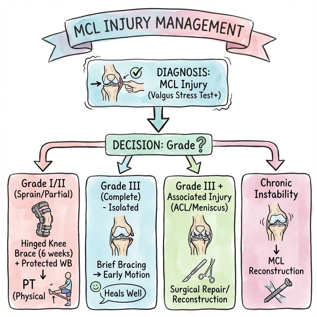
MCL Injury Management
Treatment Pathway
RICE, brace in extension. Examine for associated injuries. Consider MRI if Grade III or associated injury suspected.
Hinged brace allowing ROM. Early physiotherapy. Weight bearing as tolerated. Return to sport 2-6 weeks.
Hinged brace 6-8 weeks. Protected weight bearing initially. Supervised rehabilitation. Most heal.
ACL reconstruction if indicated. MCL usually heals during ACL recovery. Surgery if Grade III with laxity at 0 degrees.
Surgical Technique
MCL Repair Techniques
Suture anchors or bone tunnels to restore origin.
Screw with soft tissue washer.
Primary repair with non-absorbable sutures if acute.
Augmentation with graft may be added for severe injuries.
When repairing or reconstructing MCL, avoid overtensioning which leads to loss of flexion and lateral compartment overload. Tension at 20-30 degrees flexion with slight valgus.
Complications
- Cause
- Prolonged immobilization
- Prevention
- Early ROM
- Management
- Physiotherapy, MUA if severe
- Cause
- Inadequate healing
- Prevention
- Appropriate bracing duration
- Management
- Late reconstruction
- Cause
- Hematoma
- Prevention
- Early ROM, avoid NSAIDs acutely
- Management
- Usually asymptomatic
- Cause
- Surgery
- Prevention
- Careful dissection
- Management
- Neuroma management
Early ROM is key to preventing stiffness. Even Grade III injuries benefit from hinged bracing allowing motion rather than cast immobilization. Stiffness is the enemy.
Postoperative Care
Rehabilitation Protocol
Hinged brace locked initially. Toe touch weight bearing. Quad sets, SLR.
Progressive ROM in brace. Increase weight bearing. Gentle strengthening.
Full ROM. Progressive resistance. Proprioception. Wean brace.
Sport-specific training. Functional testing. Full return when stable.
Conservative: Faster progression, early ROM encouraged. Surgical (reconstruction): More protected initially, similar final timeline. Both aim for full ROM and strength before return to sport.
Outcomes and Prognosis
Outcomes by Grade
Excellent outcomes. Return to sport 1-2 weeks. Minimal long-term sequelae.
Good outcomes. Return to sport 4-6 weeks. Bracing during sport initially helpful.
Most heal with bracing. Some chronic laxity may persist but often asymptomatic.
Outcomes depend on addressing all pathology. ACL reconstruction with MCL healing typical.
Guidelines, Registries & Global Practice
- The MCL is the most commonly injured knee ligament worldwide
- Peak incidence in young, active populations and contact/collision and pivoting sports (football/soccer, rugby, American football, skiing, wrestling)
- Higher reported rates in males, largely reflecting sport-participation patterns
- Skiing classically produces combined valgus-external-rotation ACL/MCL injuries
- Most are low-grade (I to II) and managed entirely in primary or sports-medicine care
- No dedicated national MCL registry exists; isolated MCL injury rarely reaches an implant/arthroplasty registry
- Relevant registry signal is indirect: combined ACL/MCL is captured within national ACL reconstruction registries (Scandinavian, UK NLR, others)
- Those datasets reinforce ACL reconstruction with non-operative MCL care as the dominant combined-injury pathway
- Surgical MCL/POL work concentrates in multiligament and knee-dislocation cohorts
Side-by-Side Guidance (where emphasis differs)
- First-line
- Functional brace and early ROM for isolated injury
- Surgical emphasis
- Selective: chronic instability, distal avulsion, multiligament
- First-line
- Bracing and graded rehab; physiotherapy-led
- Surgical emphasis
- PMC and multiligament patterns to specialist units
- First-line
- Non-operative for isolated; ROM-preserving
- Surgical emphasis
- Repair/augment in dislocation and combined trauma
- First-line
- Conservative isolated MCL; reconstruct chronic laxity
- Surgical emphasis
- Anatomic sMCL/POL reconstruction for failed conservative
High- vs Limited-Resource Practice Variation
- Well-resourced settings: ready access to MRI and hinged functional bracing, supervised physiotherapy and graded return-to-sport testing; specialist reconstruction available for the minority who need it.
- Limited-resource settings: diagnosis relies more on careful clinical valgus stress testing (which is sufficient for most isolated injuries); improvised or non-hinged bracing and home-based rehab are used; MRI and reconstruction are reserved for combined/multiligament injury. Because isolated MCL injury heals well without surgery, clinical-examination-led conservative care travels well across resource settings.
MCL injuries are common viva topics. Know the conservative treatment algorithm, when NOT to operate, how to manage combined injuries, the LaPrade medial-knee anatomy and the grading system.
Controversies and Areas of Uncertainty
Most surgeons allow the MCL to recover full extension and resolve acute valgus laxity before ACL reconstruction (typically a few weeks), citing arthrofibrosis risk with very early combined surgery. The optimal window remains debated, and some advocate earlier reconstruction once motion is restored.
For acute distal (tibial-sided / Stener-equivalent) sMCL avulsions and complete posteromedial corner disruption, primary repair has historically had higher failure rates than for lateral-side injuries. There is growing interest in repair with suture augmentation versus primary reconstruction, but high-level comparative data are lacking.
Landmark series support non-operative care even for complete isolated tears, yet a subset develops chronic valgus or anteromedial rotatory instability. Identifying which complete tears (e.g. distally based, multi-structure POL involvement) will fail bracing is unresolved.
Side-to-side valgus opening on stress radiographs helps grade severity, but published cut-offs distinguishing isolated sMCL from combined sMCL plus POL injury vary between studies, limiting a single agreed threshold.
MCQ Practice Points
Q: What is the primary function of the sMCL? A: Primary static restraint to valgus stress - in classic sectioning studies the sMCL provides roughly three-quarters of valgus restraint at about 25 degrees of flexion, far more than at full extension.
Q: Why test MCL at 30 degrees flexion? A: Isolates the MCL. At 0 degrees, posteromedial capsule and cruciates also contribute. Laxity at 0 degrees indicates more severe injury.
Q: Why does MCL heal better than ACL? A: Extraarticular location and good blood supply. Not bathed in synovial fluid. Forms healing scar tissue.
Q: How should combined ACL/MCL injury be treated? A: ACL reconstruction, MCL heals conservatively. Exception is Grade III MCL with laxity at 0 degrees may need surgical MCL.
Q: What is Pellegrini-Stieda lesion? A: Calcification at MCL femoral origin from chronic injury with hematoma calcification. Visible on X-ray.
Q: What is the MCL Stener equivalent? A: sMCL displaces over pes anserinus preventing healing. Indication for surgical reduction.
Exam Viva Scenarios
Practise clinical reasoning and management decisions out loud
“A 22-year-old rugby player sustains a valgus blow to his knee during a tackle. He has medial knee pain and swelling. Examination shows Grade II laxity at 30 degrees with a firm endpoint. Lachman is negative. How would you manage this?”
“A 28-year-old skier with a twisting fall presents with a swollen knee. Examination shows Grade III MCL laxity and positive Lachman with pivot shift. MRI confirms ACL rupture and Grade III MCL tear. What is your treatment plan?”
“A 35-year-old presents with ongoing medial knee instability 6 months after an MCL injury that was treated conservatively. He has failed prolonged rehabilitation. Valgus stress shows Grade II laxity at 30 degrees. What would you recommend?”
Grading (Valgus at 30 degrees)
- Grade I: Tender, firm endpoint, no laxity
- Grade II: Laxity with endpoint
- Grade III: Laxity without endpoint
- Compare to contralateral side
Examination Key Points
- Test at 30 degrees: Isolates MCL
- Test at 0 degrees: PMC if unstable here
- Always check ACL (Lachman, pivot)
- Palpate along MCL course
Treatment Algorithm
- Grade I-II: Conservative (90%+ heal)
- Grade III isolated: Usually conservative
- Combined ACL/MCL: ACL surgery, MCL heals
- Surgery: chronic instability, Stener, multilig
Anatomy Pearls
- sMCL: primary valgus restraint
- Femoral origin just proximal-posterior to medial epicondyle (LaPrade)
- dMCL: part of posteromedial capsule
- Warren layers: 3 layers medial knee
Return to Sport
- Grade I: 1-2 weeks
- Grade II: 4-6 weeks
- Grade III: 8-12 weeks
- Protective brace may help initially
Evidence Base and Key Studies
Non-Operative Treatment of Complete (Grade III) Isolated MCL Tears
- Comparative study of operative versus non-operative care of complete isolated MCL tears
- Non-operatively treated knees achieved results comparable to surgically repaired knees
- Functional treatment avoided the morbidity and stiffness of surgery and casting
- Helped establish non-operative care as standard for isolated complete MCL injury
Combined ACL/Grade III MCL: Operative vs Non-Operative MCL (RCT)
- Level 1 RCT: 47 patients with combined ACL + grade III MCL, all had early ACL reconstruction
- MCL treated operatively (n=23) versus non-operatively (n=24)
- No significant difference in stability, ROM, muscle power, Lysholm or IKDC at ~2 years
- AP stability excellent in both groups
Early Operative MCL Repair Slows Recovery (Companion RCT)
- Same RCT cohort analysed for ROM and quadriceps power
- Flexion deficit greater after combined ligament repair at 6, 12 and 36 weeks
- Quadriceps power deficit larger in the repair group at 52 weeks (30.7% vs 20.5%)
- Differences resolved by 104 weeks
Early Functional Rehabilitation of Isolated Grade III MCL in Athletes
- Prospective 5-year follow-up of 35 athletes with isolated grade III MCL sprains
- Treated with lateral hinged brace and early ROM, no immobilisation or surgery
- Mean HSS knee score 45.9 of 50 at mean 5.3 years
- Results comparable to surgery or immobilisation with faster return to sport