Four-Part Concept | Blood Supply Critical | PROFHER Shapes Treatment
- Neer classification based on 4 parts: head, greater tuberosity, lesser tuberosity, shaft
- Displacement criteria: greater than 1cm translation or over 45° angulation
- Arcuate artery (from anterior circumflex) is main blood supply to head - at risk in displaced fractures
- PROFHER trial: No difference between operative and non-operative treatment at 2 years
- 85% are minimally displaced and treated non-operatively with good outcomes
- “Greater tuberosity displacement greater than 5mm in active patients warrants surgery
- “Elderly 4-part fracture = reverse shoulder arthroplasty (RSA) is gold standard
- “Valgus-impacted 4-part fractures have better blood supply - may be fixable
- “Axillary nerve at risk - assess deltoid and lateral shoulder sensation
Arcuate artery is terminal branch of anterior circumflex humeral artery. Enters at bicipital groove, runs in spiral to head. Disrupted in displaced fractures = AVN risk.
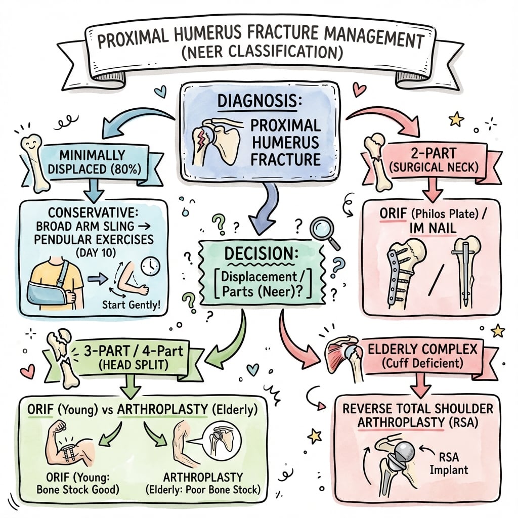
greater than 1cm displacement or over 45° angulation defines a displaced part. Count displaced parts, not fracture lines. 85% are 1-part (non-displaced).
Landmark UK trial: No functional difference at 2 years between surgical and non-surgical treatment for displaced fractures. Changed practice significantly.
Axillary nerve runs 5-7cm below acromion. Test deltoid contraction and regimental badge sensation. At risk with anterior dislocation and surgery.
- Fracture Pattern
- 1-part (non-displaced)
- Key Consideration
- 85% of all proximal humerus fractures
- Treatment
- Sling, early ROM at 2 weeks
- Fracture Pattern
- 2-part surgical neck
- Key Consideration
- greater than 1cm displacement or over 45° angulation
- Treatment
- ORIF with plate or nails
- Fracture Pattern
- 2-part GT displacement greater than 5mm
- Key Consideration
- Affects rotator cuff function
- Treatment
- ORIF with screws/suture
- Fracture Pattern
- 3-part or valgus-impacted 4-part
- Key Consideration
- Head viability more likely
- Treatment
- ORIF if reducible
- Fracture Pattern
- Displaced 3-part or 4-part
- Key Consideration
- High AVN risk, poor bone quality
- Treatment
- Reverse shoulder arthroplasty
- Fracture Pattern
- Any displaced pattern
- Key Consideration
- PROFHER supports non-op
- Treatment
- Consider non-operative
Overview and Epidemiology
Proximal humerus fractures are the third most common fracture in the elderly (after hip and distal radius). The PROFHER trial has fundamentally changed treatment approach - the majority can be treated non-operatively with equivalent outcomes to surgery.
- Bimodal distribution: young males (high-energy), elderly females (low-energy)
- Peak incidence: 60-90 years
- Female predominance increases with age
- Strongly associated with osteoporosis
- Low-energy fall onto outstretched hand (elderly) - 80%
- High-energy trauma (young) - MVA, sports
- Pathological fractures in metastatic disease
- Associated injuries: rotator cuff, brachial plexus
Anatomy and Blood Supply
The arcuate artery (ascending branch of anterior circumflex humeral artery) provides the main blood supply to the humeral head. It enters the bone at the intertubercular groove and runs superiorly. Disruption leads to AVN - risk increases with displacement and number of parts.
- Origin
- Axillary artery
- Course
- Wraps anterior to surgical neck
- Clinical Significance
- Gives arcuate artery - main supply
- Origin
- Anterolateral ascending branch
- Course
- Enters bicipital groove, spirals to head
- Clinical Significance
- Terminal vessel - no collaterals
- Origin
- Axillary artery
- Course
- Through quadrangular space with axillary nerve
- Clinical Significance
- Minor head supply, greater tuberosity supply
- Origin
- Subclavian continuation
- Course
- Behind pectoralis minor
- Clinical Significance
- At risk in fracture-dislocations
- Articular segment (Head): Blood supply concern
- Greater tuberosity: Supraspinatus, infraspinatus, teres minor attach
- Lesser tuberosity: Subscapularis attachment
- Shaft: Pectoralis major, deltoid, latissimus attach
- Supraspinatus: Pulls GT superiorly
- Pectoralis major: Pulls shaft medially
- Subscapularis: Internally rotates LT
- Deltoid: May displace shaft laterally
The axillary nerve exits the quadrangular space and wraps around the surgical neck 5-7cm below the acromion. Always document deltoid function and regimental badge sensation. Incidence of injury is 5-10% in fractures, higher with dislocations.
AAPAProximal Humerus Blood Supply
Hook:AAPA: Anterior gives Arcuate - Posterior helper - AVN if Absent!
Classification Systems
Neer Classification (1970)
Based on 4 anatomical segments and displacement criteria.
- Description
- No fragment meets displacement criteria
- Blood Supply
- Intact
- Treatment Tendency
- Non-operative
- Description
- One fragment displaced (usually surgical neck or GT)
- Blood Supply
- Usually preserved
- Treatment Tendency
- ORIF if indicated
- Description
- Two fragments displaced (head + one tuberosity attached)
- Blood Supply
- At risk
- Treatment Tendency
- ORIF or arthroplasty
- Description
- All fragments separated (head isolated)
- Blood Supply
- High AVN risk
- Treatment Tendency
- Arthroplasty preferred
Count displaced parts, not fracture lines. A fracture can have multiple lines but if only one segment is displaced greater than 1cm or over 45°, it is a 2-part fracture. The head-split pattern and anatomic neck fractures have highest AVN risk.
HGLSNeer Four-Part Classification
Hook:HGLS: Head Gets Less blood Supply when parts are displaced - count the separated parts!
Clinical Assessment
- Mechanism: FOOSH (low-energy), MVA (high-energy)
- Arm position: Held adducted, supported by other hand
- Pre-injury function: Activity level, dominant hand
- Comorbidities: Osteoporosis, diabetes, rotator cuff disease
- Look: Swelling, ecchymosis (tracks to chest/arm), deformity
- Feel: Crepitus, localized tenderness
- Move: Severely limited by pain
- Neurovascular: Axillary nerve, brachial plexus, pulses
Axillary nerve assessment is mandatory: Test deltoid contraction (arm abduction against resistance) and sensation over regimental badge area (lateral arm). Document before and after any manipulation or surgery.
- How to Test
- Deltoid contraction, regimental badge sensation
- Positive Finding
- Weak abduction, numbness lateral arm
- Injury Rate
- 5-10% in fractures, higher with dislocation
- How to Test
- Motor and sensory exam all distributions
- Positive Finding
- Variable deficits multiple nerves
- Injury Rate
- Rare in isolated fractures
- How to Test
- Radial pulse, capillary refill, Doppler
- Positive Finding
- Absent pulse, cool pale hand
- Injury Rate
- Rare - fracture-dislocation risk
- How to Test
- Elbow flexion (biceps), lateral forearm sensation
- Positive Finding
- Weak flexion, numbness
- Injury Rate
- Rare
In high-energy trauma, assess for ipsilateral clavicle fracture (floating shoulder), scapula fracture, rib fractures, and pulmonary injury. In elderly low-energy falls, consider other fragility fractures and need for bone health assessment.
- Distinguishing Features
- Localised tenderness, crepitus, ecchymosis tracking to chest/arm
- Key Investigation
- Trauma series radiographs (AP, scapular Y, axillary)
- Pitfall to Avoid
- Missing a concomitant dislocation on inadequate views
- Distinguishing Features
- Squared-off shoulder, arm held abducted/externally rotated, empty glenoid
- Key Investigation
- AP and axillary radiographs
- Pitfall to Avoid
- Reducing before documenting axillary nerve function
- Distinguishing Features
- Arm locked in internal rotation, light-bulb sign on AP, often after seizure or electrocution
- Key Investigation
- Axillary or CT imaging (light-bulb sign easily missed)
- Pitfall to Avoid
- Over-reliance on AP view alone - classically missed
- Distinguishing Features
- Tenderness and step at the ACJ, pain on cross-body adduction
- Key Investigation
- AP/Zanca view of the ACJ
- Pitfall to Avoid
- Attributing all shoulder pain to the humerus
- Distinguishing Features
- Weakness in abduction/external rotation, normal bony architecture
- Key Investigation
- Ultrasound or MRI
- Pitfall to Avoid
- Labelling a missed greater-tuberosity fracture as a cuff tear
- Distinguishing Features
- Minimal or no trauma, prior pain, lytic lesion on radiograph
- Key Investigation
- Radiograph plus staging imaging if suspicious
- Pitfall to Avoid
- Internal fixation without considering biopsy/staging
Investigations
Imaging Protocol
Three views essential: True AP (Grashey), Scapular Y (Lateral), Axillary. These form the trauma series and allow assessment of all four parts and dislocation status.
Modified axillary view taken with patient leaning back over cassette - avoids need to abduct arm. Useful in acute trauma with limited mobility.
Recommended for all operative cases. 3D reconstructions help understand fracture pattern, articular involvement, head viability. Essential for 3-part and 4-part fractures.
Rarely indicated acutely. May help assess rotator cuff in subacute phase or evaluate blood supply to head (contrast enhancement).
- Technique
- 40° oblique to cassette
- What It Shows
- Glenohumeral joint space
- Key Assessment
- Head location, displacement, dislocation
- Technique
- 90° to Grashey
- What It Shows
- Lateral view of scapula
- Key Assessment
- AP displacement, dislocation direction
- Technique
- Beam through axilla
- What It Shows
- Glenoid and head relationship
- Key Assessment
- Dislocation, GT/LT displacement
- Technique
- Patient leaning back
- What It Shows
- Modified axillary
- Key Assessment
- When axillary not possible
Always get CT for: 3-part and 4-part fractures, head-split patterns, fracture-dislocations, pre-operative planning. CT with 3D reconstruction is superior for understanding complex patterns and identifying head viability in valgus-impacted fractures.

Management Algorithm
Non-Operative Management
Indications:
- 1-part (minimally displaced) fractures - 85% of all cases
- Elderly low-demand patients with displaced fractures (PROFHER evidence)
- Significant medical comorbidities precluding surgery
- Head-split or severely comminuted fractures in non-surgical candidates
Non-Operative Protocol
Collar and cuff or sling. Pendulum exercises begin immediately. Elbow, wrist, hand ROM.
Begin passive and active-assisted shoulder ROM. Supine exercises initially. X-ray at 2 weeks.
Progress to active ROM and strengthening. Most fractures clinically healed by 6-8 weeks.
Full strengthening program. Return to activities as tolerated. Some stiffness may persist.
The PROFHER trial showed no difference in functional outcomes between surgical and non-surgical treatment for displaced proximal humerus fractures at 2 years. This has shifted practice toward more conservative management, especially in elderly patients.
Surgical Technique
ORIF with Locking Plate - Comprehensive Technique:
- Infection: 1-2% superficial, 0.5% deep
- Axillary nerve injury: 5-10% (document preop status)
- AVN: 3-14% (3-part), 15-35% (4-part)
- Screw penetration: 14% - may need removal
- Hardware failure/reoperation: 10-15%
- Stiffness: Very common - physio critical
- Proximal humerus locking plate (system of choice)
- Locking and cortical screws - multiple lengths
- K-wires: 1.6mm and 2.0mm for temporary fixation
- Heavy sutures: No. 2 or 5 FiberWire/Ethibond
- Bone hook and elevator for reduction
- C-arm positioned from contralateral side
Patient Positioning:
Setup Checklist
30-45° trunk elevation. Head secured in padded headrest. Body shifted toward operative edge of table. Arm freely draped on arm board or mayo stand.
Avoid excessive lateral neck flexion - stretches plexus. Head in neutral rotation, supported centrally.
C-arm enters from contralateral side. Confirm AP, axillary, and Velpeau views achievable. Test imaging BEFORE draping.
Prep from nipple to neck, axilla to midline posteriorly. Free drape arm to allow full manipulation.
Blood pressure can drop significantly in beach chair position (cerebral hypoperfusion). Keep MAP above 70mmHg, and avoid sudden position changes.
The tables list "ORIF plate or IMN" but never develop intramedullary nailing, which examiners expect you to be able to compare with the locking plate:
- Best indications: two-part surgical-neck fractures, and surgical-neck fractures with diaphyseal extension or a segmental shaft component (the nail spans the whole bone). Less suited to complex 3/4-part tuberosity-dominant patterns, which a plate controls better.
- Entry point is the key modern point: contemporary straight nails enter through the top of the humeral head, just medial to the rotator-cuff footprint/articular margin, sparing the supraspinatus insertion. Older curved/bent nails entered laterally through the supraspinatus tendon, contributing to rotator-cuff pain and weakness - a recognised drawback of the technique. Whichever nail is used, the cuff/entry must be repaired.
- Pros: load-sharing intramedullary device, smaller exposure, biologically friendly (less periosteal stripping), good for shaft-extending patterns. Cons: cuff-related shoulder pain, risk of malreduction/varus if entry is eccentric, and tuberosity fixation is harder than with a plate.
- Evidence: randomised and registry comparisons show broadly similar functional outcomes for nail vs locking plate in two-part surgical-neck fractures, with differing complication profiles (cuff symptoms with nails; screw perforation with plates) - choice is pattern- and surgeon-driven.
Exam point: offer intramedullary nailing for two-part surgical-neck fractures (especially with shaft extension) using a straight nail with a medial-to-the-cuff-footprint entry point to spare the supraspinatus; outcomes are comparable to locking plates, so the decision is fracture-pattern and surgeon dependent.
The troubleshooting table mentions "allograft strut" and "augment with cement" in passing, but these osteoporotic-fixation adjuncts are worth developing because medial-column failure and varus collapse/screw cut-out are the dominant failure modes of locked plating in poor bone:
- Endosteal fibular strut allograft: a segment of fibular allograft is placed inside the medullary canal across the surgical neck to restore the medial column and provide an internal buttress for the head fragment. It markedly increases construct stiffness, resists varus collapse, and gives the locking screws something solid to purchase - most useful in medial comminution / loss of medial calcar support in osteoporotic patients.
- Cement (screw-tip) augmentation: PMMA or calcium-phosphate injected around the screw tips in the head improves screw purchase and resists cut-out in osteoporotic bone; calcium phosphate additionally supports metaphyseal voids. Used with fenestrated or cannulated screws or via separate injection.
- The unifying principle: the medial calcar/medial column is the key to avoiding varus collapse - restore it (anatomic medial reduction + inferomedial calcar screw, plus a fibular strut and/or cement when the bone is too poor to hold) rather than relying on lateral locking screws alone. If augmentation cannot give a stable construct, convert to RTSA.
Exam point: in the osteoporotic surgical-neck/3-part fracture, augment the locked plate by restoring the medial column - inferomedial calcar screw, an endosteal fibular strut allograft for medial comminution, and/or cement (PMMA/calcium-phosphate) screw augmentation - to prevent the varus collapse and screw cut-out that drive fixation failure.
DRAGSSurgical Indications
Hook:DRAGS: Displaced fractures DRAG young active patients to surgery if bone is good!
Arthroplasty Options
Reverse shoulder arthroplasty (RSA) has largely replaced hemiarthroplasty for fractures. RSA provides reliable pain relief and function regardless of tuberosity healing, while hemiarthroplasty outcomes depend heavily on tuberosity healing.
- Indications
- Young patient, good rotator cuff, good bone
- Pros
- Preserves native glenoid, revision possible
- Cons
- Outcomes depend on tuberosity healing, unpredictable
- Indications
- Elderly, rotator cuff deficient, 4-part fractures
- Pros
- Reliable outcomes, less dependent on tuberosities
- Cons
- Glenoid revision difficult, scapular notching
- Indications
- Fracture with pre-existing OA (rare)
- Pros
- Address arthritis simultaneously
- Cons
- Complex surgery, rarely indicated acutely
In both hemiarthroplasty and RSA, tuberosity reconstruction is critical. Tuberosities should be fixed around the prosthesis using heavy non-absorbable sutures in a tension-band configuration. Tuberosity malunion or non-union is the most common cause of poor outcomes after shoulder arthroplasty for fracture.
Complications
- Incidence
- 15-35% (4-part)
- Risk Factors
- Displacement, head vascularity
- Management
- Close monitoring, arthroplasty if symptomatic
- Incidence
- Most common
- Risk Factors
- Non-op treatment, inadequate reduction
- Management
- Osteotomy if symptomatic, prevention key
- Incidence
- 5-10%
- Risk Factors
- Surgical neck fx, osteoporosis, smoking
- Management
- Bone graft and fixation or arthroplasty
- Incidence
- Common
- Risk Factors
- Prolonged immobilization, adhesive capsulitis
- Management
- Prevention with early ROM, may need MUA or release
- Incidence
- 5-10%
- Risk Factors
- Dislocation, surgical approach
- Management
- Most recover - observe 3-6 months
- Incidence
- Variable
- Risk Factors
- Plate or GT malposition
- Management
- Hardware removal, tuberosity osteotomy
- Incidence
- Variable
- Risk Factors
- Technical error
- Management
- Remove offending screws
AVN risk correlates with head ischemia: anatomic neck fractures (highest risk), head-split fractures, 4-part fractures (15-35%), 3-part fractures (3-14%). Valgus-impacted patterns have lower risk due to preserved medial soft tissue hinge.
MANSComplications
Hook:MANS: Malunion and AVN are the main concerns, Nerve injury and Stiffness complete the picture!
Postoperative Care and Rehabilitation
ORIF Rehabilitation Protocol
Sling immobilization. Elbow, wrist, hand ROM. Pendulum exercises.
Begin passive and active-assisted ROM. Forward flexion in supine, ER to neutral.
Active ROM in all planes. X-ray confirmation of healing. Discontinue sling.
Progressive rotator cuff and deltoid strengthening. Return to activities 3-6 months.
Outcomes and Prognosis
- Shoulder Function
- Good to excellent
- Complications
- Minimal
- Notes
- 85% of fractures, reliable outcomes
- Shoulder Function
- Moderate
- Complications
- Stiffness, malunion
- Notes
- PROFHER supports in elderly
- Shoulder Function
- Variable
- Complications
- Hardware issues, AVN
- Notes
- Best in young with good bone
- Shoulder Function
- Unpredictable
- Complications
- Tuberosity dependent
- Notes
- Falling out of favor
- Shoulder Function
- Reliable
- Complications
- Scapular notching
- Notes
- Current gold standard for 4-part elderly
Key factors affecting outcome: Age (older = more stiffness), Initial displacement (correlates with soft tissue injury), Bone quality, Patient activity level, and Tuberosity healing (for arthroplasty). Function at 1-2 years is best predictor of long-term outcome.
Guidelines, Registries & Global Practice
Global Epidemiology
Proximal humeral fractures account for roughly 4-5% of all fractures and are the third most common fragility fracture in older adults after the proximal femur and distal radius. According to PubMed-indexed epidemiology (Court-Brown et al., Injury 2018), their incidence is rising across high-income populations as longevity increases, with a marked female predominance from low-energy falls and a smaller cohort of younger patients sustaining high-energy injuries.
Side-by-Side Guidance and Evidence
- Region
- UK / international
- Core Recommendation
- No functional benefit of surgery over non-operative care for most displaced surgical-neck fractures
- Evidence Base
- Level I (RCT and high-certainty systematic review)
- Region
- USA
- Core Recommendation
- Non-operative care reasonable for many displaced patterns; reserve arthroplasty for selected elderly 3/4-part fractures
- Evidence Base
- Consensus informed by RCT evidence
- Region
- UK
- Core Recommendation
- Multidisciplinary fragility-fracture pathway, shared decision-making, default to non-operative for older low-demand patients
- Evidence Base
- Guideline / quality standard
- Region
- International
- Core Recommendation
- Restore medial column and head viability for fixation; RTSA where head not reconstructable
- Evidence Base
- Expert principles plus cohort data
- Region
- Europe
- Core Recommendation
- Individualised: physiological age, bone quality and pattern over chronological age; rising RTSA use for fracture
- Evidence Base
- Registry and cohort evidence
Registry Evidence and Practice Variation
- AOANJRR (Australia), NJR (UK), AJRR (USA): all record rising RTSA use for fracture
- RTSA shows lower cumulative revision than hemiarthroplasty for fracture indications
- Registries capture real-world implant survival beyond trial populations
- Surgery rates vary widely between health systems despite Level I evidence favouring non-operative care
- High-income settings: locking plate or RTSA; resource-limited settings rely on closed treatment or external fixation
- PROFHER-2 (ongoing, RTSA vs HA vs non-surgery) will further refine choice in 3/4-part fractures
Bone Health and Medications
- Low-energy fracture over age 50 triggers bone-health assessment (DEXA, vitamin D)
- Anti-resorptives (bisphosphonates, denosumab) and anabolic agents per local formularies
- These agents are widely subsidised and available across most national health systems; calcium and vitamin D supplementation as indicated
- Fracture liaison services reduce re-fracture risk globally
- Falls assessment and frailty optimisation alongside fracture care
- Structured smoking-cessation support (counselling, nicotine replacement, pharmacotherapy) aids bone healing
Any proximal humerus fracture from a low-energy mechanism in a patient over 50 should trigger bone health assessment - DEXA, vitamin D, and consideration of anti-resorptive therapy through a fracture liaison service. This is a consistent recommendation across international guidelines for secondary fracture prevention.
Viva Scenarios
Exam Viva Scenarios
Practise clinical reasoning and management decisions out loud
“A 75-year-old woman presents after a fall at home. She has pain and inability to move her left shoulder. X-rays show a displaced 4-part proximal humerus fracture with the head in valgus position. She is otherwise healthy and independent with ADLs.”
“A 45-year-old competitive recreational tennis player falls during a match. He has a displaced 3-part fracture (greater tuberosity and surgical neck displaced) with moderate osteopenia. He is very keen to return to sport.”
“A 60-year-old presents after high-speed MVA. On examination, the shoulder is squared-off with absent axillary nerve function. X-rays show a 3-part fracture-dislocation with the head posteriorly dislocated. CT confirms significant glenoid bone loss from a Hill-Sachs reverse lesion.”
MCQ Practice Points
Q: What is the main blood supply to the humeral head? A: The arcuate artery (ascending branch of the anterior circumflex humeral artery) provides 80% of blood supply. It enters at the intertubercular groove and is at risk in displaced fractures.
Q: In Neer classification, what defines a 'displaced part'? A: greater than 1cm translation or over 45° angulation. Count displaced parts (not fracture lines) - there are 4 anatomical parts: head, greater tuberosity, lesser tuberosity, and shaft.
Q: What is the AVN rate in 4-part proximal humerus fractures? A: 15-35% for 4-part fractures, 3-14% for 3-part fractures. Valgus-impacted 4-part fractures have lower AVN risk due to preserved medial hinge.
Q: What did the PROFHER trial demonstrate? A: No significant difference in functional outcomes (Oxford Shoulder Score) at 2 years between surgical and non-operative treatment for displaced proximal humerus fractures. Cost-effectiveness favored non-operative treatment.
Q: Which nerve is most commonly injured in proximal humerus fractures? A: Axillary nerve (5-10% incidence). It wraps around the surgical neck 5-7cm below the acromion. Test deltoid contraction and regimental badge sensation.
Q: For a 75-year-old with a displaced 4-part fracture, what is the preferred arthroplasty option? A: Reverse shoulder arthroplasty (RSA). RSA provides more reliable outcomes than hemiarthroplasty because function is less dependent on tuberosity healing.
Key Anatomy
- 4 parts: Head, Greater tuberosity, Lesser tuberosity, Shaft
- Arcuate artery (from anterior circumflex) = main blood supply
- Axillary nerve 5-7cm below acromion - test deltoid and sensation
- Pectoralis major displaces shaft medially
Neer Classification
- Count DISPLACED parts (greater than 1cm or over 45°)
- 1-part = non-displaced = 85% of fractures = non-op
- 2-part = one displaced segment = consider ORIF if young/active
- 3-part = two displaced = ORIF vs arthroplasty
- 4-part = all separated = RSA in elderly
Treatment Algorithm
- 1-part: Sling and early ROM - excellent outcomes
- 2-part GT greater than 5mm: ORIF in active patients
- 2-part surgical neck: ORIF if young, consider non-op if elderly
- 3-part: ORIF if good bone and young, RSA if elderly
- 4-part: RSA preferred over hemiarthroplasty in elderly
Surgical Pearls
- Deltopectoral approach - cephalic vein laterally
- Plate 5-8mm below GT tip to avoid impingement
- Calcar screw improves stability
- Check screw penetration with fluoroscopy AP, axillary, Velpeau views
- Tuberosity repair with heavy sutures critical for function
Complications
- AVN: 15-35% in 4-part, 3-14% in 3-part
- Malunion: Most common complication overall
- Stiffness: Early ROM prevents adhesive capsulitis
- Axillary nerve injury: Document before surgery, most recover
Evidence Base
PROFHER Trial - Surgery vs Non-Surgery
- Pragmatic multicentre RCT of 250 adults (mean age 66 years) with displaced fractures involving the surgical neck. No significant difference in the Oxford Shoulder Score averaged over 2 years (39.07 surgical vs 38.32 non-surgical; difference 0.75 points, 95% CI -1.33 to 2.84; P=0.48). No difference in complications, secondary surgery, or mortality.
Cochrane Review - Interventions for Proximal Humeral Fractures
- 47 trials, 3179 participants. High-certainty evidence of no clinically important difference between surgery and non-surgery in shoulder function at 1 year (SMD 0.10, 95% CI -0.07 to 0.27) or 2 years, with a higher risk of additional surgery after operative treatment (RR 2.06). For RTSA versus hemiarthroplasty, only very low-certainty evidence of minimal between-group functional difference, but a lower complication and reoperation rate favouring RTSA.
Hertel Criteria - Predictors of Humeral Head Ischaemia
- Prospective intraoperative study of 100 intracapsular fractures. The best predictors of head ischaemia were a short calcar/metaphyseal head extension (less than 8mm), a disrupted medial hinge, and an anatomic-neck pattern. When all three were present the positive predictive value for ischaemia reached 97%.
Neer Classification Reliability
- 50 fractures reviewed by five observers on two occasions. Mean interobserver reliability coefficient (kappa) was 0.48-0.52 (moderate), and intraobserver reproducibility averaged 0.66. Simplifying the system did not improve agreement.
Locking Plate Complications
- Prospective multicentre observational study of 187 patients. Complications occurred in 52 of 155 patients (34%) at 1 year; the commonest was intraoperative screw perforation of the head (14%). 40% of complications were technique-related; 19% underwent unplanned second surgery.
Global Fracture Epidemiology in Older Adults
- Prospective database analysis 10 years apart confirming the proximal humerus as one of the classic fragility fractures whose incidence is rising in patients aged 65 years or over, with proximal humeral fractures among those increasing in older males. Driven by ageing populations and improved longevity.
Australian Registry Data (AOANJRR)
- Registry data on shoulder arthroplasty performed for acute fracture show lower cumulative revision rates for reverse total shoulder arthroplasty than for hemiarthroplasty across follow-up. Tuberosity-dependent hemiarthroplasty has progressively been replaced by RTSA for fracture indications.