Associated Injuries Critical | Glenoid Fractures Key | Floating Shoulder Concept
- High-energy injury - always look for associated thoracic and shoulder girdle injuries
- Floating shoulder = scapula neck + clavicle fracture disrupts superior shoulder suspensory complex
- Glenoid fractures are the most important - articular involvement determines outcome
- Most body fractures heal well conservatively - surgery for articular/neck displacement
- Ideberg classification for glenoid fossa fractures guides surgical decision-making
- “80-95% have associated injuries - ribs, lung, clavicle, brachial plexus
- “Scapulothoracic dissociation = devastating - high mortality, look for it
- “Glenoid step more than 4mm or fragment more than 25% = surgical indication
- “Lateral border offset more than 20mm or angulation more than 40 degrees = ORIF neck
80-95% have associated injuries. Prioritize ATLS assessment. Rib fractures (52%), pulmonary contusion (47%), clavicle fractures (23%), brachial plexus injury (12%). High mortality rate (10-15%).
Scapula neck + clavicle fracture = double disruption of superior shoulder suspensory complex. Creates instability. Often requires surgical stabilization of clavicle or both.
Ideberg classification guides treatment. Step off more than 4mm, fragment more than 25% of surface, or instability = surgical indication. These affect long-term shoulder function.
Lateral scapula displacement on CXR. Associated with vascular injury, brachial plexus avulsion, massive soft tissue trauma. High mortality - often forequarter amputation needed.
- Key Finding
- Less than 1cm displacement
- Treatment
- Sling, early ROM, physio
- Key Finding
- Less than 1cm medialization, less than 40 degrees angulation
- Treatment
- Conservative - sling, early motion
- Key Finding
- More than 2cm medialization OR more than 40 degrees angulation
- Treatment
- ORIF via posterior approach
- Key Finding
- Clavicle + scapula neck fracture
- Treatment
- Clavicle ORIF (minimum) plus or minus scapula
- Key Finding
- Step more than 4mm, fragment more than 25%
- Treatment
- ORIF for articular congruity
- Key Finding
- Lateral scapula displacement on CXR
- Treatment
- Life-threatening - angiography, stabilization
SCAPULASCAPULA - Associated Injuries
Hook:SCAPULA fractures mean look for all these injuries - high-energy trauma signature
GOSSGOSS - Glenoid Neck Surgery Indications
Hook:GOSS criteria help decide when neck fractures need surgery
SSSCSSSC - Superior Shoulder Suspensory Complex
Hook:The SSSC is a bone-ligament ring - 2 disruptions = floating shoulder = instability
4-25-40-204-25-40-20 Rule
Hook:4mm step, 25% fragment, 40 degrees angulation, 20mm offset - four surgical thresholds
Overview and Epidemiology
Scapula fractures are uncommon injuries that typically result from high-energy trauma. The scapula is protected by thick muscle coverage and significant force is required to fracture it.
- Motor vehicle accidents - most common (50-70%)
- Falls from height
- Direct blow (crush injuries)
- Sports (uncommon - typically low energy)
The scapula fracture itself is often less important than the associated injuries. Always perform a thorough trauma assessment.
A scapula fracture indicates massive energy transfer. Maintain high suspicion for associated injuries. Mortality rate is 10-15%, primarily from associated thoracic and head injuries rather than the scapula fracture itself.
Traditionally, scapula fractures were treated almost universally conservatively with good results. The shift to surgical treatment in selected cases is based on better understanding of outcomes with significantly displaced glenoid neck and articular fractures.
Anatomy and Biomechanics
Bony anatomy:
- Body - flat triangular bone, thin (2-7mm)
- Spine - posterior ridge, divides supraspinatus from infraspinatus
- Acromion - lateral extension of spine, articulates with clavicle
- Coracoid process - anterior projection, muscle and ligament attachments
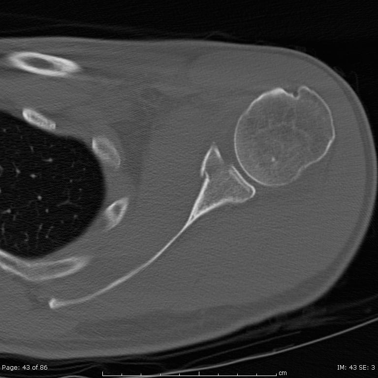
- Glenoid fossa - articular surface for humeral head
- Glenoid neck - transition between glenoid and body
Superior Shoulder Suspensory Complex (SSSC):
The Superior Shoulder Suspensory Complex is a bone-ligament ring that suspends the upper extremity from the axial skeleton. Components: glenoid process, coracoid, CC ligaments, clavicle (distal), AC joint, acromion. Single disruption = stable. Double disruption = floating shoulder = unstable.
- Rotator cuff: supraspinatus, infraspinatus, teres minor, subscapularis
- Scapulohumeral: deltoid (partial), coracobrachialis, biceps (long head)
- Axioscapular: trapezius, levator scapulae, rhomboids, serratus anterior
- Scapulothoracic: pectoralis minor, omohyoid
- Suprascapular nerve (in suprascapular notch - injury in coracoid fractures)
- Axillary nerve (quadrangular space)
- Subscapular nerves and vessels
- Brachial plexus runs anterior to scapula
Classification Systems
Anatomic Classification (most practical)
- Frequency
- 50-60%
- Typical Management
- Conservative (thick muscle coverage aids healing)
- Frequency
- 25%
- Typical Management
- Conservative vs ORIF depending on displacement
- Frequency
- 10%
- Typical Management
- ORIF if displaced more than 4mm or more than 25% involvement
- Frequency
- 8%
- Typical Management
- Conservative unless significantly displaced
- Frequency
- 3-7%
- Typical Management
- ORIF if displaced more than 1cm or associated AC dislocation
- Frequency
- Rare
- Typical Management
- Usually with body fractures
Glenoid fractures are most important prognostically - they affect glenohumeral joint function. Glenoid neck fractures matter when significantly displaced or combined with clavicle injury (floating shoulder).
A two-piece acromion on a film of a sore shoulder is a classic ISAWE/viva trap - is it an acute acromial fracture or an os acromiale (an unfused acromial ossification centre)?
- What it is: the acromion ossifies from up to four centres (from posterior to anterior: basi-acromion, meta-acromion, meso-acromion, pre-acromion), normally fusing by about the mid-twenties. Failure of fusion - most commonly at the meso-meta junction (meso-acromion) - leaves a persistent synchondrosis, the os acromiale, present in a few percent of people and bilateral in most.
- Why it matters: it is a recognised cause of subacromial impingement and rotator cuff tears (an unstable mobile fragment tilts down on the cuff with deltoid pull), and it is the commonest acromial fracture mimic.
- Telling it from a fracture: an os acromiale has smooth, rounded, well-corticated margins at a constant anatomical site (the acromial mid-portion), is usually bilateral and symmetric, and is best seen on the axillary view; an acute fracture has sharp, irregular, non-corticated edges, focal tenderness, a high-energy history, and is unilateral. Marrow oedema/MRI helps if symptomatic.
- Management: incidental, asymptomatic os acromiale - leave alone; symptomatic (painful synchondrosis or impingement) - excision of a small pre-acromion fragment, or open reduction and internal fixation with bone graft for a larger mobile meso-acromion.
Exam point: a bilateral, smoothly corticated separate ossicle at the acromial mid-portion is an os acromiale (meso-acromion commonest), not an acute fracture - confirm on the axillary view and the contralateral side before calling it a fracture.
Clinical Presentation and Assessment
Primary Survey: Scapula fractures are high-energy injuries. ATLS protocol is mandatory.
Complete the primary survey before focusing on the scapula fracture. Life-threatening chest injuries, hemorrhage, and neurological injuries take priority.
History:
- Mechanism (MVA, fall from height, direct blow)
- Associated symptoms (chest pain, dyspnea, neurological symptoms)
- Pre-injury function
- Hand dominance
Physical examination:
- Significance
- Local hematoma
- Associated Condition
- Body/neck fracture
- Significance
- Significant displacement
- Associated Condition
- Displaced glenoid neck
- Significance
- Scapulothoracic dissociation
- Associated Condition
- Life-threatening injury
- Significance
- Pneumothorax
- Associated Condition
- Rib fractures, lung injury
- Significance
- Vascular injury
- Associated Condition
- Subclavian/axillary injury
- Significance
- Brachial plexus injury
- Associated Condition
- High-energy trauma
Associated injuries to look for:
- Rib fractures (52%)
- Pulmonary contusion (47%)
- Pneumothorax (38%)
- Clavicle fractures (23%)
- Brachial plexus injury (12%)
- Humeral fractures (11%)
- Spine fractures (8%)
- Subclavian artery injury (rare but serious)
- Discriminating features
- High-energy mechanism, posterior tenderness, painful but congruent glenohumeral joint
- Confirming investigation
- AP/Y-view radiograph plus CT
- Discriminating features
- Haemarthrosis, articular step, possible subluxation
- Confirming investigation
- CT with 3D reconstruction
- Discriminating features
- Flail arm, absent pulse, gross lateral scapular displacement on CXR
- Confirming investigation
- CXR scapular index plus CT angiography
- Discriminating features
- Locked internal rotation, light-bulb sign, often post-seizure/electrocution
- Confirming investigation
- Axillary lateral or CT - empty glenoid
- Discriminating features
- Anterolateral tenderness, deformity at surgical neck
- Confirming investigation
- AP and scapular-Y radiographs
- Discriminating features
- Point tenderness over AC joint, step deformity, painful cross-body adduction
- Confirming investigation
- AP / Zanca view weighted radiograph
- Discriminating features
- Weakness rather than bony tenderness, lower-energy mechanism
- Confirming investigation
- Ultrasound or MRI
- Discriminating features
- No acute fracture; long thoracic or spinal accessory nerve palsy
- Confirming investigation
- Clinical examination plus EMG if persistent
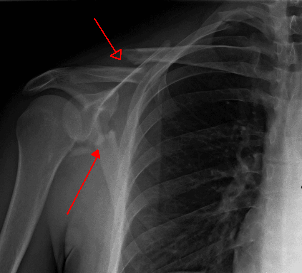
Look at the chest X-ray for lateral displacement of the scapula compared to the contralateral side. This indicates massive soft tissue disruption, likely vascular injury, and brachial plexus avulsion. High mortality. May require forequarter amputation.

Investigations
- Chest X-ray often first to show scapula fracture
- Look for lateral scapula displacement (scapulothoracic dissociation)
- Associated rib fractures, pneumothorax
- True AP scapula (Grashey view) - 30-40 degree posterior oblique
- Scapula Y-view (lateral) - shows glenoid neck and body
- Axillary lateral - glenoid rim fractures
The glenopolar angle is measured on the true AP or Y-view. Draw a line through the most superior and inferior glenoid points. Draw a second line from the inferior glenoid to the most superomedial angle of the scapula. Normal = 30-45 degrees. Less than 20 degrees suggests significant displacement requiring surgery.
CT imaging:
Essential for:
- All glenoid fractures (articular surface assessment)
- Surgical planning for displaced neck fractures
- Complex/comminuted patterns
- Assessment of lateral border offset
3D CT reconstruction:
- Excellent for visualizing fracture pattern
- Helps plan surgical approach and fixation strategy
CT angiography:
Indications:
- Scapulothoracic dissociation
- Expanding hematoma
- Absent or diminished pulses
- Suspected subclavian/axillary injury
Management
Conservative management:
The majority of scapula fractures (80-90%) are treated conservatively with good outcomes.
- Sling immobilization for comfort
- Ice, analgesia
- Address associated injuries first
- Gentle pendulum exercises as pain allows
- Wean from sling
- Active assisted ROM
- Progress as pain allows
- Avoid extremes of motion
- Once radiographic callus visible
- Progressive resistance exercises
- Rotator cuff strengthening
- Full ROM and strength expected
- Sport-specific rehabilitation
- Most return to pre-injury function
Surgical indications:
- Indication for Surgery
- Step more than 4mm, fragment more than 25% of surface, subluxation
- Indication for Surgery
- Angulation more than 40 degrees, medialization more than 20mm, glenopolar angle less than 20 degrees
- Indication for Surgery
- Double SSSC disruption - stabilize at minimum the clavicle
- Indication for Surgery
- Displaced fractures impinging on rotator cuff, reducing subacromial space
- Indication for Surgery
- Displacement more than 1cm, associated with AC joint disruption
- Indication for Surgery
- Rarely surgical - consider if lateral border more than 20mm displaced
The table says "fix the coracoid if displaced or with an AC dislocation" - the Ogawa classification explains the logic, and it is the examinable framework for coracoid fractures:
- Ogawa Type I - posterior (proximal) to the coracoclavicular (CC) ligament attachment: the fracture is behind the conoid/trapezoid insertions, so the CC ligaments stay attached to the distal fragment. This detaches the coracoid strut from the clavicle/SSSC linkage and behaves like a double disruption of the superior shoulder suspensory complex (often with an associated AC joint separation or distal clavicle fracture) - it is unstable and is the type that warrants ORIF.
- Ogawa Type II - anterior (distal) to the CC ligament attachment (the coracoid tip): the CC ligaments remain bridging clavicle-to-scapula, so the SSSC ring is intact - this is a stable avulsion usually treated non-operatively.
So an apparently "minor" coracoid fracture combined with what looks like an AC separation is really a Type I unstable pattern (the "double lesion") - which is why displacement and an associated AC injury push you toward fixation.
Exam point: classify a coracoid fracture by its relation to the CC ligament attachment - Type I (posterior/proximal to it) is unstable and often needs ORIF, Type II (the distal tip, anterior to it) is stable and treated conservatively; suspect a Type I "double lesion" whenever a coracoid fracture coexists with an AC joint disruption.
Modified Judet Approach (most common)
- Patient lateral or prone
- Incision along scapular spine, curves distally along lateral border
- Develop interval between infraspinatus and teres minor
- Excellent exposure of glenoid neck, body, spine
- Can extend for glenoid fossa
Key structures:
- Suprascapular nerve (protected by staying below spine)
- Infraspinatus and teres minor (preserve vascularity)
- Axillary nerve (inferior limit)
The Judet approach provides excellent access to the scapula body and neck.
Fixation methods:
- 3.5mm reconstruction plates (can be contoured)
- Lag screws for simple patterns
- Locking plates for osteoporotic bone
- Suture anchors for small rim fragments

For floating shoulder, the traditional approach is to fix the clavicle first. This is technically easier and often provides sufficient stability. If residual scapula displacement persists after clavicle fixation, address the scapula neck. Some advocate fixing both primarily.
Surgical Technique
Patient Positioning
- Beanbag support at 30-45 degrees
- Arm supported in traction or on mayo stand
- Allows anterior and posterior access
- C-arm from cephalad direction
- Alternative for posterior-only approach
- Better visualization of medial border
- Limited anterior access
Positioning depends on approach requirements and associated injuries.
Complications
- Incidence
- 5-15%
- Prevention/Management
- Early motion protocols, physiotherapy
- Incidence
- Variable
- Prevention/Management
- Rarely symptomatic for body; affects glenoid function
- Incidence
- Rare (less than 1%)
- Prevention/Management
- Rich blood supply protects; treat with bone graft if symptomatic
- Incidence
- 5-10% surgical
- Prevention/Management
- Careful retraction, identify nerve
- Incidence
- 10-20% glenoid fx
- Prevention/Management
- Related to articular step-off; minimize displacement
- Incidence
- 1-2%
- Prevention/Management
- Standard precautions, prophylactic antibiotics
- Most common complication
- Related to prolonged immobilization and associated injuries
- Prevention: early motion when safe
- Primarily in glenoid fossa fractures
- Related to residual step-off
- Emphasizes importance of anatomic reduction for articular fractures
The suprascapular nerve passes through the suprascapular notch and supplies supraspinatus and infraspinatus. Injury causes external rotation and abduction weakness. Protect during posterior approaches by staying inferior to the scapular spine.
Long-term outcomes:
- Body fractures: generally excellent outcomes regardless of treatment
- Neck fractures: good outcomes if displacement parameters respected
- Glenoid fractures: outcomes correlate with reduction quality
Postoperative Care and Rehabilitation
Post-ORIF protocol:
- Sling for comfort and protection
- Gentle pendulum exercises
- No active elevation or external rotation
- Wound management
- Wean sling as comfort allows
- Active assisted ROM
- Progress to active ROM
- Avoid loaded activities
- Full active ROM expected
- Progressive strengthening
- Rotator cuff rehabilitation
- Light functional activities
- Confirm radiographic healing
- Sport-specific training
- Return to full activities typically 4-6 months
- High-impact activities may take longer
Key rehabilitation principles:
- Balance protection of repair with early motion
- Rotator cuff function critical for outcome
- Address associated injuries (rib, clavicle) in rehab plan
- Patient education about expected timeline
Outcomes and Prognosis
Outcomes by fracture type:
- Outcome
- Excellent
- Notes
- Conservative treatment adequate
- Outcome
- Good
- Notes
- Conservative if meets displacement criteria
- Outcome
- Good
- Notes
- With surgical stabilization
- Outcome
- Good-Excellent
- Notes
- Key is anatomic reduction
- Outcome
- Fair-Poor
- Notes
- Develops arthritis
Prognostic factors:
- Associated injuries (head, chest) - major determinant of mortality
- Articular reduction quality - major determinant of shoulder function
- Patient age and bone quality
- Rehabilitation compliance
Most patients with scapula body fractures return to full function. Glenoid fossa fractures have variable outcomes depending on reduction quality. The presence of glenohumeral subluxation at presentation is a poor prognostic factor.
Guidelines, Registries & Global Practice
Global epidemiology
- Figure
- ~10 per 100,000 per year
- Source population
- Two Swedish counties, Ideberg 1995
- Figure
- Less than 1%
- Source population
- Pooled case series, Zlowodzki/Cole 2006
- Figure
- 3-5%
- Source population
- Pooled case series, Zlowodzki/Cole 2006
- Figure
- ~30%
- Source population
- Ideberg 1995
- Figure
- ~90% (range 80-95%)
- Source population
- Zlowodzki/Cole 2006
- Figure
- Peak in young men from high-energy trauma; older women from low-energy falls
- Source population
- Ideberg 1995
Scapula fractures are uncommon worldwide and almost always a marker of high-energy transfer; injury patterns therefore track regional trauma epidemiology (road-traffic and motorcycle trauma in much of Asia, Africa and Latin America; falls and vehicle crashes in higher-income settings).
Guidance, side by side
There is no single dedicated international guideline for scapula fractures; management is anchored on the AO Foundation principles plus society trauma-system standards. Recommendations are largely concordant because the evidence base is uniformly low (level IV).
- Position on scapula fractures
- Non-operative for most body/neck fractures; ORIF for displaced intra-articular glenoid, large lateral-border displacement/angulation, low glenopolar angle, or double SSSC disruption; posterior (Judet) approach standard
- Evidence level
- Expert consensus / level IV
- Position on scapula fractures
- No condition-specific clinical practice guideline; managed within polytrauma and shoulder-trauma principles, ATLS-first
- Evidence level
- Consensus
- Position on scapula fractures
- No scapula-specific BOAST; governed by BOAST polytrauma and open-fracture standards and trauma-network triage to a major trauma centre
- Evidence level
- Consensus / standard of care
- Position on scapula fractures
- No scapula-specific guidance; covered by NICE major-trauma pathways (NG39/NG40) emphasising ATLS, imaging and network transfer
- Evidence level
- Consensus
- Position on scapula fractures
- Reflects AO principles; emphasises CT for glenoid and surgical-threshold measurement
- Evidence level
- Expert consensus
Across AO, AAOS, BOA and EFORT the consensus is identical at the level that examiners test: complete ATLS first, image associated injuries, treat most fractures non-operatively, and reserve surgery for displaced articular glenoid fractures, grossly displaced necks (glenopolar angle 22 degrees or less, displacement 20mm or more, angulation 40-45 degrees or more) and unstable double SSSC disruptions.
Registry evidence
Scapula fractures are not implant-survival procedures, so the major arthroplasty registries (NJR, AJRR, AOANJRR, SHAR, Norwegian, NZJR) do not track them directly. Epidemiology instead comes from national trauma databases: a US National Trauma Data Bank analysis of 9,453 scapular fractures (Baldwin et al., J Trauma 2008) showed that, after adjusting for injury severity, upper-extremity, thoracic and pelvic-ring injuries remained significantly associated with scapula fracture, while many other "associations" reflected overall injury severity rather than the scapula fracture itself.
Global practice variation
- High-resource settings: ready CT and 3D reconstruction, sub-specialist shoulder/trauma surgeons, and trauma-network transfer make selective ORIF (Judet or single lateral-column approach) routine for displaced patterns.
- Limited-resource settings: CT may be scarce and most fractures are managed non-operatively with sling and early motion, which is supported by the evidence for the majority of body and minimally displaced neck fractures.
- Scapulothoracic dissociation is a universal surgical emergency requiring immediate vascular assessment regardless of setting; outcomes depend on access to vascular surgery and ICU support.
For any board, be ready to justify management on biomechanical thresholds and evidence level rather than a national pathway. State that the evidence is predominantly level IV, that most fractures heal well non-operatively, and that operative thresholds (glenoid step greater than 4mm or fragment greater than 25%, glenopolar angle 22 degrees or less, lateral-border displacement 20mm or more, double SSSC disruption) are consensus-based and consistent across AO, AAOS, BOA and EFORT.
MCQ Practice Points
Q: What percentage of patients with scapula fractures have associated injuries? A: 80-95%. Scapula fractures are high-energy injuries. Rib fractures are most common (52%), followed by pulmonary contusion (47%) and clavicle fractures (23%).
Q: What is the superior shoulder suspensory complex (SSSC)? A: A bone-ligament ring connecting the upper extremity to the axial skeleton. Components: glenoid, coracoid, CC ligaments, clavicle, AC ligaments, acromion. Two disruptions = floating shoulder = unstable.
Q: What Ideberg type is an anterior glenoid rim fracture? A: Type Ia. Type Ib is posterior rim. These rim fractures are often associated with shoulder instability and may require treatment directed at the instability rather than just the fracture.
Q: What glenoid articular step-off is an indication for ORIF? A: More than 4mm step-off or more than 25% articular surface involvement. These thresholds are based on data showing increased rates of post-traumatic arthritis with larger incongruities.
Q: On chest X-ray, what finding suggests scapulothoracic dissociation? A: Lateral displacement of the scapula compared to the contralateral side. This indicates complete disruption of the scapulothoracic connection with likely vascular injury and brachial plexus avulsion.
Clinical Decision Scenarios
Practise clinical reasoning and management decisions out loud
“A 35-year-old motorcyclist is brought to ED after high-speed accident. GCS 15, hemodynamically stable. Chest X-ray shows multiple left rib fractures and a scapula fracture. CT shows a glenoid neck fracture with 15mm medialization. How do you manage this patient?”
“A 42-year-old presents after falling from scaffolding. X-rays show a displaced midshaft clavicle fracture and CT reveals a glenoid neck fracture with 25mm medialization and glenopolar angle of 15 degrees. How do you approach this 'floating shoulder' injury?”
“A 28-year-old is brought in after a motorcycle vs truck collision. There is massive left shoulder swelling, the arm is flail, and there is no radial pulse. Chest X-ray shows the left scapula displaced 4cm lateral to the chest wall. What is your diagnosis and immediate management?”
KEY FACTS
- 1% of all fractures - high-energy mechanism required
- 80-95% have associated injuries - ATLS protocol mandatory
- 10-15% mortality (from associated chest/head injuries)
- Most body fractures (50-60%) treated conservatively
- Peak age 25-40, M:F ratio 2:1
- MVA most common mechanism (50-70%)
CLASSIFICATION
- Anatomic: Body (50-60%), Neck (25%), Glenoid fossa (10%), Processes (8%)
- Ideberg (glenoid): I (rim), II (transverse), III (oblique), IV (horizontal), V (combined), VI (comminuted)
- SSSC: double disruption = floating shoulder = unstable
- Ada-Miller (neck): Type I (anatomic neck), Type II (through body)
- Body fractures: usually heal well with conservative treatment
SURGICAL THRESHOLDS (4-25-40-20 Rule)
- Glenoid fossa: step more than 4mm OR fragment more than 25% of surface
- Glenoid neck: angulation more than 40 degrees OR medialization more than 20mm
- Glenopolar angle less than 20 degrees (normal 30-45 degrees)
- Floating shoulder: fix clavicle minimum, consider scapula if residual displacement
- Humeral head subluxation = surgical indication
- GH instability with rim fracture = consider Bankart repair
FLOATING SHOULDER
- Double SSSC disruption (usually clavicle + scapula neck fracture)
- Creates unstable glenoid segment - weight causes medialization
- Treatment: stabilize clavicle FIRST (easier, restores length)
- Reassess scapula after clavicle fixation under fluoroscopy
- Add scapula ORIF if persistent displacement more than 20mm
- Some advocate fixing both primarily - evidence mixed
SCAPULOTHORACIC DISSOCIATION
- Lateral scapula displacement on CXR (compare to contralateral)
- Complete soft tissue disruption - massive energy transfer
- Associated vascular injury (subclavian/axillary), brachial plexus avulsion
- High mortality (10-20%) and amputation rate (20%)
- Immediate CT angiography, vascular surgery consultation
- May need forequarter amputation if limb non-salvageable
ASSOCIATED INJURIES (Remember SCAPULA)
- Spine fractures (cervical/thoracic) - 8%
- Clavicle fractures - 23% (creates floating shoulder)
- Arterial injury (subclavian) - rare but serious
- Pulmonary contusion - 47% (most common thoracic)
- Upper extremity nerve (brachial plexus) - 12%
- Lateral rib fractures - 52% (most common association)
SURGICAL APPROACHES
- Modified Judet (posterior): along spine/lateral border
- Interval: infraspinatus-teres minor (preserves cuff)
- Protect suprascapular nerve (stay below spine)
- Deltopectoral (anterior): for isolated anterior rim
- Positioning: lateral decubitus 30-45° or prone
- Fixation: 3.5mm recon plates, lag screws, locking plates
TRAPS AND PEARLS
- Always complete ATLS trauma assessment first
- Look for associated chest injuries - ribs, pneumothorax
- Glenoid fractures affect long-term function most
- Body fractures heal well conservatively
- Modified Judet = standard posterior approach
- Glenopolar angle less than 20° = surgical indication
Evidence Base
- Pooled 520 fractures from 22 case series: scapula fractures comprise less than 1% of all fractures and 3-5% of shoulder-girdle fractures.
- Approximately 90% of patients had associated injuries.
- Both operatively and non-operatively treated fractures achieved largely good functional results when treated by appropriate indication; operative infection and secondary-surgery rates were low.
- Glenoid fossa and scapular neck fractures were the patterns most commonly treated operatively.
- About 25% had a concomitant clavicle or acromioclavicular injury.
- Plate-and-screw fixation through a posterior (Judet) approach was most common; complication rate was low.
- Good to excellent functional results in approximately 85% of operatively treated cases at a mean of 49.9 months.
- Floating shoulder (ipsilateral scapular neck plus clavicle fracture) is not inherently unstable.
- Mean Constant score 76 for the conservatively treated group versus 71 for the operative group.
- Caudal (inferior) dislocation of the glenoid was the key poor-prognostic finding: Constant 42 with it versus 85 without.
- In the absence of caudal glenoid displacement, conservative treatment gave a good functional outcome.
- 22 patients had ORIF more than 3 weeks (mean 30 days) after injury for displaced intra-articular fractures, glenohumeral medialisation, angular deformity, or double SSSC lesions.
- Mean DASH score 14 at a mean follow-up of 27 months.
- 13 of 16 followed patients returned to previous work and recreation without restriction.
- No wound complications, infection, or nonunion occurred.
- Annual incidence of scapular fractures was 10 per 100,000 inhabitants over a 10-year period across two Swedish counties.
- 30% of scapular fractures involved the glenoid cavity.
- The most common intra-articular pattern was the anterior chip (rim) fracture, associated with shoulder dislocation in about two-thirds of cases.
- This series underpins the Ideberg glenoid-fossa classification used to guide surgical planning.
- Operative thresholds used: medial/lateral displacement of 20mm or more, angulation of 45 degrees or more, double SSSC disruption, or glenopolar angle of 22 degrees or less.
- Scapular neck angulation corrected from a mean 38.7 degrees pre-operatively to 3.6 degrees; mean post-operative glenopolar angle 35.4 degrees.
- Mean DASH score 11.4 and mean Subjective Shoulder Value 88.9; 10 of 11 returned to pre-injury work.
- No infections or neurovascular injuries with a less invasive single-column approach.