Superior Labrum Anterior to Posterior | Biceps Anchor | Type II Critical
- Type II is the CLINICALLY SIGNIFICANT SLAP tear - biceps anchor is detached
- O-Brien test and Speed test are key provocative tests
- Overhead athletes (throwers) are high-risk population
- SLAP repair outcomes declining in literature - tenotomy/tenodesis rising
- Age over 40: consider biceps tenotomy/tenodesis over SLAP repair
- “MR arthrography is gold standard for SLAP diagnosis
- “Peel-back test: arthroscopic confirmation (ABER position)
- “High failure rate of SLAP repair in throwing athletes
- “Type II can be subdivided: anterior, posterior, combined
Type II SLAP = biceps anchor detached. This is the clinically significant lesion. Types I and III can be debrided. Type IV involves biceps tendon itself.
Throwers have posterosuperior SLAP tears from peel-back mechanism in late cocking. Often associated with GIRD (glenohumeral internal rotation deficit) and posterior capsular tightness.
Recent literature shows high failure rates of SLAP repair, especially in patients over 40. Trend toward biceps tenotomy or tenodesis with comparable outcomes and faster recovery.
Under 40: Consider SLAP repair in motivated patient. Over 40: Biceps tenotomy or tenodesis preferred. Degenerative labral changes common over 40.
- Pathology
- Degenerative fraying
- Treatment
- Debridement only
- Role of Biceps
- Anchor intact - leave
- Pathology
- Biceps anchor detached
- Treatment
- Repair or biceps procedure
- Role of Biceps
- Anchor must be addressed
- Pathology
- Bucket handle tears
- Treatment
- Excise bucket handle
- Role of Biceps
- Anchor intact - leave
- Pathology
- Bucket handle into biceps
- Treatment
- Excise +/- repair or tenotomy
- Role of Biceps
- Biceps tendon involvement
- Pathology
- Combined lesions
- Treatment
- Address each component
- Role of Biceps
- Variable - case dependent
AGESLAP Repair vs Biceps Procedure Decision
Hook:AGE guides SLAP repair vs biceps procedure decision!
Overview and Epidemiology
SLAP repairs peaked then declined in the 2000s-2010s as studies showed high failure rates, especially in patients over 40 and throwers. Trend now toward biceps tenotomy or tenodesis which provides reliable pain relief with faster recovery.
- Originally described in 1990 (Snyder)
- Peak age 26-50 years
- Male greater than female
- Overhead athletes overrepresented
- Increasing diagnosis with MRI/arthroscopy
- Compression: Fall on outstretched hand, arm forward and abducted
- Traction: Sudden pulling force on arm
- Peelback: Throwing motion (late cocking) in throwers
- Degenerative: Age-related in Type I
- Repetitive overhead: Cumulative microtrauma
Pathophysiology and Mechanisms
Superior Labral Anatomy
10 o-clock to 2 o-clock position on glenoid.
LHB tendon inserts at supraglenoid tubercle, blending with superior labrum.
Less firmly attached than anterior labrum - may appear loose on arthroscopy.
Relatively avascular zone (10-12 o-clock) - limits healing.
Sublabral foramen (absent anterosuperior labrum) and Buford complex (cord-like MGHL) are normal variants that should NOT be repaired. True SLAP tears show: fraying, instability of biceps anchor, extension toward biceps, and positive peel-back test.
The Thrower's Shoulder: Internal Impingement, GIRD and the "Dead Arm"
In the overhead athlete the posterior (Type IIB) SLAP is the end of a recognised pathological cascade (Burkhart/Morgan, the "disabled throwing shoulder"):
- Scapular dyskinesis (the "SICK" scapula) and repetitive late-cocking load lead to a posteroinferior capsular contracture.
- This causes GIRD (glenohumeral internal rotation deficit) — loss of internal rotation compared with the contralateral side, with a posterosuperior shift of the humeral-head contact point.
- The shifted contact produces posterosuperior (internal) impingement — the undersurface of the rotator cuff and greater tuberosity abut the posterosuperior glenoid/labrum in abduction-external rotation.
- The vertical biceps vector then peels back the posterosuperior labrum → Type IIB SLAP, often with undersurface cuff fraying.
- The clinical endpoint is the "dead arm" — sudden loss of throwing velocity and control.
GIRD management is rehabilitation-first: the sleeper stretch and cross-body adduction stretch restore internal rotation and resolve symptoms in the large majority. Arthroscopic posteroinferior capsular release is reserved for GIRD that remains symptomatic despite a dedicated stretching programme (≈3 months). Addressing the GIRD/scapula is essential — operating on the labrum while ignoring the contracture is a classic cause of failure.
Classification Systems
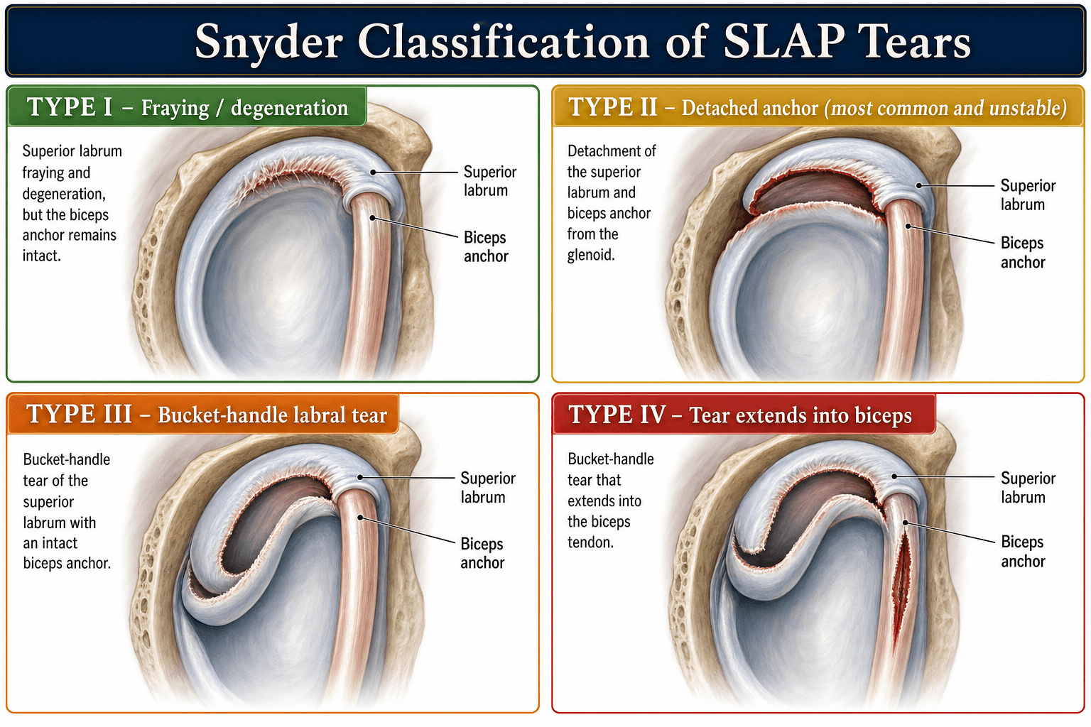
Original Snyder Classification (Types I-IV)
- Pathology
- Fraying of superior labrum
- Treatment
- Debridement
- Key Point
- Anchor intact - no repair
- Pathology
- Biceps anchor detached from glenoid
- Treatment
- Repair or biceps procedure
- Key Point
- MOST IMPORTANT clinically
- Pathology
- Bucket handle tear of labrum
- Treatment
- Excise bucket handle
- Key Point
- Anchor intact - preserve it
- Pathology
- Bucket handle extending into LHB
- Treatment
- Excise +/- repair/tenotomy
- Key Point
- Biceps tendon split
FBBBSLAP Classification
Hook:FBBB - Know Type II (Biceps anchor) is the KEY clinical SLAP!
Clinical Assessment
- Mechanism: Compression, traction, throwing
- Pain: Anterior/deep shoulder, with overhead activity
- Clicking/popping: Common complaint
- Sport level: Overhead athlete critical
- Weakness: May report with overhead activity
- O-Brien test: Pain with forward flex/adduct/IR, relieved with ER
- Speed test: Resisted forward flexion
- Anterior slide: Hand on hip, force applied
- Biceps load test: ABER with biceps contraction
- GIRD: Check internal rotation deficit
Arm at 90° forward flexion, 10-15° adduction, thumb down (IR). Resist downward pressure. Then repeat with palm up (ER). Positive: Pain with IR, relieved with ER. Most commonly used clinical test for SLAP.
Key Examination Findings
Check for GIRD (internal rotation deficit greater than 20° compared to opposite side), posterior capsular tightness, scapular dyskinesis.
SLAP tears often associated with rotator cuff pathology - assess thoroughly.
Differential Diagnosis
The "deep anterior shoulder pain with overhead activity" presentation overlaps with several conditions. No single SLAP test is reliable in isolation, so the differential must be worked through clinically and on imaging.
- Discriminating Features
- Pain over bicipital groove, point tender, Speed/Yergason positive
- Best Test
- Ultrasound or MRI of groove; diagnostic LHB block
- Discriminating Features
- Painful arc, weakness on cuff testing, night pain
- Best Test
- MRI; often coexists with SLAP
- Discriminating Features
- Posterior pain in late cocking, GIRD, throwers
- Best Test
- ABER MR arthrography; arthroscopy
- Discriminating Features
- Apprehension, prior dislocation, relocation positive
- Best Test
- MR arthrography (anteroinferior labrum)
- Discriminating Features
- Point tenderness over AC joint, cross-body pain
- Best Test
- Targeted exam, AC joint injection
- Discriminating Features
- No fraying, stable biceps anchor, asymptomatic
- Best Test
- Arthroscopic recognition - do NOT repair
SOAPSLAP Clinical Tests
Hook:SOAP up the shoulder tests for SLAP diagnosis!
Investigations
MR Arthrography (Gold Standard)
82-98% for Type II SLAP.
Intra-articular gadolinium increases labral visualization.
Contrast extension between labrum and glenoid at superior labrum.
Increases sensitivity for peel-back lesions.
Superior to standard MRI for labral pathology.
Arthroscopic ABER positioning: With arm in abduction and external rotation (throwing position), the biceps vector becomes vertical and peels the posterosuperior labrum off the glenoid. Positive peel-back = Type II SLAP (especially IIB in throwers).


Management Algorithm
SLAP Type-Based Treatment
Decision Pathway
Degenerative fraying. Anchor intact. Simple debridement of frayed tissue. No repair needed.
Biceps anchor detached. Options: SLAP repair (under 40, motivated), biceps tenotomy (over 40, low demand), or tenodesis (over 40, active).
Bucket handle tear. Anchor intact. Excise the displaced bucket handle fragment. Preserve the anchor.
Bucket handle into biceps. If less than 30% biceps involvement: excise bucket handle. If greater than 30%: biceps tenotomy or tenodesis.
Surgical Technique
Arthroscopic SLAP Repair
Surgical Steps
Confirm SLAP classification. Perform peel-back test. Assess biceps tendon quality.
Debride frayed tissue. Decorticate glenoid rim to bleeding bone. Mobilize the labrum.
1-3 suture anchors at superior glenoid rim. Place at 10-11 o-clock and 12-1 o-clock. Avoid suprascapular nerve on superior anchor.
Pass sutures through labrum. Tie to restore labral bumper and biceps anchor stability.
Avoid placing anchors too posterior (risk suprascapular nerve at spinoglenoid notch) or too medial on glenoid neck (inadequate purchase). Anchors should be on the glenoid rim at the articular margin.
Complications
- Procedure
- SLAP repair
- Incidence
- Common
- Management
- Aggressive PT, avoid in throwers
- Procedure
- SLAP repair
- Incidence
- Common in throwers
- Management
- Revision or biceps procedure
- Procedure
- Tenotomy
- Incidence
- ~23% (vs ~7% tenodesis)
- Management
- Cosmetic - counsel preop
- Procedure
- Repair
- Incidence
- Rare
- Management
- Avoid superior/posterior anchor
- Procedure
- Any
- Incidence
- Variable
- Management
- Address all pathology, consider revision
Overhead throwing athletes return unreliably after SLAP repair. In a systematic review (Thayaparan 2019), pitchers returned to sport at only 57.5% versus 87.1% for non-pitching athletes. Peel-back forces during late cocking stress the repair. Many surgeons now favour biceps tenodesis for throwers with Type II SLAP.
SLAP-Repair-Specific Complications
Beyond the table above, two repair-specific issues are examinable:
- Postoperative glenoid chondrolysis — a devastating, rapid loss of glenohumeral articular cartilage in a young patient after labral surgery. Recognised associations are intra-articular pain-pump catheters (especially bupivacaine), thermal/radiofrequency devices, and prominent or bioabsorbable (PLLA) anchors. It presents as pain and progressive stiffness within months. Prevention is the key message: avoid intra-articular pain pumps, minimise thermal energy, and ensure anchors are seated below the articular surface.
- Hardware and nerve complications — proud or backed-out anchors and bioabsorbable-anchor reactions (synovitis, cyst formation) can damage cartilage or cause persistent pain; a posteriorly placed superior anchor risks the suprascapular nerve at the spinoglenoid notch. Over-tightening the repair (especially with too many anchors) is a frequent cause of postoperative stiffness, which is poorly tolerated by throwers.
Postoperative Care
Rehabilitation Protocol (SLAP Repair)
Sling immobilization. Elbow/hand exercises. Gentle pendulums only. No active biceps.
Wean sling. Passive to active-assisted ROM. Avoid ABER position. No resisted biceps.
Full AROM. Isometric cuff strengthening. Begin light biceps activity.
Progressive resistance. Sport-specific training. No throwing until 6 months minimum.
Biceps procedures have faster recovery. Tenotomy: sling 1-2 weeks, no restrictions by 6 weeks. Tenodesis: protect biceps 6 weeks, no heavy biceps loading 3 months. Both return to full activity faster than SLAP repair.
Outcomes and Prognosis
Procedure-Specific Outcomes
Overall return to sport around 70% (Thayaparan 2019), but pitchers return at only ~58%. Outcomes are poorer in throwers, older patients, and workers-compensation cases. Postoperative stiffness is the most common concern.
Level I evidence (Belk 2021) shows equivalent pain and functional scores; cosmetic (Popeye) deformity is the key difference - 23.3% after tenotomy versus 6.8% after tenodesis.
Comparable pain relief to tenotomy with a much lower cosmetic deformity rate; preferred in younger, active, or cosmetically concerned patients, and an effective salvage for failed SLAP repair (McCormick 2014, Boileau 2009).
Return to Sport
6-9 months for non-throwing sports. Throwing athletes may take 9-12 months with variable return to prior level.
Often faster return (3-4 months).
Guidelines, Registries & Global Practice
Global Epidemiology
SLAP lesions account for a small proportion of all shoulder arthroscopies (around 6% in large series; Erickson 2016) but are over-represented in overhead athletes - baseball pitchers, volleyball and tennis players, swimmers and javelin throwers. Peak presentation is in active adults roughly 20-50 years, with a male predominance reflecting sport and occupational exposure. Isolated SLAP tears are uncommon; most coexist with rotator cuff or biceps pathology, particularly over 40, where degenerative superior labral change is frequently incidental.
Guidelines & Society Positions (Side by Side)
- Emphasis
- Evidence-based, shared decision-making
- Practical Position
- No isolated SLAP CPG; supports trial of non-operative care and procedure choice by age/demand
- Emphasis
- Stepwise, rehab-first
- Practical Position
- Physiotherapy and activity modification before surgery; tenodesis favoured in older patients
- Emphasis
- Technique and classification fidelity
- Practical Position
- Distinguish true SLAP from normal variants; address concomitant pathology
- Emphasis
- Athlete-centred
- Practical Position
- Individualised approach in throwers; caution about repair outcomes
There is no dedicated randomised-trial-based clinical practice guideline specific to SLAP repair; recommendations are consensus- and cohort-driven. The consistent global theme across societies is a rehabilitation-first approach and a shift from labral repair toward biceps tenodesis, especially with increasing age.
Registry & Database Signals
- Surgeon practice databases (US): Both single-practice (Erickson 2016) and the nationwide ABOS examination database (Cvetanovich 2020) show a significant fall in SLAP repair volume and a rise in biceps tenodesis over the last decade, with tenodesis concentrated in patients over 35.
- SLAP repair has no implant-survivorship registry equivalent to arthroplasty (NJR / AOANJRR / SHAR); evidence comes from cohort series and the database trends above.
High- vs Limited-Resource Practice Variation
- MR arthrography and ABER sequences readily available
- Arthroscopic repair, tenodesis and tenotomy all offered
- Structured sports-physiotherapy and return-to-throwing programmes
- Procedure choice driven by age, sport and patient preference
- Diagnosis often clinical plus standard MRI or even plain films to exclude bony pathology
- Biceps tenotomy favoured - cheap, fast, no implants, minimal rehab burden
- Repair reserved for young high-demand athletes where anchors and rehab are accessible
- Greater reliance on activity modification and analgesia
SLAP tears are a common viva topic. Know the Snyder classification (especially Type II), clinical tests (O-Brien, peel-back), and the global trend toward biceps tenotomy/tenodesis. Be prepared to discuss treatment in overhead athletes and why outcomes are variable.
Controversies and Areas of Uncertainty
The biggest unresolved question. Registry data show a clear practice shift to tenodesis, and Boileau (2009) reported far better sport return after tenodesis - but his tenodesis group was older, confounding the comparison. Whether a motivated patient under 30 with an isolated Type II tear does better with repair (preserving native anatomy) or primary tenodesis remains unsettled.
The biomechanical role of the biceps anchor in glenohumeral stability is debated. If its contribution is minor, sacrificing it (tenotomy/tenodesis) carries little functional cost - which would favour the move away from repair. Cadaveric and clinical data remain conflicting.
Superior labral signal and a meniscoid attachment are common in asymptomatic shoulders and increase with age. A "SLAP" seen on imaging or scope is not necessarily the pain generator - over-treatment of normal variants and age-related change is a real risk.
No approach reliably returns elite pitchers to prior performance. Whether to repair, add posterior capsular release for GIRD, or proceed directly to tenodesis is individualised and controversial, and honest counselling about uncertain return is essential.
MCQ Practice Points
Q: Which SLAP type is the most clinically significant? A: Type II - biceps anchor is detached from the glenoid. This requires surgical treatment (repair or biceps procedure). Types I and III can usually be debrided.
Q: How is a Type I SLAP treated? A: Debridement only. Type I is degenerative fraying with intact biceps anchor. No repair is needed - simply debride the frayed tissue.
Q: What does a positive peel-back test indicate? A: Type II SLAP tear (especially Type IIB). In ABER position, the biceps vector becomes vertical and peels the posterosuperior labrum off the glenoid.
Q: How is the O-Brien test performed? A: Arm at 90° forward flexion, 10-15° adduction, thumb down (IR). Resist downward pressure. Repeat with palm up (ER). Positive: pain with IR, relieved with ER.
Q: What is the sublabral foramen? A: Normal anatomical variant - detachment of anterosuperior labrum at 1-3 o-clock position. Should NOT be repaired. Distinguished from SLAP by stable biceps anchor.
Q: Why is SLAP repair less recommended in patients over 40? A: Degenerative labral and biceps changes reduce healing potential, and registry data (Cvetanovich 2020) show tenodesis is preferentially performed in patients over 35. Biceps tenotomy or tenodesis gives more reliable pain relief in this group.
Exam Viva Scenarios
Practise clinical reasoning and management decisions out loud
“A 28-year-old recreational volleyball player presents with right shoulder pain during overhead serving. O-Brien test is positive. MR arthrography shows a Type II SLAP lesion with intact biceps tendon. How would you manage this?”
“A 35-year-old had SLAP repair 18 months ago but has persistent anterior shoulder pain with overhead activities. MRI shows the repair appears intact. What is your approach?”
“A 22-year-old elite baseball pitcher has posterior shoulder pain during the cocking phase of throwing. MR arthrography shows a posterosuperior Type IIB SLAP tear. Examination also reveals 25° GIRD. What is your treatment plan?”
Classification (Snyder I-IV)
- Type I: Fraying - debride only
- Type II: Biceps anchor detached - MOST IMPORTANT
- Type III: Bucket handle - excise, anchor intact
- Type IV: Bucket into biceps - excise +/- tenotomy
Clinical Tests (SOAP)
- Speed test: Resisted forward flexion
- O-Brien test: Forward flex/adduct/IR vs ER
- Anterior slide: Hand on hip, force applied
- Peel-back: Arthroscopic ABER position
Type II Decision Factors
- Under 40: Consider SLAP repair
- Over 40: Biceps tenotomy or tenodesis
- Throwing athlete: High failure rate - consider tenodesis
- Degenerative biceps: Tenotomy or tenodesis
Imaging
- MR arthrography: Gold standard
- Look for contrast at labral-glenoid junction
- ABER sequence increases sensitivity
- Peel-back test confirms at arthroscopy
Outcomes
- SLAP repair: ~70% return to sport overall
- Pitchers return at only ~58% (vs ~87% non-pitchers)
- Tenotomy: Popeye deformity ~23% vs ~7% tenodesis
- Tenodesis: Equivalent pain/function, less deformity
Normal Variants (Do NOT repair)
- Sublabral foramen (1-3 o-clock)
- Buford complex (cord-like MGHL)
- Meniscoid labrum (loose attachment)
- Distinguish by stable biceps anchor
Evidence Base and Key Studies
Original Description of SLAP Lesions (Snyder Classification)
- Retrospective review identifying 27 SLAP lesions among more than 700 shoulder arthroscopies
- Defined the four original types (I-IV); injury runs posterior to anterior including the biceps anchor
- Most common mechanism was a compression force from a fall onto an outstretched arm
- No preoperative imaging test reliably defined the lesion - diagnosis was arthroscopic
Declining SLAP Repair, Rising Biceps Tenodesis
- 619 SLAP repairs among 9,765 shoulder arthroscopies across four surgeons, 2004-2014
- Percentage of SLAP repairs fell significantly over the decade (P less than .001)
- Number and percentage of biceps tenodeses rose significantly over the same period
- Most SLAP repairs were performed for Type II tears; mean patient age fell over time
ABOS Database - Nationwide Decline in SLAP Repair
- 9,908 cases from the American Board of Orthopaedic Surgery part-II examination database, 2012-2017
- Significant decline in SLAP repair rate over the study period (P less than .001)
- Patients receiving biceps tenodesis were significantly more likely to be over 35 years
- SLAP repair remained concentrated in younger patients
Return to Sport After SLAP Repair (Pitchers Fare Worst)
- Systematic review of 22 studies, 944 patients undergoing arthroscopic SLAP repair
- Overall return to sport 69.6%; return to previous level of play 69.0%
- Mean time to return to sport 8.9 months
- Pitchers returned at only 57.5% versus 87.1% for non-pitching athletes
Biceps Tenodesis as Alternative to Type II SLAP Repair (Boileau)
- Cohort of 25 patients with isolated Type II SLAP lesions: 10 SLAP repair vs 15 biceps tenodesis
- Return to previous sport: 87% after tenodesis vs only 20% after SLAP repair (P = .01)
- 60% of repair patients were dissatisfied (persistent pain or failure to return to sport)
- Four failed SLAP repairs were successfully salvaged with subsequent biceps tenodesis
Biceps Tenodesis for Failed Type II SLAP Repair
- Prospective series of 42 patients undergoing open subpectoral tenodesis after a failed Type II SLAP repair
- Significant improvement in ASES, SANE and WOSI scores and in range of motion (P less than .0001)
- 81% returned to active duty and sport at mean 3.5-year follow-up
- Single transient musculocutaneous neurapraxia; mean patient age 39 years
Tenotomy vs Tenodesis - Cosmesis and Biomechanics (Hsu)
- Systematic review of clinical and biomechanical studies on biceps tenotomy versus tenodesis
- Higher incidence of cosmetic (Popeye) deformity after tenotomy than tenodesis
- Tenodesis associated with a higher likelihood of residual bicipital pain
- All included clinical studies were Level IV - no consensus could be reached on the superior technique
Tenotomy vs Tenodesis - Level I Meta-Analysis (Belk)
- Meta-analysis of 5 Level I randomised controlled trials, 468 patients (236 tenodesis, 232 tenotomy)
- Cosmetic deformity 6.8% after tenodesis vs 23.3% after tenotomy (P less than .001)
- No difference in Constant-Murley, VAS pain or ASES scores between groups
- Comparable complication and functional outcomes at mean 23-month follow-up