High Stress Zone | FEAR Deformity | Reduce Before Reaming | CMN Gold Standard
- Proximal fragment FEAR: Flexed (iliopsoas), Externally rotated, Abducted (glutes), Rotated
- REDUCE BEFORE REAMING - nail follows reamer path, cannot correct malreduction
- Cephalomedullary nail is gold standard (not SHS, not plate)
- Long nail preferred - protects entire femur from stress riser
- Atypical fractures - bisphosphonates over 5 years, lateral beaking, check contralateral
- “Russell-Taylor Type II = piriformis involved = trochanteric entry nail required
- “Blocking (Poller) screws placed in concavity of deformity to guide nail
- “Varus malunion is most common error - accept slight valgus, NEVER varus
- “Atypical fractures: bilateral in 30%, prodromal thigh pain, stop bisphosphonates
Proximal fragment is Flexed (iliopsoas), Externally rotated (short rotators), Abducted (gluteus medius/minimus). Match leg position to proximal fragment for reduction.
CRITICAL principle: The nail follows the reamer path. If you ream before reducing, you will lock in the malreduction. Always confirm reduction on AP/lateral BEFORE reaming.
Bisphosphonates over 5 years = risk of atypical fracture. Look for: transverse pattern, lateral cortex beaking, minimal trauma. Always image contralateral femur.
Varus malunion leads to implant failure and nonunion. Accept slight valgus, NEVER varus. Use blocking screws if needed for coronal plane control.
- Key Action
- Long CMN, reduce before reaming
- Implant Choice
- Trochanteric entry CMN, long nail
- Key Action
- Check for atypical, image contralateral
- Implant Choice
- CMN + bone graft, stop bisphosphonates
- Key Action
- Cannot use piriformis entry
- Implant Choice
- Trochanteric entry CMN required
- Key Action
- STOP - do not accept this
- Implant Choice
- Blocking screws, reposition, re-reduce
- Key Action
- Risk of complete fracture
- Implant Choice
- Prophylactic nailing
FEARProximal Fragment Deformity
Hook:FEAR the proximal fragment - it pulls into Flexion, ER, and Abduction!
LATERAL BEAKAtypical Fracture Features
Hook:Look for the LATERAL BEAK - hallmark of atypical fracture!
BLOCKINGReduction Technique Aids
Hook:BLOCKING screws are your friend for coronal plane control!
PIRIFORMISRussell-Taylor Classification
Hook:PIRIFORMIS intact = Type I, involved = Type II (trochanteric entry)!
Overview and Epidemiology
The subtrochanteric region extends from the lesser trochanter to 5cm distally. This is the zone of highest mechanical stress in the entire femur - transition from cancellous to cortical bone with maximum bending moment.
Demographics
- Young adults: High-energy trauma (MVA, motorcycle, falls from height)
- Elderly: Low-energy falls, pathological, or atypical fractures
- 10-15% of proximal femur fractures
- Increasing incidence of atypical fractures (bisphosphonate awareness)
- Male predominance in young, female in elderly
Understanding epidemiology guides clinical suspicion for different fracture etiologies.
Anatomy and Biomechanics
Subtrochanteric Region
- Superior: Lesser trochanter
- Inferior: 5cm below lesser trochanter (or to isthmus)
- Bone type: Transitional - cancellous to cortical
- Iliopsoas: Lesser trochanter - FLEXES proximal fragment
- Gluteus medius/minimus: Greater trochanter - ABDUCTS proximal fragment
- Short external rotators: Intertrochanteric - EXTERNALLY ROTATES proximal fragment
- Adductors: Linea aspera - ADDUCTS distal fragment
The proximal fragment is controlled by the gluteal abductors and iliopsoas. Since the fracture has disrupted lever arm, these muscles pull the proximal fragment into:
- Flexion: 30-60 degrees (iliopsoas)
- External rotation: Short rotators
- Abduction: Gluteus medius/minimus
To reduce: Match the leg to the proximal fragment (flex hip, abduct, externally rotate slightly)
Classification Systems
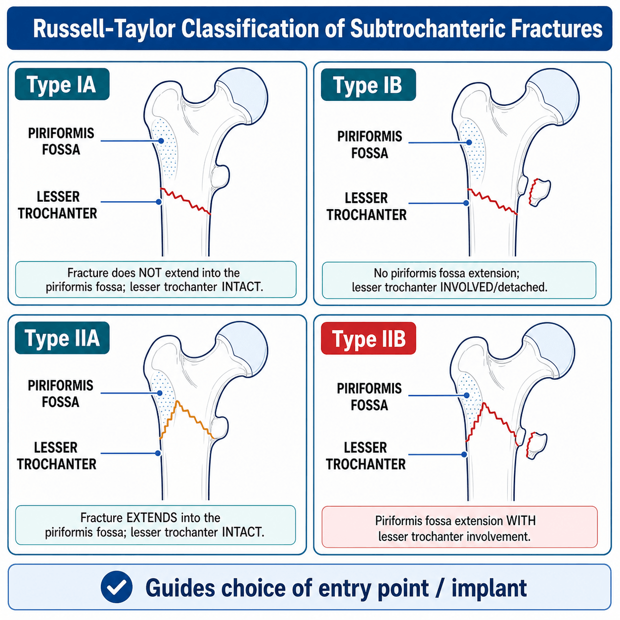
Russell-Taylor Classification
The Russell-Taylor classification determines nail entry point based on fracture extension:
- Piriformis Fossa
- Intact
- Lesser Trochanter
- Intact
- Entry Point
- Piriformis entry possible
- Piriformis Fossa
- Intact
- Lesser Trochanter
- Fractured
- Entry Point
- Piriformis entry possible
- Piriformis Fossa
- Involved
- Lesser Trochanter
- Intact
- Entry Point
- Trochanteric entry required
- Piriformis Fossa
- Involved
- Lesser Trochanter
- Fractured
- Entry Point
- Trochanteric entry required
Type I = Piriformis fossa intact = Can use piriformis entry nail Type II = Piriformis involved = MUST use trochanteric entry nail
Most modern nails are designed for trochanteric entry, making this distinction less critical clinically but still important for exams.
Differential Diagnosis
- Discriminating Features
- Comminution, spiral/oblique pattern, significant trauma, FEAR proximal-fragment deformity
- Key Action
- Long cephalomedullary nail, reduce before reaming
- Discriminating Features
- Transverse pattern, lateral cortex beaking, minimal trauma, prodromal thigh pain, bisphosphonate over 5 years
- Key Action
- Image contralateral femur, stop antiresorptive, bone-health workup
- Discriminating Features
- Lytic lesion, known malignancy, weight loss/night sweats, cortical destruction
- Key Action
- Staging imaging and biopsy before definitive fixation
- Discriminating Features
- Fracture line from distomedial to proximolateral; behaves biomechanically like subtrochanteric
- Key Action
- Intramedullary nail (not sliding hip screw)
- Discriminating Features
- Between greater and lesser trochanter, no shaft extension distal to lesser trochanter
- Key Action
- Sliding hip screw or cephalomedullary nail per pattern
- Discriminating Features
- Proximal to intertrochanteric line, risk of avascular necrosis
- Key Action
- Arthroplasty or fixation depending on age/displacement
History
History Taking
- Motor vehicle accident
- Motorcycle crash
- Fall from height
- Sporting injury
- Associated injuries common
- Minimal trauma (fall from standing)
- Spontaneous fracture
- Prodromal thigh pain (weeks to months before)
- Bisphosphonate history (ask duration specifically)
- Glucocorticoid use
- Known malignancy
- Weight loss, night sweats
- Previous radiation
- Metabolic bone disease history
Thorough history helps distinguish fracture etiology and guides workup.
Examination
Physical Examination
- Shortened limb
- External rotation deformity
- Thigh swelling (significant blood loss)
- Unable to weight bear
- Ecchymosis (may be delayed)
- Proximal fragment position assessment difficult clinically
- Note rotational deformity
- Check for angular deformity
- Assess soft tissue condition (open vs closed)
- Distal pulses (dorsalis pedis, posterior tibial)
- Sciatic nerve function
- Compartment assessment
- Motor/sensory exam of foot
- ATLS for high-energy
- Ipsilateral injuries (floating knee)
- Spine clearance
- Chest/abdomen in polytrauma
Complete assessment identifies associated injuries requiring attention.
Investigations
Radiographic Assessment
- Full-length femur (AP and lateral) - MUST see hip and knee
- AP pelvis - for comparison and proximal extent
- Contralateral femur - if atypical suspected
- Fracture pattern and comminution
- Proximal and distal extent
- Piriformis fossa involvement (Russell-Taylor)
- Canal diameter for nail sizing
- Evidence of atypical features (lateral beaking)
- Complex fracture patterns
- Surgical planning for difficult cases
- Proximal extension assessment
- Pathological fracture evaluation
- Stress fracture evaluation
- Incomplete contralateral fracture assessment
- Pathological lesion characterization
- If metastatic disease suspected
Imaging selection depends on clinical suspicion and surgical planning needs.

Management
Core Management Principles
REDUCE BEFORE REAMING
The nail follows the path of the reamer. If you ream in malreduction, the nail will hold that malreduction permanently. Always confirm reduction on AP and lateral fluoroscopy BEFORE reaming.
- Anatomic alignment (length, rotation, axis)
- Stable fixation allowing early mobilization
- Preserve biology where possible
- Address underlying cause (if pathological or atypical)
- Match leg position to proximal fragment (flex, abduct, slight ER)
- Hip flexion 30-60 degrees on fracture table
- Abduct leg to match abducted proximal fragment
- Use blocking screws for coronal plane control
- Accept slight valgus, NEVER varus
Core principles guide all management decisions for subtrochanteric fractures.
The peritrochanteric/subtrochanteric femur is a common site of metastatic and impending pathological fracture, and the management diverges from a simple trauma nail:
- Suspect pathology with a low-energy mechanism, antecedent pain (a prodrome of activity-related thigh/hip pain), a lytic/destructive lesion, or a known/possible primary; in any such case do NOT just nail it.
- Biopsy/stage before fixation if the primary is unknown - nailing an undiagnosed primary bone sarcoma (or seeding the canal) can convert a curable tumour into an unsalvageable limb. Get the diagnosis (and exclude myeloma/renal/other primaries) first.
- Protect the WHOLE femur with a long cephalomedullary nail - in metastatic disease the bone distal (and proximal) to the lesion is also at risk, so a long load-sharing nail prophylactically spans the femur rather than leaving a stress riser.
- The construct must outlast the patient/bone: pathological bone may never unite, so durable fixation (and often cement augmentation) matters more than waiting for biological healing.
- Give post-operative radiotherapy to the fixed segment to control local tumour, and manage bone health/systemic disease with oncology.
Exam point: a pathological subtrochanteric fracture needs diagnosis/biopsy before fixation (if primary unknown), a long whole-femur cephalomedullary nail, a durable (often cemented) construct that does not rely on union, and post-operative radiotherapy - the impending-fracture scoring is covered under metastatic bone disease.
Surgical Technique
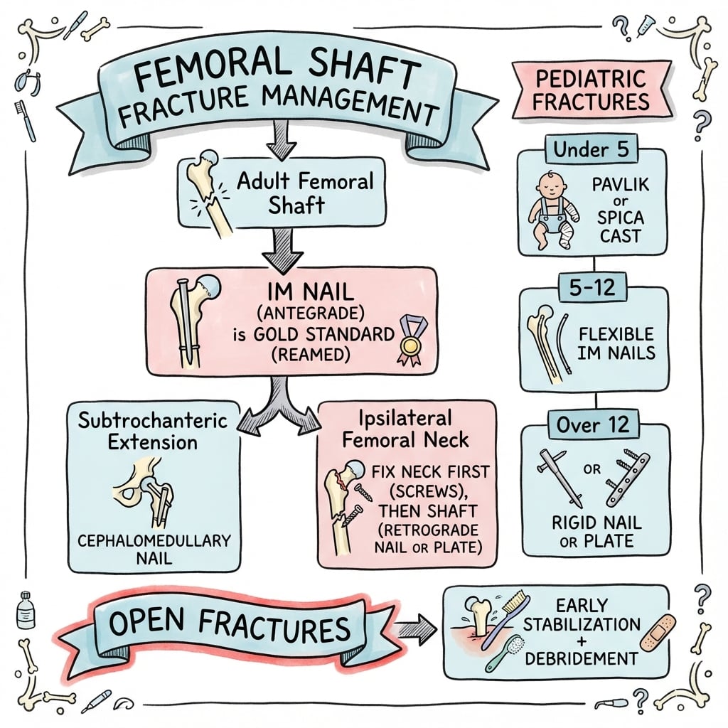
Cephalomedullary Nailing - Gold Standard
- Fracture table (supine) preferred
- Alternative: Lateral decubitus for better reduction control
- Radiolucent table with manual traction acceptable
- Trochanteric entry: Most common, suitable for all patterns
- Piriformis entry: Only for Russell-Taylor Type I
- Entry point determines nail trajectory
- Position patient with hip flexed 30-60 degrees
- Abduct leg to match proximal fragment position
- Reduce fracture under fluoroscopy
- CONFIRM REDUCTION ON AP AND LATERAL BEFORE REAMING
- Make incision over entry point
- Open cortex with awl
- Pass guidewire across fracture with reduction held
- Ream in 2mm increments to 1-1.5mm above nail diameter
- Insert nail maintaining reduction
- Lock proximally (lag screw to femoral head)
- Lock distally (static for comminuted, dynamic if simple)
The reduce-before-reaming principle is paramount for successful outcomes.

Complications
Intraoperative Complications
- Varus alignment (NEVER accept)
- Procurvatum (flexion deformity)
- Rotational malreduction
- Prevention: Reduce BEFORE reaming
- Entry point fracture
- Distal fracture during nail insertion
- Prevention: Adequate reaming, correct entry point
- May need additional fixation
- Lag screw malpositioning
- Short nail creating stress riser
- Inadequate distal locking
Intraoperative complications are largely preventable with careful technique.

Postoperative Care
Immediate Postoperative (Days 0-14)
- Check surgical wounds at 48 hours
- Remove drains at 24-48 hours when output is below 50mL
- Wound inspection at 2 weeks for suture removal
- Chemical prophylaxis for minimum 4-6 weeks
- LMWH preferred (enoxaparin 40mg daily)
- Mechanical prophylaxis with TED stockings and intermittent pneumatic compression
- Physiotherapy commencing day 1 postoperatively
- Weight-bearing status dependent on fracture pattern and fixation stability
- Most CMN fixation allows touch weight-bearing or weight-bearing as tolerated
- Comminuted or unstable patterns may require protected weight-bearing for 6-8 weeks
- Multimodal analgesia regimen
- Wean opioids as tolerated
- Consider regional blocks for enhanced recovery
Early mobilization reduces complications and improves outcomes in elderly patients.
Outcomes and Prognosis
Union Rates
- Union Rate
- 90-95%
- Complication Rate
- 10-15%
- Notes
- Gold standard, load-sharing
- Union Rate
- 80-85%
- Complication Rate
- 20-25%
- Notes
- Historical data
- Union Rate
- 70-80%
- Complication Rate
- 30-40%
- Notes
- Higher failure, reserved for salvage
Prognostic Factors
- Simple fracture pattern
- Adequate reduction achieved
- Long cephalomedullary nail
- Young, healthy patient
- Good bone quality
- Comminuted pattern
- Varus malreduction
- Atypical fracture (delayed healing)
- Open fracture
- Osteoporosis
Atypical Fracture Outcomes
Atypical bisphosphonate-associated fractures have delayed healing compared to standard subtrochanteric fractures. Consider:
- Bone grafting at time of fixation
- Teriparatide postoperatively
- Longer protected weight-bearing
- Extended follow-up for union assessment
Guidelines, Registries & Global Practice
Global Epidemiology
Subtrochanteric fractures account for approximately 10-15% of proximal femoral fractures, with a bimodal distribution: high-energy injuries in young men and low-energy, osteoporotic or atypical fractures in older women. Atypical femoral fractures are strongly associated with antiresorptive therapy but remain rare in absolute terms.
- Magnitude
- RR 47.3 (95% CI 25.6-87.3)
- Source
- Schilcher, NEJM 2011 (Sweden)
- Magnitude
- 5 per 10,000 patient-years
- Source
- Schilcher, NEJM 2011
- Magnitude
- 3.2-50 per 100,000 person-years
- Source
- ASBMR Task Force 2014
- Magnitude
- Approx 70% per year since last use
- Source
- Schilcher, NEJM 2011
According to PubMed, these figures derive from the Swedish nationwide cohort of Schilcher and colleagues (DOI) and the second ASBMR Task Force report (DOI).
Guideline & Society Positions
- Position
- Cephalomedullary (intramedullary) nail is the preferred construct; anatomical reduction before reaming; long nail to avoid distal stress riser
- Basis
- Expert consensus + systematic review (Grade B)
- Position
- Extracapsular fractures with subtrochanteric involvement should be fixed with an intramedullary nail rather than a sliding hip screw
- Basis
- Evidence-based guideline
- Position
- Early surgery, full-length femoral imaging, anatomical reduction and rehabilitation within an orthogeriatric pathway
- Basis
- Standards of care
- Position
- Screen low-energy subtrochanteric fractures for atypical features; stop antiresorptive, image contralateral femur, optimise bone health, consider teriparatide
- Basis
- Consensus / Level IV
Registry & Practice Variation
National hip-fracture registries and arthroplasty/trauma registries (including the AOANJRR in Australia, the National Hip Fracture Database in the UK, and equivalent Scandinavian registries that generated the atypical-fracture risk estimates above) consistently show cephalomedullary nailing as the dominant fixation method for subtrochanteric and reverse-oblique patterns. Practice variation persists in entry point (trochanteric versus piriformis), routine use of cerclage/open reduction adjuncts, and weight-bearing protocols.
MCQ Practice Points
Q: What is the classic proximal fragment deformity in subtrochanteric fractures and why?
A: FEAR - Flexed (iliopsoas on lesser trochanter), Externally rotated (short external rotators), Abducted (gluteus medius/minimus on greater trochanter). The distal fragment is adducted by the adductors.
Q: What is the critical principle regarding reduction and reaming in subtrochanteric fractures?
A: REDUCE BEFORE REAMING. The nail follows the path of the reamer. If you ream a malreduced fracture, the nail locks in that malreduction. Always confirm reduction on AP and lateral fluoroscopy before reaming.
Q: What determines the nail entry point in Russell-Taylor classification?
A: Piriformis fossa involvement. Type I (piriformis intact) = piriformis entry possible. Type II (piriformis involved) = trochanteric entry required.
Q: What are the key features of an atypical bisphosphonate-associated fracture?
A: Transverse or short oblique pattern, lateral cortex beaking/thickening, minimal trauma mechanism, associated with over 5 years bisphosphonate use. Must image contralateral femur (bilateral in 28%).
Q: Where do you place blocking screws to prevent varus malunion?
A: On the medial side of the distal fragment (in the concavity of the deformity). This narrows the canal medially and forces the nail/wire to track more laterally, preventing varus.
Exam Viva Scenarios
Practise clinical reasoning and management decisions out loud
“A 28-year-old male presents after motorcycle accident with isolated subtrochanteric femur fracture. X-rays show comminuted fracture with the proximal fragment appearing flexed and abducted. Neurologically intact.”
“A 72-year-old woman on alendronate for 9 years presents with sudden-onset thigh pain while walking. X-rays show an incomplete lateral cortex fracture with cortical thickening at the subtrochanteric region.”
“You are nailing a subtrochanteric fracture. After reaming, you notice the fracture has displaced into varus. The consultant asks how this happened and what you would do.”
Definition and Location
- Lesser trochanter to 5cm distally
- Highest stress zone of entire femur
- Transition from cancellous to cortical bone
- Bimodal: young trauma vs elderly osteoporotic
Proximal Fragment Deformity (FEAR)
- Flexed 30-60 degrees (iliopsoas)
- Externally rotated (short rotators)
- Abducted (gluteus medius/minimus)
- Match leg position to proximal fragment
Russell-Taylor Classification
- Type I = Piriformis intact = Piriformis entry OK
- Type II = Piriformis involved = Trochanteric entry
- A = Lesser trochanter intact
- B = Lesser trochanter fractured
Key Surgical Principles
- CMN is gold standard (not plate)
- REDUCE BEFORE REAMING
- Long nail preferred (protects entire femur)
- Blocking screws for alignment control
- Accept slight valgus, NEVER varus
Atypical Fractures
- Bisphosphonates over 5 years
- Transverse pattern with lateral beaking
- Minimal or no trauma
- Stop bisphosphonates, check contralateral
- Consider teriparatide, bone graft
Blocking Screw Placement
- Place in short fragment
- Place in concavity of deformity
- Medial for varus tendency
- Anterolateral for procurvatum
Evidence Base
Intramedullary vs Extramedullary Fixation: Systematic Review
- Systematic review of 3 Level I and 9 Level IV studies. Grade B evidence that intramedullary implants reduce operative time and reduce fixation failure compared with extramedullary devices for subtrochanteric fractures.
- Pooled relative risk favoured intramedullary fixation for failure of fixation.
ASBMR Task Force: Atypical Femoral Fractures (Second Report)
- Revised diagnostic case definition: transverse orientation became central, the periosteal/endosteal stress reaction (beaking) was upgraded from a minor to a major feature, and minimal comminution was permitted.
- Absolute risk of atypical femoral fracture in bisphosphonate users is low (3.2 to 50 cases per 100,000 person-years), rising to roughly 100 per 100,000 person-years with long-term use; risk declines after the drug is stopped.
- Inconsistent evidence that teriparatide advances healing.
Bisphosphonate Use and Atypical Femoral Shaft Fractures
- Swedish nationwide study: among women aged 55+ with subtrochanteric/shaft fractures, the age-adjusted relative risk of atypical fracture with bisphosphonate use was 47.3 (95% CI 25.6-87.3), but the increase in absolute risk was only 5 cases per 10,000 patient-years.
- Risk rose with duration of use and fell by approximately 70% per year after withdrawal.
Malreduction and Nonunion Risk
- In 102 traumatic subtrochanteric fractures treated with cephalomedullary nails, varus angulation greater than 10 degrees was strongly associated with malunion, nonunion and implant failure (p less than 0.0001).
- Overall union rate was 95%; open reduction to avoid varus was not associated with higher complication rates.
Blocking (Poller) Screws for Reduction Control
- Prospective series of 21 proximal/distal metaphyseal tibial fractures: blocking (Poller) screws supplemented small-diameter nail fixation with all fractures uniting and mean varus-valgus alignment within 1 degree.
- Companion biomechanical work demonstrated blocking screws reduce construct deformation by 25-57% in short-fragment metaphyseal models.
Teriparatide and Atypical Fracture Healing (Meta-analysis)
- Meta-analysis of 6 studies (214 atypical femoral fractures): teriparatide was associated with lower delayed union (OR 0.24) and nonunion (OR 0.21) and a healing time approximately 1.7 months shorter.
- No significant difference in reoperation rate.