Article summary
A masterclass in spinopelvic parameters. From Pelvic Incidence to the SRS-Schwab classification, understand the mathematics of modern deformity correction.
Educational content is reviewed for source visibility, editorial coherence, and correction readiness.
No individual clinician credential is claimed unless a named person is shown.
Verify before clinical use; this is not medical advice or a substitute for local guidance.
Adult Spinal Deformity: The Complete Guide to Sagittal Balance
Adult Spinal Deformity (ASD) is arguably one of the most intellectually demanding topics in orthopaedic surgery training. For decades, the foundation of our surgical education focused almost exclusively on the coronal plane—chasing the perfect Cobb angle in adolescent idiopathic scoliosis. However, over the last twenty years, the field of spinal surgery has undergone a Copernican revolution. We now recognize that the sagittal plane is the primary driver of pain, disability, and health-related quality of life (HRQOL) in the adult population.
Adult Spinal Deformity is not just a cosmetic issue; it is a functional and physiological catastrophe. As patients lose their lumbar lordosis due to degenerative cascade or iatrogenic flatback, they are forced into a forward-pitched posture. To keep their gaze horizontal and their head centered over their feet, they must recruit compensatory mechanisms: retroverting their pelvis, extending their hips, and bending their knees. This relentless compensatory effort is metabolically exhausting.
For trainees deep in fellowship exam preparation (whether facing the FRACS, FRCS, ABOS Part II, or equivalent board exams), mastering the nuances of sagittal balance is non-negotiable. This comprehensive guide provides a masterclass breakdown of the spinopelvic parameters, compensation mechanisms, classification systems, and surgical strategies that define modern deformity practice.
The Physiology of Posture: The "Cone of Economy"
Jean Dubousset, a titan of spinal deformity, introduced the elegant concept of the "Cone of Economy." In a normally balanced spine, the body maintains an upright posture with minimal muscular effort. The center of gravity falls perfectly within a narrow, inverted cone extending upward from a narrow base of support (the feet).
- Balanced Posture: The skeleton bears the mechanical load. Paraspinal and lower extremity muscle activity is minimal. Energy expenditure is optimized.
- Imbalanced Posture: As the trunk leans forward (positive sagittal balance), the center of gravity shifts anteriorly toward the edge of the cone. The body must recruit the posterior tension band—the paraspinal muscles, gluteals, and hamstrings—firing them constantly to prevent falling forward.
This sustained isometric contraction leads to profound muscle fatigue, intractable mechanical back pain, and eventually, the inability to stand upright or walk even short distances.
When a patient loses lumbar lordosis, the body initiates a predictable, top-down chain of compensatory mechanisms to maintain a horizontal gaze and keep the head over the pelvis:
- Spinal Compensation: Hyperextension of the cervical spine (cervical hyperlordosis) and reduction of thoracic kyphosis.
- Pelvic Compensation: Pelvic retroversion (rotating the pelvis backward to bring the spine more vertical).
- Lower Limb Compensation: Hip extension, knee flexion, and ankle dorsiflexion.
Clinical Pearl: When you see a patient in the clinic standing with flexed knees, they aren't necessarily dealing with knee osteoarthritis—they are often masking a severe underlying sagittal plane deformity!
Spinopelvic Parameters: The Physics of the Spine
To plan a surgical correction, you must accurately measure the deformity. These radiographic parameters are the foundational language of spine surgery. Understanding them is crucial not just for your exams, but for executing safe, effective surgical interventions.
1. Pelvic Incidence (PI) - The Genetic Fingerprint
The Pelvic Incidence is a morphological parameter. It is a fixed, anatomical measurement defined by the shape of the pelvis, and once skeletal maturity is reached, it does not change with patient positioning or posture (excluding SI joint dissociation).
- Definition: The angle between a line perpendicular to the sacral plate at its midpoint and a line connecting this midpoint to the center of the bi-femoral head axis.
- Normal Range: 50° ± 10° (ranges from roughly 35° to 85°).
- Significance: PI is the cornerstone of sagittal balance because it dictates the exact amount of Lumbar Lordosis (LL) a specific patient needs.
- Low PI (<45°): Requires less lordosis. These patients naturally have a flatter back and a shorter curve of lordosis.
- High PI (>60°): Requires significant, sweeping lordosis to remain balanced. A high PI patient is highly susceptible to iatrogenic flatback if fused in insufficient lordosis.
2. Pelvic Tilt (PT) - The Compensation
The Pelvic Tilt is a positional parameter. It measures the degree to which the patient has rotated their pelvis backward (retroversion) to compensate for a forward-leaning trunk.
- Definition: The angle between the vertical plumb line and the line connecting the midpoint of the sacral plate to the bi-femoral head axis.
- Normal Range: < 20°.
- Clinical Pearl: A high PT (>25°) is a red flag. It means the patient is actively recruiting their hip extensors and retroverting their pelvis to stand upright. It is the hallmark of "hidden" deformity. If you only look at the spine and ignore a high PT, you will underestimate the required surgical correction.
3. Sacral Slope (SS)
The Sacral Slope is also a positional parameter, representing the horizontal platform upon which the spine rests.
- Definition: The angle between the horizontal line and the sacral plate.
- The Golden Formula: PI = PT + SS
- This geometric equation is fundamental. Because PI is a fixed constant for the patient, any increase in PT (pelvic retroversion to compensate for deformity) must result in a mathematically equivalent decrease in SS (the sacrum becomes more vertical).
4. Lumbar Lordosis (LL) and Roussouly Typology
- Measurement: The Cobb angle from the superior endplate of L1 to the superior endplate of S1.
- Distribution: Lordosis is not evenly distributed. In a normal spine, approximately 66% (two-thirds) of the lumbar lordosis is located in the lower lumbar spine (L4-S1).
- Exam Tip: Failing to restore this focal, distal lordosis during an L4-S1 fusion is the most common cause of iatrogenic flatback syndrome. Fusing L4-S1 completely straight will guarantee adjacent segment disease above the construct.
5. Sagittal Vertical Axis (SVA)
- Definition: The horizontal distance between a plumb line dropped from the center of C7 and the posterior superior corner of the sacrum (S1).
- Normal: < 5 cm. (Glassman et al. famously demonstrated in 2005 that positive SVA > 5cm is the most reliable radiographic predictor of poor clinical health status).
- Significance: Positive SVA means the patient's trunk is falling forward. However, always remember that SVA can be artificially reduced by the patient bending their knees or retroverting their pelvis during the standing X-ray.
The SRS-Schwab Classification System
Developed by the Scoliosis Research Society (SRS) and Dr. Frank Schwab, this classification system is universally tested in surgical education because it perfectly correlates radiographic parameters with Health-Related Quality of Life (HRQOL) scores (like the ODI and SF-36).
It consists of a descriptive coronal curve type and three critical sagittal modifiers.
Coronal Curve Types
- Type T: Thoracic Major Curve (>30°, apex T9 or higher).
- Type L: Lumbar or Thoracolumbar Major Curve (>30°, apex T10 or lower).
- Type D: Double Major Curve (both >30°).
- Type N: No major coronal deformity (coronal curve <30°).
The Sagittal Modifiers (The "0, +, ++" System)
These are the exact targets you must calculate during your preoperative planning.
| Modifier | Parameter Focus | Grade 0 (Normal) | Grade + (Moderate) | Grade ++ (Marked Deformity) |
|---|---|---|---|---|
| PI minus LL | Mismatch | < 10° | 10° - 20° | > 20° |
| Global Alignment | SVA | < 4 cm | 4 - 9.5 cm | > 9.5 cm |
| Pelvic Tilt | PT | < 20° | 20° - 30° | > 30° |
Classic Teaching Goal: Historically, the goal of surgery was to return the patient to "Grade 0" in all three modifiers. However, this has recently been updated based on the patient's age (see Age-Adjusted Alignment below).
Surgical Planning: The Mathematics of Correction
When evaluating a patient for deformity correction, how do you determine exactly how much lordosis to surgically create? The most widely utilized formula in fellowship exam preparation is:
Target LL = PI ± 9° (Practically, aiming for LL = PI is a safe and reliable target).
Case Example for the Exams: A 60-year-old female presents with severe back pain and forward pitch.
- Her fixed Pelvic Incidence (PI) = 65°.
- Her current measured Lumbar Lordosis (LL) = 15°.
- Calculation: PI-LL Mismatch = 65° - 15° = 50° of mismatch.
- Surgical Goal: To achieve an LL of ~65°, you need to surgically create 50° of new lordosis.
How do we generate 50° of Lordosis?
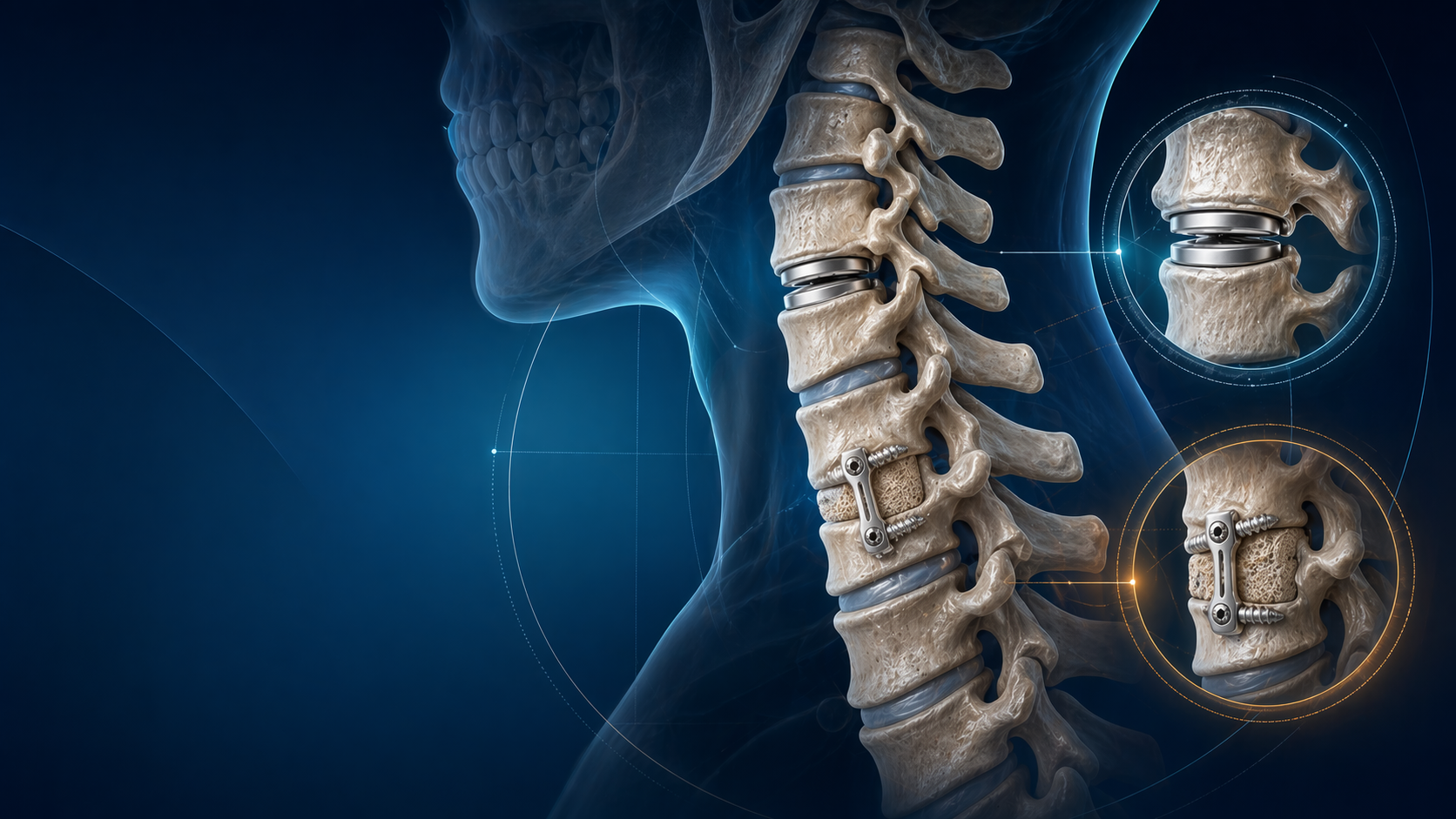
Deformity surgeons utilize different grades of osteotomies to physically shorten the posterior column and hinge the spine backward. The Schwab Osteotomy Classification grades these releases by the anatomical amount of bone resected:
- Grade 1 (PCO): Partial Facetectomy. Removal of the inferior articular process. Yields minimal focal lordosis but provides flexibility.
- Grade 2 (SPO/Ponte): Complete facet resection (superior and inferior) plus removal of the ligamentum flavum, effectively detaching the posterior elements from level to level. Yields ~5-10° of lordosis per level when compressed.
- Grade 3 (PSO - Pedicle Subtraction Osteotomy): A powerful 3-column osteotomy. Involves a wedge resection of the posterior elements, pedicles, and the posterior vertebral body, leaving the anterior cortex intact as a hinge. Yields ~30° of lordosis at a single level. Highly morbid, with significant blood loss.
- Grade 4 (BDBO - Bone-Disc-Bone Osteotomy): Resection of a wedge through the vertebral body and the adjacent disc space.
- Grade 5 (VCR - Vertebral Column Resection): Complete removal of a vertebral segment (body, pedicles, posterior elements) and the adjacent discs. Used for severe, rigid, sharp kyphotic deformities. Yields maximum correction but carries the highest risk of neurological injury.
- Grade 6: Multiple VCRs.
Complications: The Steep Price of Correction
Adult deformity surgery is a massive physiological undertaking. Complication rates in large corrections can exceed 40-50%, and understanding how to prevent them is a primary focus of fellowship training and board examinations.
Proximal Junctional Kyphosis (PJK) and Failure (PJF)
PJK occurs when the spinal segments immediately above the fused construct fail, tumbling forward into kyphosis.
- Radiographic Definition (PJK): A proximal junctional sagittal Cobb angle > 10° and at least 10° greater than the preoperative measurement.
- Clinical Failure (PJF): When PJK is accompanied by a fracture of the Upper Instrumented Vertebra (UIV), pullout of the UIV screws, or neurologic deficit requiring revision surgery.
- Risk Factors: Over-correction of sagittal balance (pushing an elderly patient into too much lordosis), utilizing a rigid PSO, osteoporosis, or stopping the construct at the "apex" of the thoracic kyphosis (e.g., stopping at T7 instead of going to T2 or T3).
- Prevention Strategies:
- Achieving a "soft landing" using transition rods (e.g., changing from 6.0mm CoCr to 5.5mm Titanium at the top of the construct).
- Using sublaminar tethers or hooks at the UIV+1 to dissipate stress.
- Prophylactic vertebroplasty (cement augmentation) of the UIV and UIV+1.
- Strict adherence to age-adjusted alignment targets.
Pseudarthrosis and Implant Failure
- Pathology: Failure of the bone to fuse leads to micromotion. Over time, metal fatigues, resulting in rod fracture (most commonly seen at the lumbosacral junction or across a PSO site).
- Prevention: Robust sacropelvic fixation is absolutely mandatory for long constructs crossing the lumbar spine. You must protect S1 screws with pelvic fixation (Iliac screws or S2-Alar-Iliac [S2AI] screws). Furthermore, employing multi-rod constructs (3-rod or 4-rod techniques across the lumbosacral junction and osteotomy sites) significantly reduces the rate of rod fracture.
Clinical Trap: One of the most common pitfalls for trainees is aiming for the exact same rigid alignment parameters in a 75-year-old as they would in a 25-year-old.
Virgin Lafage and the International Spine Study Group (ISSG) revolutionized our understanding by proving that natural sagittal alignment shifts forward with age. An asymptomatic 75-year-old naturally has a higher PT, a higher SVA, and a larger PI-LL mismatch than a young adult.
If you surgically force a 75-year-old spine into "perfect" Grade 0 alignment (SVA = 0cm, PI-LL = 0°), you will overcorrect them. Their body will fight the rigid construct, almost guaranteeing a catastrophic PJK at the top of the fusion.
For elderly patients, tolerate mild positive balance. A target SVA of 5-7cm and a PI-LL mismatch of 10-15° is often perfectly appropriate and greatly reduces the risk of proximal junctional failure.
Conclusion
Adult Spinal Deformity is fundamentally a three-dimensional biomechanical problem that requires a four-dimensional solution—the fourth dimension being the patient's age and inevitable tissue aging over time. The spinopelvic parameters (PI, PT, SS, LL, and SVA) are not arbitrary numbers to memorize; they are the biomechanical blueprint that dictates a patient's ability to stand, walk, and live without intractable pain.
Mastering the calculation of the PI-LL mismatch, understanding the compensatory role of Pelvic Tilt, and appropriately planning osteotomies while respecting age-adjusted goals is the hallmark of a safe and sophisticated spine surgeon. Keep these principles at the forefront of your mind as you navigate your surgical education and tackle complex viva cases in your upcoming fellowship exams.
Landmark References for Trainees
- Glassman, S. D., et al. (2005). "The impact of positive sagittal balance in adult spinal deformity." Spine. (The landmark paper establishing SVA > 5cm as the primary driver of poor clinical outcomes).
- Schwab, F., et al. (2012). "Adult spinal deformity-postoperative standing imbalance: how much can you tolerate? An overview of key parameters in assessing alignment and planning surgical reconstruction." Spine. (Established the PI-LL < 10° rule).
- Lafage, R., et al. (2016). "Age-Adjusted Alignment Goals Have the Potential to Reduce PJK." Spine. (The critical paper introducing the concept that older patients require less aggressive sagittal correction).
- Terran, J., et al. (2013). "The SRS-Schwab adult spinal deformity classification: assessment and clinical correlations based on a prospective operative and nonoperative cohort." Neurosurgery.
- Roussouly, P., et al. (2005). "Biomechanical analysis of the spino-pelvic organization and adaptation in pathology." European Spine Journal. (Essential reading on the distribution of lumbar lordosis based on sacral slope).
Share this article
Useful for a journal club, study list, or teaching session.