Article summary
How to build a fast, reliable system for reading and describing radiographs and scans under exam pressure.
Educational content is reviewed for source visibility, editorial coherence, and correction readiness.
No individual clinician credential is claimed unless a named person is shown.
Verify before clinical use; this is not medical advice or a substitute for local guidance.
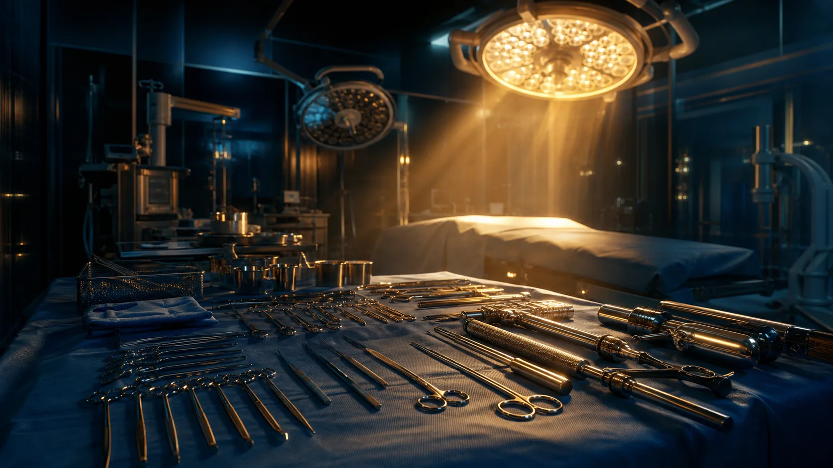
Picture this: you are standing in front of a glowing screen in a silent exam hall. The cursor blinks, the clock is ticking, and you are staring at a complex pelvic radiograph. In a high-stakes orthopaedic exam, simply recognising a fracture is never enough; you must decode the image, construct a blisteringly accurate differential diagnosis, and articulate your findings with the calm precision of a seasoned consultant. Building a fast, reliable system for reading and describing scans under pressure is the difference between passing with confidence and unravelling when the examiner pushes for more detail.
Establish Your Routine and Stick to It
When the adrenaline is pumping in a viva or a written exam, your cognitive bandwidth narrows. The most dangerous thing you can do is abandon your systematic approach in a frantic bid to spot the obvious abnormality. The eye is naturally drawn to the glaring pathology—a grossly displaced distal radius or a pathological hip—but if you comment on that immediately without checking the rest of the film, you reveal a fatal flaw in your radiographic discipline. Examiners are testing your routine as much as your knowledge.
Under exam conditions, you must default to a regimented algorithm. Think of your routine as a mental safety harness. If you don't put it on the moment you look at the film, you are free-climbing without a rope. The routine prevents you from making careless errors, such as missing a subtle fracture because you were blinded by a large effusion, or failing to check the surrounding soft tissues for surgical emphysema.
Your first step should always be to confirm the patient and the image. The moment the image appears, state aloud: "This is a radiograph of [patient detail] taken on [date]." Then, confirm the radiographic view. Is it anteroposterior (AP) or posteroanterior (PA)? Is it a true lateral? Examiners frequently include oblique views dressed up as laterals to test your spatial awareness. Always assess the adequacy of the film. For a long bone, ensure you can see the joint above and the joint below. Never attempt to describe a tibial shaft fracture if the knee and ankle are cut off; you must actively state that the film is inadequate and request a repeat or a wider view.
Decoding Plain Radiographs: The ABCDEF System
For plain films, you need a reliable, sequential framework. The ABCDEF system is a robust favourite that ensures you scour every pixel of the image in a repeatable order. Under exam pressure, moving through this checklist should become entirely automatic.
A: Adequacy and Alignment
Begin with adequacy, checking the penetration, rotation, and anatomical coverage. Next, look at the alignment. Trace the cortical margins of every bone. In joints, check the congruity of the articular surfaces. For the spine, follow the anterior vertebral line, the posterior vertebral line, the spinolaminal line, and the spinous processes. If a line is broken or stepped, you have an alignment issue.
B: Bones
Examine the bones methodically. Start at the top right of the image and sweep your eyes along every cortex, looking for any disruption. Look for changes in bone density—focal areas of sclerosis or lucency. Pay attention to the trabecular pattern. Disruption of the normal cancellous honeycomb architecture is a classic indicator of an occult impaction fracture, such as a subtle tibial plateau split.
C: Cartilage and Joints
Assess the joint spaces. Are they preserved? In an exam setting, a narrowed joint space combined with subchondral sclerosis and osteophytes paints a clear picture of osteoarthritis. Look for intra-articular bodies or gas. A vacuum phenomenon in the joint space is a normal finding, but you must state it confidently so the examiner knows you have not mistaken it for something sinister.
D: Distribution of Disease
If you spot a lesion or a pattern of degeneration, analyse its distribution. Is it affecting the joint symmetrically? Does it involve the metacarpophalangeal joints or the distal interphalangeal joints? This step is crucial for narrowing your differential diagnosis from a generic "arthropathy" to a specific condition like rheumatoid arthritis or gout.
E: Everything Else (Soft Tissues)
Do not ignore the soft tissues. An elbow fat pad sign, a displaced pronator quadratus line, or soft tissue swelling can be the only clue to a devastating occult fracture. Always explicitly state your soft tissue findings, even if they are normal. It proves you have looked.
F: Foreign Bodies and Further Imaging
Finally, check for surgical emphysema, projectiles, or retained metalwork. Then, conclude your description by stating what further imaging you require. If you suspect a scaphoid fracture but the initial films are clear, your final statement should be a request for a CT scan or cross-sectional imaging.
Advanced Cross-Sectional Imaging: Making MRI and CT Work for You
When the examiner transitions to magnetic resonance imaging (MRI) or computed tomography (CT), the rules of the game change. You are no longer looking at shadows on a flat plane; you are navigating a three-dimensional digital map. The most critical mistake trainees make with cross-sectional imaging is trying to look at everything at once.
When presented with an MRI, never attempt to interpret it without first identifying the sequences and the plane of the scan. You will quickly drown in a sea of pixels if you do not know what the different shades of grey represent.
Know Your Sequences
You must instantly distinguish between T1-weighted, T2-weighted, and PD (proton density) sequences. Do not fall into the trap of relying solely on the tiny text on the scan, which is often illegible on a projector or absent in a viva booklet.
Instead, rely on fluid. Water is dark on T1 and bright on T2. Fat is bright on T1 and darker on T2, especially with fat suppression techniques. Bone marrow follows fat: it is bright on T1. If you see a dark marrow lesion on a T1 sequence adjacent to a normal bright marrow background, you are looking at marrow oedema, marrow replacement, or a tumour.
A practical shortcut for spine imaging is the "match" principle. If you see a lesion on a sagittal image, flick between the T1 and T2 sequences. If the signal intensity matches the cerebrospinal fluid (dark on T1, bright on T2), you are likely dealing with a fluid-filled lesion, such as a Tarlov cyst or synovial cyst. If it matches fat, it is a benign haemangioma.

The Art of the Verbal Description
Your eyes might see a posterior wall fracture, but if your mouth says "a break in the back bit of the hip socket", you will fail the station. The verbal description of imaging is a core surgical skill, and examiners are listening for precise, unambiguous language. You must narrate the film as if you are guiding a blindfolded colleague through a minefield.
Always start laterally and move medially. Describe the proximal fragment and its relationship to the distal fragment. State the displacement, the angulation, and the shortening.
Describing Displacement
Use standard anatomical terms. Do not say the bone has "moved over". Say it is "displaced medially by fifty percent of the shaft width". If there is no displacement, confidently state that the fracture is "in anatomical alignment".
Describing Angulation
This is where countless candidates stumble. The direction of the fracture apex dictates the direction of the angulation. If the apex of the fracture points anteriorly, the fracture is angulated anteriorly. Do not look at the direction the distal fragment is pointing; look at the point of the angle. Use the terms varus and valgus accurately. Varus means the distal segment is angled towards the midline; valgus means it is angled away from the midline.
Describing Rotation
Rotational deformities are notoriously difficult to assess on a single two-dimensional film. In the lower limb, look at the profile of the femoral condyles or the malleoli, and compare the position of the proximal and distal fragments. If you cannot definitively assess rotation, state that clearly: "Rotation cannot be fully assessed on this single AP view, and clinical correlation is required."
Naming the Fracture
Once you have described the specifics, tie it together with a recognised eponymous or classification name, but only if you are absolutely certain. Naming a fracture shows a high level of knowledge, but naming it incorrectly shows dangerous ignorance. If the fracture is a displaced, comminuted, intra-articular fracture at the distal radius with dorsal angulation, confidently declare it a "dorsally angulated, intra-articular distal radius fracture".
The "Aunt Minnie" vs. The Pattern Recognition Dance
In radiology, an "Aunt Minnie" is a condition with a presentation so pathognomonic that you can diagnose it at a glance. If your Aunt Minnie walks into a room, you do not need a genetic test to know it is her; you just know. In orthopaedic exams, spotting an Aunt Minnie early can secure you a pass mark instantly, but leaning on them is a dangerous game.
Examiners expect you to reach your diagnosis through a deductive pattern recognition dance, not by guessing. If you see a lytic lesion in the distal femur of a young patient, do not instantly guess a giant cell tumour. Walk the examiner through your thought process.
First, state the margins: are they well-defined or permeative? Permeative margins suggest an aggressive process, such as a malignancy or infection. Second, note the zone of transition. A narrow zone of transition implies a benign, slow-growing process, whereas a wide zone of transition implies a malignant, rapidly growing process. Third, look for matrix calcification. Chondroid matrix looks like "rings and arcs" or "popcorn", whereas osteoid matrix looks like "cloud-like" or "elephant trunk" densities.
By methodically analysing the margins, the transition zone, and the matrix, you guide the examiner to your conclusion alongside you. Even if you ultimately misdiagnose the lesion as an aneurysmal bone cyst instead of a fibrous dysplasia, your systematic approach will earn you the marks for safe, logical surgical reasoning. Examiners fail candidates who guess and get it wrong; they pass candidates who reason and get it wrong, provided the logic was sound.
Handling the Viva Pressure Cooker
The viva voce examination is a uniquely stressful psychological beast. You are sitting opposite a consultant who is showing you an image specifically chosen for its complexity or ambiguity. The pressure to speak quickly and fill the silence can lead to rambling, errors, and the utter destruction of your carefully rehearsed system.
The most valuable tool in your viva arsenal is silence. When the image appears, take a breath. Two full seconds of silence feels like an eternity to you, but to the examiner, it simply looks like a careful, considered surgeon assessing a film. Use that pause to run through the first few steps of your routine before you open your mouth.
The Power of the Direct Statement
Examiners frequently present images that are ambiguous or require a specific management pathway. When asked for your definitive answer, do not hedge. "I think this might be..." is weak. "This is a..." is strong. Even if you are uncertain, stating your best guess with conviction allows the examiner to correct you and move on, or to agree and award you the marks. Hedging forces the examiner to prompt you repeatedly, wasting precious time.
If the examiner asks a specific question, answer it directly. Do not launch into your ABCDEF routine if the examiner points to a lesion and asks, "What does this look like?" Answer the question first, then offer to describe the rest of the film. "This appears to be a sclerotic lesion in the proximal femur. Would you like me to describe the rest of the film?" This demonstrates excellent examination technique and respect for the examiner's time.
Managing the Follow-up Question
Be prepared for the "what next" question. Once you have identified a fracture or a lesion, the examiner will immediately ask for your management plan. Have a standard framework ready. Start with the patient: are they haemodynamically stable? Then address the limb: is the neurovascular status intact? Then address the imaging: do you need a CT scan to further characterise this? Finally, address the definitive management, be it conservative care, splintage, or surgical fixation.
Building Your Personal Image Bank
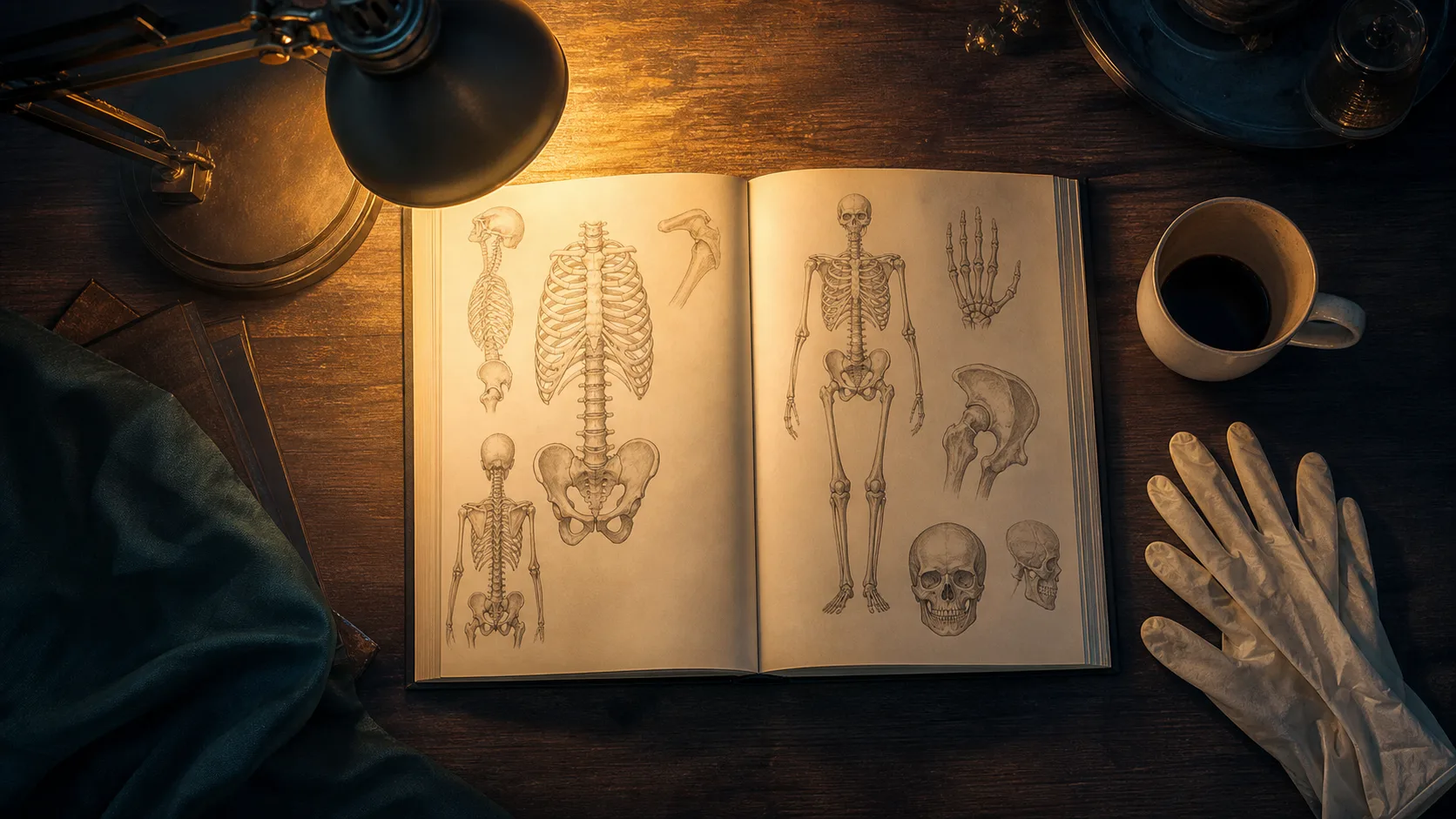
No amount of theoretical reading can substitute for the raw visual mileage required to master orthopaedic imaging. Your brain is a pattern recognition engine, and you need to feed it a vast, varied diet of radiographs, CTs, and MRIs to build a reliable internal database.
Do not rely solely on the images provided in your core orthopaedic textbooks. These are often classic, textbook-perfect examples that bear little resemblance to the ambiguous, slightly rotated, or poorly penetrated films you will face in an exam.
Start collecting your own image bank. Whenever you see an interesting case in clinic, on the ward, or in trauma theatre, screenshot the imaging (ensuring you strictly remove all patient identifiers and adhere to your local hospital's data governance policies). Save these images into a structured folder system on your computer. Create categories for trauma, arthropathies, paediatric conditions, and tumours.
Once a week, sit down with your image bank and run a rapid-fire review session. Force yourself to describe the film aloud, exactly as you would in a viva. Time yourself. If you spend more than ten seconds identifying the sequence of an MRI or the angulation of a fracture, flag the image for further review.
Leveraging Digital Platforms for Daily Practice
In addition to your personal collection, utilise the wealth of established digital resources designed for radiology and orthopaedic revision. There are several high-quality, widely recognised online platforms that provide extensive databases of annotated orthopaedic images. These platforms allow you to scroll through hundreds of cases, test your diagnostic skills, and compare your descriptions against those of expert radiologists and surgeons.
Integrate these platforms into your daily revision schedule. Treat them like spaced-repetition flashcards. Exposure to a massive volume of normal, variant, and abnormal imaging will sharpen your eye and accelerate your interpretation speed. When you sit your exam, you want the act of diagnosing to feel as natural and automatic as riding a bike.
Mastering orthopaedic imaging is ultimately a test of discipline, vocabulary, and visual experience. Stick relentlessly to your routine, articulate your findings with anatomical precision, and trust the systematic process you have built over thousands of practice films. When the screen lights up in that exam hall, you will not just be looking at shadows—you will be commanding them.
Share this article
Useful for a journal club, study list, or teaching session.