Article summary
A comprehensive review of the great debate in arthroplasty. Mechanical Alignment vs Kinematic Alignment vs Restricted Kinematic Alignment. Evidence, techniques, and the robotic revolution.
Educational content is reviewed for source visibility, editorial coherence, and correction readiness.
No individual clinician credential is claimed unless a named person is shown.
Verify before clinical use; this is not medical advice or a substitute for local guidance.
Kinematic vs Mechanical Alignment in TKA: The Great Arthroplasty Debate
For nearly four decades, the dogma of Total Knee Arthroplasty (TKA) was elegantly simple: "Make it straight." The survivorship of the implant was the absolute king, and mechanical alignment was the unshakeable law governing orthopaedic surgery training. But as implant materials improved—particularly with the advent of highly cross-linked polyethylene—and catastrophic wear ceased to be the primary mode of failure, surgeons began confronting a more nuanced and frustrating problem: "Why are 20% of my patients with radiographically 'perfect' X-rays still deeply unhappy?"
This fundamental question ignited what is arguably the most significant debate in modern joint replacement surgery: Mechanical Alignment (MA) versus Kinematic Alignment (KA). For trainees preparing for fellowship exams, understanding this paradigm shift is no longer optional; it is a critical requirement. Examiners expect a sophisticated grasp of not just how to cut the bone, but why we cut the bone the way we do.
This comprehensive guide dissects the philosophies, the landmark evidence, the biomechanical rationale, and the emergence of restricted alignment concepts that serve as the new middle ground in surgical education.
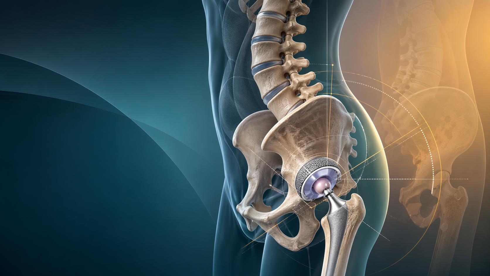
Visual Element: Resection plane visualizer. A split-screen diagram. Left: Mechanical Alignment (Cuts perpendicular to mechanical axis, large soft tissue release). Right: Kinematic Alignment (Cuts parallel to joint line, soft tissue preservation).
The Mechanical Axis Dogma: History and Rationale
The Philosophy: Pioneered and popularized by John Insall in the 1980s, the primary goal of Mechanical Alignment is to create a neutral hip-knee-ankle (HKA) axis, effectively aiming for 0° of coronal alignment.
The systematic approach is rigid and standardized:
- The Femur: Cut perpendicular (90°) to the mechanical axis of the femur. Because the anatomic axis of the femur typically sits at a valgus angle to the mechanical axis, this usually dictates a distal femoral cut of 5° to 7° of valgus relative to the anatomic axis.
- The Tibia: Cut perpendicular (90°) to the mechanical axis of the tibia.
- Gap Balancing: Because these systematic cuts often ignore the patient's native joint line obliquity, the soft tissues (specifically the collateral ligaments) must be incrementally released until the flexion and extension gaps are perfectly rectangular and equal.
The Logic of the Era: In the era of conventional, easily oxidizable polyethylene, eccentric loading was a death sentence for a knee replacement. A neutral limb distributes the mechanical load evenly across the medial and lateral compartments of the tibial polyethylene tray, minimizing edge-loading, sub-surface delamination, and catastrophic premature wear. For survival, mechanical alignment was an absolute necessity.
The Problem: Humans Are Not Robots The inherent flaw in universal mechanical alignment is that it assumes neutral alignment is the "normal" state for all humans. It is not.
Landmark research by Bellemans et al. (2012) on Constitutional Varus demonstrated that up to 32% of men and 17% of women have a native, healthy alignment of 3° of varus or more. Forcing a patient who has lived 60 years in 5° of constitutional varus into 0° of mechanical neutral is an architectural violation. It requires:
- Significantly changing their native joint line obliquity.
- Extensively releasing the Medial Collateral Ligament (MCL) to balance the newly created, non-anatomical gaps.
- Altering the native patellofemoral tracking kinematics.
Kinematic Alignment (KA): The Restoration Philosophy
The Philosophy: Championed largely by Stephen Howell, Kinematic Alignment entirely discards the pursuit of a universal 0° HKA axis. Instead, KA aims to resurface the knee, meticulously restoring the patient's precise pre-arthritic constitutional anatomy and kinematic axes.
KA is built on restoring the three fundamental kinematic axes of the knee:
- Primary Transverse Axis: The axis around which the tibia extends and flexes on the femur.
- Secondary Transverse Axis: The axis around which the patella extends and flexes on the femur.
- Longitudinal Axis: The axis around which the tibia internally and externally rotates on the femur.
The Technique (Calipered KA): Rather than measuring angles relative to the center of the hip or ankle, the surgeon removes a thickness of bone and cartilage exactly equal to the thickness of the implant components being inserted.
- If the distal femoral condyle of the implant is 9mm thick, the surgeon resects exactly 9mm of distal femoral bone/cartilage (compensating for 2mm of estimated cartilage wear on the arthritic side).
- The tibial cut is made to precisely match the patient's native varus or valgus and slope.
- Crucially, no ligament releases are performed.
The Logic: If you place the prosthetic joint surfaces back into the exact spatial orientation where the native joint lived before the onset of osteoarthritis, the native ligaments will be perfectly tensioned without any need for surgical release. The knee will theoretically feel "normal" because the kinematics—including physiologic roll-back and screw-home rotation—are preserved.
Mechanical alignment changes the anatomy to fit the implant. Kinematic alignment changes the implant position to fit the anatomy. In pure KA, the soft tissue envelope is the master, and the bone cuts are the servant.
Restricted Kinematic Alignment (rKA): The Pragmatic Middle Ground
While pure KA is philosophically attractive, it carries a distinct biomechanical risk. Reproducing severe native deformities—for instance, placing a tibial tray in 12° of varus to match a patient's extreme constitutional bowing—might perfectly preserve their ligaments, but it forces the implant into a hostile biomechanical environment. Extreme varus baseplates risk catastrophic medial edge loading, polyethylene failure, and aseptic loosening.
Restricted Kinematic Alignment (rKA), pioneered by Vendittoli and others, is the pragmatic compromise. It is currently the most rapidly adopted philosophy in global orthopaedic surgery training, driven heavily by the adoption of robotics.
The rKA Protocol:
- Start with KA Principles: Aim to resurface the knee and preserve the native joint line obliquity.
- Apply The Guardrails (The "Safe Zone"):
- The overall Arithmetic HKA must fall within ± 3° (some allow up to ± 5°) of neutral.
- The joint line obliquity must be maintained within 5° of neutral relative to the floor.
- The tibial baseplate varus should typically not exceed 5°.
- Adjust if Necessary: If the patient's native anatomy falls outside these guardrails, the surgeon modifies the bone cuts just enough to bring the alignment inside the safe zone boundaries, but no further. Once inside the boundary, minimal, targeted soft tissue balancing may be required.
Visual Element: The CPAK (Coronal Plane Alignment of the Knee) Classification matrix. A grid showing the 9 phenotypes of knee alignment based on Joint Line Obliquity and Arithmetic HKA.
Understanding the CPAK Classification
For fellowship exam preparation, understanding MacDessi's CPAK (Coronal Plane Alignment of the Knee) classification is mandatory. It categorizes knees into 9 phenotypes based on two variables:
- Arithmetic HKA (aHKA): Valgus, Neutral, or Varus.
- Joint Line Obliquity (JLO): Apex Distal, Neutral, or Apex Proximal.
The most common phenotype is Type I (Varus aHKA with Apex Distal JLO). The CPAK classification provides a common language for surgeons to discuss how a specific knee phenotype should be reconstructed, shifting the conversation away from simplistic "varus vs valgus" binaries.
The Robotic Revolution: Enabling Precision
It is vital to understand that pure KA and rKA are remarkably difficult to execute consistently with standard manual instruments. Traditional intramedullary and extramedullary jigs possess an inherent error margin of 1° to 3°. Furthermore, calipered manual KA relies on the surgeon accurately estimating the thickness of worn cartilage, which is highly subjective.
Robotics (such as MAKO, ROSA, and VELYS) have served as the ultimate enabler of the kinematic philosophy.
- Sub-Millimeter Precision: A robotic system allows a surgeon to plan a 3.5° varus tibial cut and execute it with an accuracy of 0.5mm and 0.5°. This prevents the accidental creation of extreme outlier alignments.
- Predictive Virtual Balancing: This is the true power of robotics. Systems allow the surgeon to stress the joint, map the soft tissue tension through the entire arc of motion, and virtually adjust the implant position on a screen before a single bone cut is made. If placing the implant in purely kinematic alignment results in an unacceptable gap imbalance, the surgeon can pivot to rKA, adjust the virtual cuts by 1°, and instantly see the predicted effect on ligament tension.
The Evidence Corner: What Does the Literature Say?
When defending an alignment philosophy in a fellowship exam, you must cite the literature. Here is the current landscape:
| Aspect | Mechanical Alignment (MA) | Kinematic Alignment (KA) |
|---|---|---|
| Survivorship | The indisputable gold standard. Excellent, proven long-term data (>20 years) from joint registries worldwide. | Comparable at mid-to-long term (10 years, Howell et al.). The feared epidemic of "catastrophic early failure" due to varus baseplates has not materialized in the literature. |
| Function (PROMs) | Reliable pain relief, but burdened by the persistent ~20% patient dissatisfaction rate. | Multiple RCTs and meta-analyses show statistically significant improvements in Patient Reported Outcome Measures (PROMs), higher "Forgotten Joint Scores," and slightly better terminal flexion. |
| Complications | Mid-flexion instability if medial releases are overly aggressive. Stiffness from over-stuffing gaps. | Potential for patellofemoral tracking issues if severe valgus deformities are left uncorrected. Theoretical risk of baseplate subsidence in massive BMI patients with extreme varus components (hence the shift to rKA). |
Key Study for Exams: Young et al. (The KA TKA Trial)
- A robust, double-blind, randomized controlled trial comparing MA versus KA.
- Result: The study found no clinically significant difference in the primary outcome measure (Oxford Knee Score) at 2 years. However, the KA group demonstrated statistically better range of motion and functional scores in early post-operative phases. This paper is frequently cited by MA purists to argue that KA does not offer a "night and day" difference, while KA advocates point to the ROM benefits.
Conclusion: The Paradigm Shift in Orthopaedic Surgery Training
We are witnessing a monumental paradigm shift in arthroplasty. The specialty is moving away from "Systematic Surgery" (forcing every distinct patient into a single geometric mold) and toward "Personalized Surgery" (tailoring the reconstruction to match the patient's unique phenotype).
- For the patient with natively straight legs (CPAK Type V): Mechanical Alignment and Kinematic Alignment achieve the exact same bone cuts.
- For the patient with constitutional varus: Kinematic and Restricted Kinematic Alignment offer a more physiological, joint-preserving reconstruction with significantly less soft tissue trauma.
As technology improves and long-term registry data for rKA matures, the debate will continue to evolve. For the orthopaedic trainee, mastering the nuances of this debate is essential for both exam success and delivering superior patient outcomes.
Related Topics for Further Review:
- Robotics and Navigation Systems in Arthroplasty
- Unicompartmental Knee Arthroplasty (The ultimate purely kinematic knee replacement)
- Polyethylene Wear Mechanisms and Material Sciences
- Diagnosing and Managing TKA Instability (Flexion vs Extension Gap mismatch)
- The Patellofemoral Joint in TKA: Resurfacing vs Retention
Share this article
Useful for a journal club, study list, or teaching session.