Article summary
A complete textbook-level review of scaphoid anatomy, classification, imaging strategies, and management of the 'Ferrari' of carpal bones.
Educational content is reviewed for source visibility, editorial coherence, and correction readiness.
No individual clinician credential is claimed unless a named person is shown.
Verify before clinical use; this is not medical advice or a substitute for local guidance.
The scaphoid (from the Greek skaphos, meaning "boat") is unequivocally the keystone of the carpus. It acts as the crucial mechanical link bridging the proximal and distal carpal rows, coordinating their complex multi-planar motion like an intricate mechanical tie-rod. However, this critical biomechanical role, combined with a notoriously precarious retrograde blood supply, makes the scaphoid one of the most problematic and unforgiving bones in the human body when injured.
Often referred to as the "Ferrari" of the carpus—highly tuned, expensive to fix, and disastrous when crashed—the scaphoid demands absolute respect from any clinician. For those in orthopaedic surgery training, mastering the scaphoid is non-negotiable. It represents a high-yield, guaranteed topic in every major fellowship exam preparation syllabus, including the FRACS, FRCS, and ABOS.
This guide serves as a comprehensive, textbook-level reference for the functional anatomy, diagnosis, classification, operative management, and salvage strategies for scaphoid fractures.
Visual Element: A detailed 3D exploded view of the carpus, highlighting the scaphoid's articulation with the radius, lunate, capitate, trapezium, and trapezoid.
1. Functional Anatomy and Biomechanics: The Foundation
To understand scaphoid fractures, you must fundamentally understand its unique anatomy. The scaphoid is a remarkable structure, covered in articular cartilage over 80% of its surface area. It articulates with five bones: the radius proximally, the trapezium and trapezoid distally, and the capitate and lunate ulnarly. This massive articular surface leaves very little bare bone for ligamentous attachment or, crucially, vascular entry.
The Blood Supply: A Retrograde Problem
The vascularity of the scaphoid is the single most important physiological factor determining the prognosis and union rate of a fracture. The landmark anatomical studies by Gelberman and Menon dictate our modern understanding of this fragile network.
- The Primary Supply: Approximately 70-80% of the scaphoid—including the entire proximal pole and the majority of the waist—is supplied by the dorsal carpal branch of the radial artery.
- The Entry Point: These vessels enter the bone through a narrow non-articular ridge on the dorsal aspect, located at the level of the scaphoid waist and extending to the distal pole.
- The Retrograde Flow: Once inside the bone, the intraosseous blood flows in a strictly retrograde fashion (from distal to proximal) to perfuse the proximal pole.
- The Volar Supply: A smaller volar branch from the radial artery enters the distal tubercle, supplying only the distal 20-30% of the bone.
- Clinical Consequence: A fracture propagating through the waist effectively acts as a guillotine, severing the intraosseous vascular conduit to the proximal pole. The proximal fragment immediately becomes ischemic or avascular, relying entirely on creeping substitution and neovascularization across the fracture site to survive. This precise anatomical quirk explains why displaced proximal pole fractures carry an almost 100% rate of avascular necrosis (AVN) without rigid fixation.
Biomechanics: The Tie-Rod of the Carpus
The scaphoid's movement is inextricably linked to wrist motion, specifically during the "dart-throwing motion" (radial extension to ulnar flexion) which is essential for most activities of daily living.
- The Flexion Moment: The scaphoid has a natural, intrinsic tendency to flex volarly due to strong compressive loading from the trapezium and trapezoid during grip and wrist extension.
- The Scapholunate Link: It is tethered to the lunate via the stout scapholunate interosseous ligament (SLIL). While the scaphoid wants to flex, the triquetrum wants to extend, and the lunate sits in the middle, balanced by these opposing forces.
- The Humpback Deformity: When the scaphoid fractures through the waist, this delicate balance is destroyed. The distal fragment flexes unrestrained (palsy-walsy), while the proximal fragment extends along with the lunate (creating a Dorsal Intercalated Segment Instability, or DISI, posture). This volar collapse at the fracture site creates the classic "humpback" deformity. Left uncorrected, this deformity alters the radiocarpal and midcarpal contact mechanics, inevitably initiating a predictable cascade of degenerative arthritis.
2. Epidemiology and Mechanism of Injury
Scaphoid fractures are overwhelmingly the most common carpal fractures, accounting for 60-70% of all carpal bone breaks.
- Patient Profile: The classic demographic is a young, active male between 15 and 30 years of age. They are common in contact sports, snowboarding, and high-energy trauma. In pediatric populations, they are rare due to the late ossification of the scaphoid, and in the elderly, distal radius fractures typically fail first.
- Mechanism of Injury: The quintessential mechanism is a Fall on an Outstretched Hand (FOOSH). Biomechanical studies demonstrate that a scaphoid fracture typically requires the wrist to be in extreme extension (greater than 95 degrees) combined with radial deviation. In this position, the scaphoid impacts squarely against the dorsal rim of the radius, creating a massive bending moment across its waist.
- Associated Injuries: In any high-energy FOOSH, a scaphoid fracture should not be viewed in isolation. Always have a high index of suspicion for concomitant injuries such as perilunate fracture-dislocations (trans-scaphoid perilunate dislocations), distal radius fractures, radial head fractures, and ligamentous injuries (especially SLIL tears).
3. Clinical Evaluation: Trusting Your Hands
The diagnosis of a "Clinical Scaphoid Fracture" commands absolute respect in the emergency department and the fracture clinic. X-rays taken immediately post-injury can be falsely negative in up to 15-20% of cases due to a lack of fracture site resorption. Therefore, a high degree of clinical suspicion dictates mandatory prophylactic treatment.
The "Triad" of Clinical Signs: A thorough clinical examination relies on three primary provocative tests. When all three are positive, the probability of a scaphoid fracture is exceedingly high.
- Anatomical Snuffbox Tenderness: (Sensitivity ~90%, Specificity ~40%). The anatomical snuffbox is bordered by the EPL dorsally and the EPB/APL volarly. To test accurately, palpate deep within the snuffbox with the patient's wrist actively placed in ulnar deviation. Ulnar deviation extracts the scaphoid from the radial fossa, making the waist palpable.
- Scaphoid Tubercle Tenderness: (Sensitivity ~87%, Specificity ~57%). Palpate volarly at the proximal wrist crease, just radial to the FCR tendon. Exquisite tenderness here often correlates with waist or distal pole fractures.
- Axial Compression / Telescoping: (Sensitivity ~80%). Apply a longitudinal compressive force down the axis of the thumb metacarpal towards the radius. This loads the trapeziometacarpal joint, driving the trapezium into the scaphoid, which compresses the fracture site and elicits deep pain.
Clinical Pearl: Pain elicited on resisted supination and pronation is an underutilized sign that can strongly indicate either an occult scaphoid fracture or a significant scapholunate ligament injury due to the torque applied across the carpus.
4. Imaging Strategy: Beyond the Standard Views
A standard AP and lateral wrist radiograph is grossly insufficient to rule out a scaphoid fracture. The complex, curved, three-dimensional shape of the scaphoid means that its longitudinal axis lies at a 45-degree angle to both the sagittal and coronal planes of the wrist.
Radiography: The Scaphoid Series
You must explicitly request a dedicated Scaphoid Series:
- PA and Lateral: To assess overall carpal alignment (Gilula's arcs) and look for DISI/VISI.
- The Ziter View (Scaphoid View): This is a PA view with the wrist in ulnar deviation and the X-ray tube angled 20° to 30° toward the elbow. Ulnar deviation brings the scaphoid into extension, parallel to the cassette, while the tube angulation shoots straight down the anatomical axis of the bone, beautifully profiling the waist.
- Semi-Pronated Oblique (45°): Excellent for visualizing the waist and evaluating for subtle cortical step-offs.
Advanced Imaging Indications in Surgical Education
- MRI (Magnetic Resonance Imaging): The undisputed Gold Standard for acute occult fractures. It approaches 100% sensitivity and specificity. T1 images show a dark fracture line; T2/STIR images demonstrate diffuse hyperintense bone marrow edema. MRI should ideally be performed within 3-5 days of injury if X-rays are negative but clinical suspicion remains. It also invaluable for assessing proximal pole vascularity and detecting associated ligament tears (SLIL/LTIL).
- CT (Computed Tomography): The modality of choice for assessing displacement, angulation, and union. CT is mandatory for preoperative planning. You must evaluate the scans in the true longitudinal and sagittal planes of the scaphoid (not the wrist). Look for an intrascaphoid angle >35° (indicating humpback deformity) or a cortical step-off >1mm. CT is also the best tool at 12 weeks to definitively confirm bony bridging and union.
5. Classification Systems: Guiding Management
Understanding classification is vital not just for fellowship exam preparation, but for making sound clinical decisions regarding stability and operative indications.
Mayo Classification (Anatomical Location)
- Distal Third (10-20%): Generally stable, excellent blood supply, high union rates.
- Middle Third / Waist (70-80%): The most common. Prone to displacement and delayed union.
- Proximal Third (10-20%): Extremely precarious blood supply. High rate of AVN. Almost all require operative fixation regardless of displacement.
Russe Classification (Direction of Fracture Line)
Based on the angle of the fracture relative to the longitudinal axis of the scaphoid. It dictates biomechanical stability.
- Horizontal Oblique: The fracture line is perpendicular to compressive forces across the carpus. Very stable. High union rate with casting.
- Transverse: Subject to a combination of shear and compressive forces. Potentially unstable.
- Vertical Oblique: The fracture line is parallel to the longitudinal compressive forces of the carpus. This creates massive shear forces that cause the fracture to slide. Inherently unstable and notoriously difficult to fix with a single screw.
Herbert Classification (Clinical & Radiographic Type)
The most comprehensive system used in modern orthopaedic surgery training.
- Type A: Stable Acute Fractures (A1 Tubercle, A2 Incomplete waist).
- Type B: Unstable Acute Fractures (B1 Distal oblique, B2 Complete/Displaced waist, B3 Proximal pole, B4 Trans-scaphoid perilunate fracture-dislocations).
- Type C: Delayed Union (radiographic widening, cyst formation after 6-8 weeks).
- Type D: Established Non-Union (sclerotic margins, pseudoarthrosis).
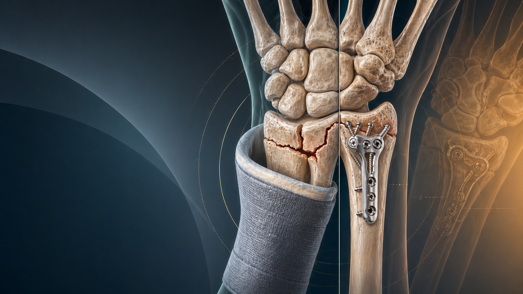
6. Treatment Strategies: Cast vs. Cut
The management of scaphoid fractures has evolved significantly over the last two decades, leaning heavily toward early operative fixation for active patients to minimize the profound socioeconomic impact of prolonged casting.
Non-Operative Management
- Indication: Strictly limited to acute, genuinely non-displaced (less than 1mm step-off on CT), stable fractures (Herbert A2, Mayo distal third/waist).
- Technique: Cast immobilization.
- Duration: Typically 8-12 weeks, but guided purely by radiographic union, not arbitrary timelines.
- The Casting Debate (Colles vs. Spica): Historically, a long-arm thumb spica was mandated to prevent forearm rotation. However, robust modern evidence (including data surrounding the SWIFFT trial) suggests that a simple below-elbow, short-arm cast (leaving the thumb entirely free) yields identical union rates for non-displaced waist fractures compared to a spica cast, with significantly better patient compliance and less post-cast stiffness. However, many conservative surgeons still prefer a short-arm thumb spica for proximal pole fractures to theoretically minimize micromotion from the thumb metacarpal.
- Clearance: A patient is only "cleared" when a CT scan demonstrates bridging trabecular bone across at least 50% of the fracture site cross-section. A pain-free clinical exam is unreliable.
Operative Management
- Absolute Indications:
- Displacement > 1mm or step-off.
- Any proximal pole fracture (due to high AVN risk).
- Vertical oblique fracture patterns (Russe).
- Intrascaphoid angle > 35° (Humpback deformity).
- Associated perilunate dislocation.
- Relative Indications: Patient preference for rapid return to work/sport, delayed presentation.
- Hardware: The gold standard is the Headless Compression Screw (e.g., Acutrak, Herbert screw). These screws are designed with variable pitch (leading threads are wider than trailing threads) to generate compression as the screw is seated, and they bury entirely beneath the articular cartilage to prevent joint destruction.
Surgical Approaches: The Orthopaedic Trainee's Dilemma
Choosing the correct approach is a frequent fellowship exam viva topic.
-
Volar Approach (Modified FCR Approach):
- Best for: Waist and Distal Pole fractures, and critically, for correcting a Humpback deformity.
- Advantage: Safely avoids the crucial dorsal blood supply entering the dorsal ridge. It allows excellent access to pack bone graft volarly to prop open a collapsed fracture.
- Technique: The incision is made directly over the Flexor Carpi Radialis (FCR) tendon. The sheath is incised, the tendon retracted ulnarly, and the floor of the sheath is incised to expose the radiocarpal capsule. The internervous plane is safe, utilizing the interval between the FCR (Median nerve) and the Radial Artery.
- Trajectory: From distal to proximal. Often requires extreme wrist extension and removal of a small lip of the trapezium to achieve the correct starting point down the central axis of the scaphoid.
-
Dorsal Approach:
- Best for: Proximal Pole fractures.
- Advantage: The proximal pole is a tiny, hemispherical fragment. Approaching it dorsally allows the surgeon to visualize the articular surface and place the screw precisely perpendicular to the fracture line, directly down the central axis of the scaphoid. Attempting to hit a small proximal pole fragment from a volar start is technically demanding and prone to eccentric screw placement.
- Risk: The dissection goes directly through the dorsal capsule and risks iatrogenic injury to the fragile dorsal carpal branch of the radial artery. Meticulous, limited capsulotomy is required.
- Technique: Usually through the 3rd and 4th extensor compartment interval (EPL and EDC).
Visual Element: Surgical illustration comparing the Volar vs Dorsal screw trajectory, overlaying the central axis of the scaphoid.
7. Complications and Salvage: When Things Go Wrong
Avascular Necrosis (AVN)
- Incidence: Occurs in 30-40% of proximal pole fractures, and nearly 100% of displaced proximal pole fractures without surgery.
- Radiographic Appearance: On X-ray, the proximal pole appears densely sclerotic (white) relative to the surrounding osteopenic carpal bones (which undergo disuse osteopenia from casting).
- MRI Diagnosis: The most sensitive modality. AVN is confirmed by a complete loss of signal intensity in the proximal pole on both T1 and T2 weighted images, indicating absent marrow perfusion.
Scaphoid Non-Union
- Definition: Failure of the fracture to demonstrate progressive healing by 6 months.
- Consequence: A scaphoid non-union is not a benign condition. The persistent humpback deformity and asynchronous movement of the carpal rows invariably lead to altered joint kinematics and degenerative wear.
- Treatment Algorithms:
- No AVN, Minimal Deformity: Percutaneous screw fixation or in situ inlay bone grafting (Matti-Russe technique using distal radius cancellous autograft).
- No AVN, Humpback Deformity: Volar wedge bone grafting (iliac crest autograft) to restore length and correct the intrascaphoid angle, combined with screw or K-wire fixation.
- AVN Present: The proximal pole is dead. Standard non-vascularized bone grafts will fail. You must bring a new blood supply to the bone. Options include:
- 1,2 ICSRA Graft (Zaidemberg Graft): A pedicled vascularized bone graft taken from the dorsal radius, based on the 1,2 Intercompartmental Supraretinacular Artery.
- Free Vascularized Medial Femoral Condyle (MFC) Graft: The modern gold standard for difficult non-unions with a necrotic proximal pole and significant bone loss. It provides robust, highly osteogenic corticocancellous bone with an independent vascular pedicle anastomosed to the radial artery.
SNAC Wrist (Scaphoid Non-Union Advanced Collapse)
SNAC wrist is the tragic, end-stage natural history of an untreated scaphoid non-union. The altered mechanics cause specific, sequential articular wear. Memorizing these stages is crucial for surgical education and exam prep.
- Stage I: Arthritis localized solely to the radial styloid and the distal pole of the scaphoid.
- Treatment: Radial styloidectomy and scaphoid fixation/grafting.
- Stage II: Arthritis progresses to the scaphocapitate joint (the midcarpal joint).
- Treatment: Proximal Row Carpectomy (PRC) or Scaphoid Excision and 4-Corner Fusion (capitate, hamate, lunate, triquetrum).
- Stage III: Periscaphoid arthritis, extending to the lunocapitate joint.
- Treatment: PRC is contraindicated here because the head of the capitate is degenerated (it cannot articulate with the lunate fossa of the radius). 4-Corner Fusion is required.
- Stage IV: Pancarpal arthritis involving the entire radiocarpal and midcarpal joints.
- Note: The radiolunate joint is miraculously preserved until the very end because the lunate and the lunate fossa are spherically congruent, preventing abnormal shear forces.
- Treatment: Total Wrist Arthrodesis (Fusion) or Total Wrist Arthroplasty (Replacement in low-demand patients).
8. Summary: The Golden Rules
The scaphoid is an unforgiving bone. A missed diagnosis, a poorly applied cast, or an inadequately reduced fracture can sentence a young, healthy patient to a lifetime of chronic pain, restricted motion, and premature osteoarthritis.
For the orthopaedic trainee, follow these absolute tenets:
- Suspect it relentlessly: Treat any patient with snuffbox or scaphoid tubercle tenderness after a FOOSH as having a scaphoid fracture until an MRI or a 2-week follow-up X-ray definitively proves otherwise.
- Respect the vascularity: Always keep Gelberman's retrograde blood supply in mind. It dictates your approach, your prognosis, and your urgency.
- Fix the bad ones: Have a low threshold to operate on proximal pole fractures, any fracture with >1mm displacement, or fractures presenting with a DISI/humpback deformity.
- Demand proof of union: Never discharge a patient based solely on a lack of pain. Demand a fine-cut CT scan aligned to the scaphoid axis to prove bridging trabecular bone before removing them from immobilization or clearing them for heavy labor.
Related topics
Share this article
Useful for a journal club, study list, or teaching session.