Ilioinguinal approach · three-window technique · Judet-Letournel classification
- Judet-Letournel classification: 5 elementary + 5 associated patterns (10 total) — be able to draw them all in the viva.
- The ilioinguinal approach has three windows: lateral (iliac wing, ASIS to SI joint), middle (pelvic brim, between iliopsoas and the external iliac vessels), and medial (superior pubic ramus to symphysis).
- The corona mortis is an aberrant vessel found in roughly 40–80% of hemipelvises (venous most common; arterial variant about 30–45%), crossing the superior pubic ramus a mean of about 40–60mm lateral to the symphysis — identify and ligate it prophylactically before avulsion.
- The lateral femoral cutaneous nerve is the most commonly injured nerve (anterolateral thigh dysaesthesia in about 12–25% with the classic ilioinguinal approach, usually transient) — look for it early under the external oblique, about 1–2cm medial to the ASIS.
- The reduction goal is anatomic: less than 2mm articular step-off, which gives 85–90% good-to-excellent outcomes (Matta); non-anatomic reduction gives only about 50%.
- Surgical indications: any roof arc less than 45 degrees, dome impaction, posterior wall greater than 40%, displacement greater than 2mm through the weight-bearing dome, or pelvic brim displacement greater than 5mm.
When & Why
Indication. A displaced anterior column (or anterior-column-dominated) acetabular fracture in a fit patient, where the articular surface is incongruent and the hip joint is at risk — managed operatively within the optimal window of 3–7 days from injury, once soft-tissue swelling and associated injuries permit. The whole rationale is to restore a congruent weight-bearing dome and prevent post-traumatic arthritis. Operative indications — Matta criteria. Operate when any one of the following is present:
- What it means
- On AP and both obliques, the angle from a vertical line through the femoral head centre to where the fracture exits the joint is under 45 degrees on any view
- Decision
- Weight-bearing dome involved — operate
- What it means
- Marginal impaction of the superior acetabulum (CT essential — often missed on plain films)
- Decision
- Operate and elevate the impaction
- What it means
- Posterior wall fragment large enough to render the hip unstable
- Decision
- Add a posterior (Kocher-Langenbeck) approach
- What it means
- Articular step-off in the weight-bearing surface
- Decision
- Operate — aim for anatomic reduction
- What it means
- Displacement at the brim compromising hip stability and column continuity
- Decision
- Operate
- What it means
- Loose bodies or femoral head impaction preventing a concentric reduction
- Decision
- Operate to remove fragments
Ilioinguinal or modified Stoppa? Both are valid anterior routes to the same column; the choice follows fracture morphology, not dogma:
Three windows give access from the iliac wing to the symphysis. Best for high anterior column injuries and when iliac wing access is needed. More soft-tissue dissection; the LFCN and external iliac vessels are at risk in their respective windows.
Single midline incision; excellent view of the quadrilateral plate and pelvic brim from below, with infrapectineal buttress plating. Meta-analysis shows better reduction with less blood loss. Preferred for low anterior column fractures, quadrilateral-plate comminution, bilateral anterior injuries and elderly medial head protrusion.
Pre-operative assessment. These are high-energy injuries (motor vehicle collision, fall from height) in the young, or low-energy fractures in the elderly. Document the mechanism, the time since injury, and associated urological, vascular and neurological injury:
Mechanism (high vs low energy); time since injury (optimal 3–7 days); associated injuries (urological, vascular, neurological); comorbidities (anticoagulation, diabetes, smoking, immunosuppression); functional baseline and expectations.
Neurovascular status of the femoral and sciatic nerves (documented baseline); Morel-Lavallée degloving lesion, abrasions, open wounds; associated chest, spine and long-bone injuries; urological injury (blood at the meatus, high-riding prostate, perineal bruising).
Imaging. The three-view Judet series plus CT defines the fracture:
- What it shows
- Overall alignment, both columns, teardrop
- Key measurement
- Matta roof arc angles (each greater than 45 degrees to spare the dome)
- What it shows
- Anterior column in profile, posterior wall en face
- Key measurement
- Anterior column displacement; posterior wall size
- What it shows
- Posterior column in profile, anterior wall en face
- Key measurement
- Posterior column alignment; anterior wall involvement
- What it shows
- Fracture pattern, comminution, fragments
- Key measurement
- Dome impaction, marginal impaction, loose bodies
- What it shows
- Quadrilateral plate, femoral head
- Key measurement
- Medial wall displacement, head congruency
On AP and both oblique views, draw a vertical line through the femoral head centre and measure the angle to where the fracture exits the articular surface. If any of the three roof arc angles is less than 45 degrees, the weight-bearing dome is involved and surgery is indicated. On CT, the superior 10mm of the acetabulum carries about 90% of the joint load.
Contraindications. Absolute: active pelvic sepsis or soft-tissue infection; severe comorbidity precluding major surgery; severe osteoporosis precluding internal fixation. Relative: delayed presentation beyond about 21 days (callus makes reduction very difficult — consider acute total hip arthroplasty); severe femoral head damage (Pipkin III/IV — consider THA); pre-existing severe hip osteoarthritis; morbid obesity; previous ipsilateral inguinal surgery. A Morel-Lavallée lesion raises infection risk and may demand a staged approach. Consent. Quote anatomic reduction (less than 2mm) giving 85–90% good-to-excellent outcomes versus about 50% if non-anatomic, and a 20–30% eventual THA conversion at 10–15 years despite a good reduction. Discuss the specific risks: LFCN dysaesthesia (about 12–25%, usually transient), femoral nerve palsy (1–3%, usually neuropraxia), vascular injury (less than 1% but catastrophic if the external iliac is injured), corona mortis bleeding, DVT/PE (5–10%), infection (2–3% superficial, less than 1% deep), heterotopic ossification (20–50% without prophylaxis), post-traumatic arthritis (20–30%) and nonunion (about 5%). Setup. General anaesthesia with an arterial line and a hypotensive technique (mean arterial pressure about 60–65 to reduce blood loss); tranexamic acid 1g at induction (repeat at 3 hours); a cell saver if blood loss over 1L is anticipated; and a Foley catheter (it decompresses the bladder — a target in the medial window). Avoid neuraxial blockade so post-operative lower-limb neurology can be assessed.
The Operation
The goal is an anatomic, congruent reduction of the weight-bearing dome through the ilioinguinal approach, fixed stably enough to allow early protected mobilisation. The exposure — the three windows — is laid out in full below as the first operative steps, and in depth on the ilioinguinal approach and modified Stoppa pages.
Operative sequence — ilioinguinal three-window technique
- Supine on a radiolucent table; small bump under the ipsilateral buttock to internally rotate the hemipelvis; arms tucked with the brachial plexus protected; Foley catheter in; all pressure points padded.
- Mark the landmarks: the ASIS (start point; the LFCN exits about 10–20mm medial), the pubic symphysis (end point; medial window limit), the inguinal ligament (ASIS to pubic tubercle — stay 2cm superior), the pubic tubercle and the iliac crest (the lateral window extends along its inner table).
- Incision: an oblique curvilinear bikini-line incision from the ASIS, curving medially 2cm above the inguinal ligament along Langer's lines, to the pubic symphysis (extend 2–3cm across the midline if needed), 12–15cm long.
- Position the C-arm on the contralateral side, swung to achieve AP, obturator oblique (45 degrees, affected side down) and iliac oblique (45 degrees, affected side up).
- Divide subcutaneous tissue and Scarpa's fascia; ligate crossing superficial epigastric vessels; expose the external oblique aponeurosis.
- Incise the external oblique aponeurosis 2cm above the inguinal ligament.
- Identify the lateral femoral cutaneous nerve early — it usually crosses 10–20mm medial to the ASIS but is highly variable, running over or through the iliacus and inguinal ligament. Protect it with a vessel loop; it is the most commonly injured nerve here (anterolateral thigh dysaesthesia in about 12–25%, usually transient). If it is truly in the way it may be sacrificed with informed consent.
- Elevate the iliacus subperiosteally from the inner table with a Cobb elevator, from the ASIS posteriorly toward the SI joint as needed.
- Pack with a laparotomy sponge to maintain exposure.
- This window gives access to the iliac wing and the superior anterior column. There is no true internervous plane here — the iliacus is simply stripped off bone.
- Develop the interval between the iliopsoas laterally (femoral nerve) and the external iliac vessels medially.
- Ligate the lateral femoral circumflex vessels that cross this window.
- Identify and ligate the corona mortis if present — an aberrant anastomosis between the external iliac/inferior epigastric and obturator systems running over the superior pubic ramus, a mean of about 40–60mm (range 30–90mm) lateral to the symphysis. A vascular corona mortis (venous, arterial or both) is found in roughly 40–80% of hemipelvises (the arterial variant about 30–45%). Ligate it prophylactically with clips or ties before it is avulsed.
- Elevate subperiosteally along the pelvic brim down to the quadrilateral plate.
- Never retract the external iliac vessels laterally (it kinks and injures them) — only gentle medial retraction.
- Detach the rectus abdominis from the pubic symphysis (tag it for repair).
- Identify the spermatic cord (male) or round ligament (female) and retract it medially with a Penrose drain.
- Elevate subperiosteally along the superior pubic ramus, staying on bone — the bladder lies 10–20mm medially.
- Expose Cooper's ligament (the pectineal line) out to the pubic tubercle; this window gives access to the inferior extension of the anterior column and the quadrilateral plate.
- Debride fracture haematoma from all three windows; assess the pattern under direct vision and confirm the CT findings (marginal impaction, loose bodies).
- Obtain the three fluoroscopy views: AP (both columns, teardrop), obturator oblique (anterior column profile, posterior wall en face) and iliac oblique (posterior column profile, anterior wall en face).
- Plan the reduction sequence. Principle: restore pelvic brim continuity first (middle window) — it is the keystone and the reference for the rest of the reduction.
- Large fragments: Farabeuf or Jungbluth clamps; a ball-spike pusher through a stab incision as a percutaneous joystick; pointed reduction forceps. Sequence: brim first (middle window), then superior fragments (lateral window), then inferior fragments (medial window).
- Comminution: do NOT use lag screws (they collapse the fragments) — use buttress or neutralisation plates, reduce the largest fragments first, accept minor cortical gaps if the articular surface is congruent, bone-graft significant defects, and use spring plates for small marginal fragments.
- Marginal impaction (often missed on plain films — CT is essential): create a cortical window or work through the fracture line, elevate the impacted articular fragments with a curved curette or bone tamp, support with cancellous graft from the iliac crest (or femoral head), and buttress with a plate to prevent re-collapse.
- Verify: palpate the articular surface directly through the windows, and confirm on all three fluoroscopy views. Goal: less than 2mm articular step-off.
- Hold the reduction with 2.0mm K-wires perpendicular to the fracture line (or 2.7mm Schanz pins for larger fragments); leave the tails long for easy removal.
- Confirm NO joint penetration on all three fluoroscopy views — a K-wire may look extra-articular on one view but penetrate the joint on another. An intra-articular wire causes chondrolysis.
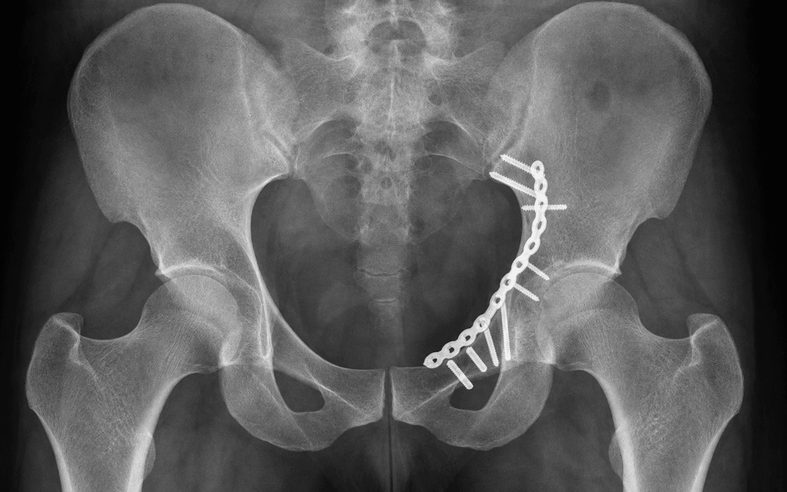
- Contour a 3.5mm reconstruction plate to the pelvic brim (inner table), running from the iliac wing, across the brim, to the superior pubic ramus; position it on bone, medial to the iliopectineal eminence.
- Screws: iliac wing bicortical (30–50mm); pelvic brim and superior ramus unicortical directed posteriorly (20–35mm).
- Place lag screws through the plate for large non-comminuted fragments; aim for a minimum of 6 cortices each side of the fracture.
- Indicated for medial wall comminution or displacement, central head protrusion, and elderly osteoporotic bone.
- Contour a 3.5mm plate from the superior ramus along the quadrilateral surface, positioned infrapectineal (below the pectineal line); direct screws posteriorly 30–40 degrees into the posterior column (avoiding the hip joint).
- It acts as a buttress preventing the femoral head from medialising.
- Remove all provisional K-wires and obtain definitive AP, obturator oblique and iliac oblique views.
- Verify: anatomic reduction of all columns (less than 2mm step-off in the weight-bearing dome), no intra-articular hardware (check each screw on all three views), adequate bicortical screw purchase, a concentric femoral head with no subluxation, and the pelvic brim restored. Save the images to the operative record.
- Haemostasis across all three windows: bone wax for iliac wing bleeding, cautery for soft-tissue bleeders, topical haemostatics for ooze; irrigate with a minimum of 3L saline.
- A large-bore (19Fr) drain in the lateral and/or middle window, routed away from vessels and nerves; remove when output is less than 30mL per 8 hours (typically 48–72 hours).
- Layered closure: rectus abdominis to pubic periosteum (0 Vicryl); external oblique over the inguinal ligament (0 Vicryl); Scarpa's fascia (2-0 Vicryl) to reduce dead space and hernia risk; dermis (3-0 Vicryl or Monocryl); skin subcuticular (4-0 Monocryl) or staples. Apply a waterproof dressing and an abdominal binder.
A vascular corona mortis is found in roughly 40–80% of hemipelvises (the venous variant most common; the arterial variant about 30–45%), crossing the superior pubic ramus a mean of about 40–60mm lateral to the symphysis. If it is avulsed during medial-window dissection it retracts into the pelvis and bleeds massively and difficultly. Actively look for it in every case and ligate it before it can be avulsed — never rely on seeing it by chance.
The external iliac vessels lie just medial to the pelvic brim. Lateral retraction kinks and injures them; use only gentle medial retraction. If you encounter sudden profuse bleeding from the middle window, apply direct pressure with a laparotomy pack, call vascular surgery early, and obtain proximal control (common iliac, above the inguinal ligament) and distal control before any repair — never clamp blindly.
Direct pelvic brim screws POSTERIORLY toward the posterior column, never medially — medially directed screws risk the external iliac vessels (10–20mm medial) and the obturator vessels. Posterior direction is safe for 30–50mm screws, and you need a minimum of 6 cortices each side of the fracture.
A screw or K-wire may look extra-articular on one view but penetrate the joint on another. Check every screw on all three fluoroscopy views (AP, obturator oblique, iliac oblique) before final tightening — an intra-articular screw causes chondrolysis and rapid arthritis.
Aftercare & Complications
Rehabilitation. Weight-bearing is pattern-dependent and always begins at toe-touch:
- Weight-bearing
- Toe-touch
- Duration
- 6–8 weeks
- Weight-bearing
- Toe-touch
- Duration
- 8–10 weeks
- Weight-bearing
- Toe-touch
- Duration
- 10–12 weeks
- Weight-bearing
- Toe-touch
- Duration
- 12 weeks
Immediate (Day 0): neurovascular check (femoral and sciatic nerves), post-operative AP and Judet radiographs, VTE prophylaxis with LMWH (enoxaparin 40mg subcutaneously) starting 12 hours post-op, PCA analgesia, and drain-output monitoring. Day 1–3: mobilise with physiotherapy at toe-touch weight-bearing; continue VTE prophylaxis; start heterotopic-ossification prophylaxis on Day 1 — indomethacin 75mg orally daily for 6 weeks (preferred), or a single 700cGy fraction within 72 hours if NSAIDs are contraindicated; remove drains when output is less than 30mL per 8 hours; wound check before discharge. Follow-up: 2 weeks (wound check, suture or staple removal), 6 weeks (clinical review and radiographs, consider advancing weight-bearing), 12 weeks (radiographs, usually progress to full weight-bearing), 6 months (radiographs, assess for AVN, HO and arthritis), 1 year (functional outcome), then annually or as symptoms dictate. Complications
- Recognition
- Anterolateral thigh numbness or dysaesthesia
- Prevention
- Identify early under the external oblique; protect with a vessel loop
- Management
- Usually neuropraxia, recovers 3–6 months; counsel the patient
- Recognition
- Weak hip flexion or knee extension post-op
- Prevention
- Stay subperiosteal (iliopsoas protects); release retractors every 30 min
- Management
- Document; NCS/EMG at 6 weeks; physiotherapy; most recover 6–12 months
- Recognition
- Sudden profuse bleeding, hypotension
- Prevention
- Never retract laterally; identify the corona mortis; stay on bone
- Management
- Direct pressure, call vascular surgery, proximal and distal control, primary repair or interposition graft, fasciotomies if ischaemia over 4–6 hours
- Recognition
- Sudden arterial bleeding from the medial window
- Prevention
- Identify and ligate prophylactically before avulsion
- Management
- Direct pressure, clip or suture ligation
- Recognition
- Clear fluid in the wound, haematuria, catheter malfunction
- Prevention
- Foley to decompress; stay subperiosteal (bladder 10–20mm medial)
- Management
- Primary two-layer repair, prolonged catheter drainage 10–14 days, urology consult, cystogram before removal
- Recognition
- Calf swelling and tenderness, dyspnoea
- Prevention
- Mechanical plus pharmacological prophylaxis extended 4–6 weeks
- Management
- Therapeutic anticoagulation
- Recognition
- Erythema, wound discharge, systemic sepsis
- Prevention
- Antibiotic prophylaxis; manage Morel-Lavallée lesions
- Management
- Superficial — antibiotics; deep — washout, debridement, retain or remove hardware
- Recognition
- Decreased range of movement, radiographic Brooker grade
- Prevention
- Indomethacin 6 weeks or single-dose radiotherapy
- Management
- Excision if functionally significant, once mature
- Recognition
- Progressive joint-space narrowing, groin pain
- Prevention
- Anatomic reduction (the strongest modifiable predictor)
- Management
- Conservative first, then THA if end-stage
- Recognition
- Groin pain at 6–12 months
- Prevention
- Timely, gentle reduction
- Management
- MRI at 6–12 months if symptomatic; THA if confirmed
- Recognition
- Persistent pain, radiographic lucency
- Prevention
- Stable fixation; bone-graft defects
- Management
- Revision fixation if symptomatic
Viva & Exam Focus
LMMLMM — the three windows and what is at risk
Clinical Decision Scenarios
Practise clinical reasoning and management decisions out loud
“A 35-year-old motorcyclist presents after a high-speed collision with a displaced anterior column acetabular fracture. CT shows 8mm displacement at the pelvic brim with quadrilateral plate involvement. How would you manage this patient?”
“Describe the corona mortis. Why is it clinically significant and how do you manage it?”
“How do you assess acetabular fracture reduction quality, and what is the evidence for anatomic reduction?”
Indications
- Displaced anterior column greater than 2mm through the weight-bearing dome
- Pelvic brim displacement greater than 5mm
- Any roof arc less than 45 degrees (Matta)
- Anterior column + posterior hemitransverse pattern
Three windows (LMM)
- LATERAL: iliac wing (ASIS to SI joint) — LFCN at risk
- MIDDLE: pelvic brim (psoas lateral, vessels medial) — corona mortis
- MEDIAL: superior pubic ramus — bladder at risk
- Each window exposes a different part of the anterior column
Critical structures + distances
- LFCN: variable, typically 10–20mm medial to ASIS (dysaesthesia about 12–25%)
- External iliac vessels: just medial to the brim — never retract laterally
- Corona mortis: mean about 40–60mm (range 30–90mm) lateral to the symphysis (vascular variant about 40–80%)
- Bladder: about 10–20mm medial to the superior ramus
Reduction principles
- Reduce the brim FIRST as the reference for the rest of the reduction
- Goal: less than 2mm articular step-off
- Verify on ALL 3 views (AP, obturator oblique, iliac oblique)
- Matta: anatomic gives 85–90% good, non-anatomic about 50%
Screw safety
- Direct screws POSTERIORLY, never medially
- Medial screws risk the external iliac vessels
- Minimum 6 cortices each side of the fracture
- Check ALL 3 fluoro views for intra-articular hardware
Corona mortis
- Aberrant vessel: external iliac/inferior epigastric to obturator system
- Vascular variant in about 40–80% of hemipelvises (arterial about 30–45%, venous predominates)
- Location: mean about 40–60mm (range 30–90mm) lateral to the symphysis
- ALWAYS identify and ligate prophylactically
Post-operative protocol
- Toe-touch weight-bearing 6–12 weeks (pattern dependent)
- VTE prophylaxis: LMWH/DOAC for 4–6 weeks
- HO prophylaxis: indomethacin 6 weeks or single-dose radiotherapy
- 20–30% proceed to THA at 10–15 years despite anatomic reduction
Examiner favourites
- Draw all 10 Judet-Letournel patterns
- Name the structures at risk in each window with distances
- Quote the Matta outcome data (85–90% versus 50%)
- Explain the corona mortis and its management
Background & Evidence
Classification — Judet-Letournel. The acetabulum is divided into an anterior and a posterior column; fractures are described as elementary (single-column or single-wall) or associated (combinations). Be able to draw all ten patterns in the viva.
- Pattern
- Anterior wall
- Typical approach
- Ilioinguinal or Stoppa
- Pattern
- Anterior column
- Typical approach
- Ilioinguinal or Stoppa
- Pattern
- Posterior wall
- Typical approach
- Kocher-Langenbeck
- Pattern
- Posterior column
- Typical approach
- Kocher-Langenbeck
- Pattern
- Transverse
- Typical approach
- Ilioinguinal or Kocher-Langenbeck (by level)
- Pattern
- T-type
- Typical approach
- Combined or single, by dominant column
- Pattern
- Posterior column + posterior wall
- Typical approach
- Kocher-Langenbeck
- Pattern
- Transverse + posterior wall
- Typical approach
- Kocher-Langenbeck (plus anterior if needed)
- Pattern
- Anterior column + posterior hemitransverse
- Typical approach
- Ilioinguinal or Stoppa
- Pattern
- Both-column
- Typical approach
- Ilioinguinal or Stoppa (plus extended if needed)
Epidemiology. Acetabular fractures are high-energy injuries of young adults (motor vehicle collisions, falls from height) and increasingly low-energy fractures of the elderly. Outcome is strongly volume-dependent — timely referral to a specialist pelvic and acetabular unit is the single most consistent recommendation across regions. Outcomes. Matta's series showed anatomic reduction (less than 2mm displacement) in 71% of hips operated within 21 days, with 76% good-to-excellent clinical results overall, closely linked to radiographic reduction; subsequent THA occurred in 6% and osteonecrosis in 3%. At minimum 10-year follow-up, THA conversion reaches about 13.8% with a further 20% of retained native hips showing established arthritis — so anatomic reduction reduces but does not eliminate long-term joint failure. Global practice. Acetabular fracture surgery is concentrated in specialist units worldwide, and outcome is strongly volume-dependent:
- Global consensus / practice variation
- Universal: anatomic reduction, less than 2mm articular step-off (Matta) — the strongest predictor of joint survival worldwide
- Global consensus / practice variation
- The historic standard is the ilioinguinal (Letournel). Many European, North American and Asian units have shifted to the anterior intrapelvic (modified Stoppa) for quadrilateral-plate and medial-wall access, supported by meta-analysis showing better reduction with less blood loss
- Global consensus / practice variation
- Growing global trend toward acute total hip arthroplasty (with or without limited fixation — 'fix and replace') for low anterior column and quadrilateral-plate fractures with marginal impaction or femoral head damage
- Global consensus / practice variation
- Mechanical plus pharmacological prophylaxis is standard; agent and duration vary by national society guidance (LMWH or DOAC, typically extended 4–6 weeks)
- Global consensus / practice variation
- Routine prophylaxis is selective for anterior-only approaches (lower HO risk than posterior or extensile); indomethacin or single-dose radiotherapy where indicated
State the principle first (anatomic reduction, specialist-unit care), then acknowledge that the approach is where practice legitimately differs — classic ilioinguinal versus anterior intrapelvic (modified Stoppa) — and justify your choice from fracture morphology, not dogma.
References
- Matta JM. Fractures of the acetabulum: accuracy of reduction and clinical results in patients managed operatively within three weeks after the injury. J Bone Joint Surg Am. 1996;78(11):1632-45. 2. Letournel E, Judet R. Fractures of the Acetabulum. 2nd ed. Springer-Verlag; 1993. 3. Giannoudis PV, et al. Operative treatment of displaced fractures of the acetabulum: a meta-analysis. J Bone Joint Surg Br. 2005;87(1):2-9. 4. Tile M, et al. Fractures of the Pelvis and Acetabulum: Principles and Methods of Management. 4th ed. Thieme; 2015. 5. Tornetta P 3rd, Matta JM. Outcome of operatively treated unstable posterior pelvic ring disruptions. Clin Orthop Relat Res. 1996;(329):186-93. 6. Shigemura T, Murata Y, Yamamoto Y, et al. Comparison between ilioinguinal approach and modified Stoppa approach for the treatment of acetabular fractures: an updated systematic review and meta-analysis. Orthop Traumatol Surg Res. 2022;108(2):103204. PMID 35066214. 7. Shazar N, Eshed I, Ackshota N, et al. Comparison of acetabular fracture reduction quality by the ilioinguinal or the anterior intrapelvic (modified Rives-Stoppa) surgical approaches. J Orthop Trauma. 2014;28(6):313-9. PMID 24100918. 8. Leite TFO, Pires LAS, Goke K, et al. Corona Mortis: anatomical and surgical description on 60 cadaveric hemipelvises. Rev Col Bras Cir. 2017;44(6):553-559. PMID 29267551. 9. Kashyap S, Diwan Y, Mahajan S, et al. The majority of corona mortis are small calibre venous blood vessels: a cadaveric study of North Indians. Hip Pelvis. 2019;31(1):40-47. PMID 30899714. 10. Magill P, McGarry J, Queally JM, et al. Minimum ten-year follow-up of acetabular fracture fixation from the Irish tertiary referral centre. Injury. 2012;43(4):500-4. PMID 22269123.
Fractures of the acetabulum: accuracy of reduction and clinical results in patients managed operatively within three weeks after the injury
- 259 patients (262 displaced acetabular fractures) operated within 21 days, mean 6-year follow-up
- Anatomic reduction achieved in 185 hips (71%); rate fell with greater fracture complexity, older age and longer injury-to-surgery interval
- Overall clinical result excellent 40%, good 36% (76% good/excellent), fair 8%, poor 16% — closely linked to radiographic reduction
- Subsequent THA in 6%, osteonecrosis in 3%; outcome worsened by femoral head injury, older age and operative complications
Comparison between ilioinguinal approach and modified Stoppa approach for the treatment of acetabular fractures: an updated systematic review and meta-analysis
- Six comparative studies pooled comparing modified Stoppa (anterior intrapelvic) with the ilioinguinal approach
- Anatomical reduction significantly more likely with the modified Stoppa approach (OR 1.75, 95% CI 1.13–2.69, p=0.01)
- Modified Stoppa had significantly shorter operative time and lower blood loss
- No significant difference in nerve injury, vascular injury, infection, heterotopic ossification, or excellent/good clinical scores
Comparison of acetabular fracture reduction quality by the ilioinguinal or the anterior intrapelvic (modified Rives-Stoppa) surgical approaches
- 225 patients: 122 ilioinguinal versus 103 anterior intrapelvic (AIP/modified Rives-Stoppa)
- Anatomic reduction 68.9% (ilioinguinal) versus 82.5% (AIP), p=0.018
- For both-column fractures, anatomic reduction 54.2% versus 79.4% favouring AIP (p=0.018)
- Complication rates similar; AIP enables posterior column and quadrilateral plate reduction and infrapectineal buttress plating from the contralateral side
Corona Mortis: anatomical and surgical description on 60 cadaveric hemipelvises
- 60 cadaveric hemipelvises dissected for the arterial corona mortis
- Arterial corona mortis present in 45% of specimens
- Mean calibre of the anastomotic branch 2.7mm and of the obturator artery 2.6mm
- Anastomoses between obturator, internal iliac, external iliac and inferior epigastric systems are common over the superior pubic ramus
Minimum ten-year follow-up of acetabular fracture fixation from the Irish tertiary referral centre
- Minimum 10-year clinical and radiographic follow-up after operative acetabular fixation
- Total hip arthroplasty conversion rate 13.8% at 10 years
- A further 20% of retained native hips showed established radiographic arthritis
- 63% of functional outcome scores were good or excellent; concurrent injury (especially sciatic nerve) was the strongest predictor of poor function