Foot & Ankle Trauma · Core Procedure · Weber B/C with a displaced posterior malleolus
- Fibula first restores length, rotation and lateral stability — the key to the ankle mortise. Measure fibular length at 12-15mm from tip to plafond and confirm rotation before plating.
- Operate on displaced trimalleolar injuries: Weber B or C with talar shift, medial clear space widening greater than 4mm, or a posterior malleolus greater than 25 percent of the plafond (or greater than 2mm of step-off).
- Fixing the posterior malleolus restores the PITFL and often stabilises the syndesmosis — reassess the syndesmosis after every component is fixed.
- The mortise view (15-20 degrees of internal rotation) is the key intraoperative check: medial clear space must equal superior clear space (both about 4mm).
- Always run a Cotton test once the malleoli are fixed; if translation is greater than 2mm, fix the syndesmosis 2-4cm above the joint with the foot in neutral dorsiflexion.
- Demand anatomic reduction — 1mm of lateral talar shift cuts tibiotalar contact area by about 42 percent.
When & Why
Indication. Operative fixation of a trimalleolar ankle fracture is indicated when the mortise is disrupted — a displaced bimalleolar or trimalleolar injury with talar shift or mortise incongruity, a Weber B or C fibula fracture with medial clear space widening greater than 4mm, a posterior malleolus greater than 25 percent of the tibial plafond (or greater than 2mm of articular step-off), an open fracture, or an irreducible fracture-dislocation with interposed soft tissue. The goal is an anatomic, stable mortise that allows early motion. Absolute indications
- Displaced trimalleolar fracture with talar shift or mortise incongruity
- Weber B or C fibula fracture with medial clear space widening greater than 4mm
- Posterior malleolus greater than 25 percent of the articular surface on CT (some argue greater than 2mm displacement at any size)
- Open fracture — emergency debridement and stabilisation
- Irreducible fracture-dislocation with interposed soft tissue Relative indications
- Posterior malleolus 20-25 percent with step-off greater than 2mm
- Syndesmotic instability persisting after fibula fixation
- Young, active patient with any displacement
- Bilateral fractures needing early mobilisation Defer immediate ORIF when
- Severe soft-tissue swelling — wait for the wrinkle sign (typically 7-14 days), or temporarily span a fracture-dislocation with an external fixator
- Fracture blisters over the planned approach — wait for re-epithelialisation
- Active infection or significant medical instability
- Peripheral vascular disease with non-healing potential, or a non-ambulatory patient with minimal symptoms (relative) Three principles frame the whole operation:
Restoring fibular length and rotation is the key to the mortise. Fix the fibula before the malleoli, because an unreduced fibula blocks reduction of the rest.
Reducing the posterior fragment restores the PITFL attachment and stabilises the syndesmosis. Fix it before deciding whether a syndesmotic screw is still needed.
One millimetre of lateral talar shift cuts tibiotalar contact area by about 42 percent. Accept nothing less than an anatomic, symmetric reduction on the mortise view.
Consent. Discuss the procedure-specific risks: post-traumatic arthritis (10-30 percent, higher with cartilage damage or residual displacement), symptomatic hardware needing removal (20-40 percent, especially medial screws), wound complications (5-10 percent), nerve injury — usually a neuropraxia that recovers (5-15 percent), syndesmotic stiffness if over-tightened or malreduced, DVT/PE (2-10 percent), nonunion (less than 5 percent with sound technique) and malunion (5-15 percent). With anatomic reduction, 80-90 percent achieve good to excellent results; patients return to walking without aids at 3-4 months and to sport or heavy labour at 6-12 months, with some permanent stiffness common but usually functional. Alternatives are non-operative care (only for stable, minimally displaced fractures), temporary external fixation for severe soft-tissue compromise, and — very rarely, for severe comminution in the low-demand elderly — primary arthrodesis. Setup. Position the patient supine with a bump under the ipsilateral hip to give 15-20 degrees of internal rotation, which brings the fibula into profile; a radiolucent triangle under the knee helps, and a lateral decubitus position is used if a direct posterior approach to the posterior malleolus is planned. Apply a thigh tourniquet at 350mmHg, exsanguinating by elevation (avoid Esmarch compression over the fracture), and free-drape the whole leg for manipulation. Position the image intensifier perpendicular to the table and confirm you can obtain AP, lateral and mortise views before prepping — the mortise view needs 15-20 degrees of internal rotation of the leg or external rotation of the C-arm. Typical instrument set | Category | Items | |----------|-------| | Reduction | Pointed reduction clamps, Weber clamp, dental pick, K-wires | | Fibula | One-third tubular plate (7-8 hole) or precontoured locking fibular plate | | Screws | 3.5mm cortical, 2.7mm cortical for lag, 4.0mm partially threaded cancellous | | Medial malleolus | 4.0mm partially threaded cancellous screws, medial malleolar plate | | Posterior malleolus | 3.5mm cortical screws, mini-fragment plate if needed | | Syndesmosis | Long 3.5mm cortical screw, suture button device (TightRope) | | General | Periosteal elevator, bone clamps, drill guide, depth gauge |
The Operation
The goal is to restore an anatomic, stable mortise through three incisions — lateral over the fibula, anteromedial at the medial malleolus, and posterolateral when the posterior malleolus needs open reduction — fixing the fibula first, then the posterior malleolus, then the medial malleolus, and finally assessing the syndesmosis. The exposure is laid out step by step below (and in depth on the posterolateral approach to the ankle and medial approach to the ankle pages).
- Where it is at risk
- Emerges through the lateral compartment 10-12cm above the lateral malleolus and crosses the fibular incision
- How to protect it
- Identify it before the lateral incision and retract anteriorly
- Where it is at risk
- Subcutaneous and anterior to the medial malleolus
- How to protect it
- Make the medial incision anterior to the malleolar apex; identify and retract
- Where it is at risk
- Subcutaneous posterolaterally, running with the short saphenous vein
- How to protect it
- Avoid posterior dissection on the lateral side; identify it in the posterolateral approach
- Where it is at risk
- Posteromedial ankle in the tarsal tunnel, between FDL and FHL
- How to protect it
- Stay on the FHL side in the posterolateral approach; never pass medial to FHL
- Where it is at risk
- In the retrofibular groove behind the lateral malleolus
- How to protect it
- Identify and retract; place the fibular plate posterolateral, not directly lateral
- Where it is at risk
- In the groove on the posterior tibia, directly under the posterior malleolus fragment
- How to protect it
- Protect it medially while fixing the posterior malleolus
Operative sequence
- Supine with a bump under the ipsilateral hip for 15-20 degrees of internal rotation; free-drape the leg for manipulation.
- Thigh tourniquet at 350mmHg, exsanguinate by elevation.
- Position the image intensifier and confirm AP, lateral and mortise views before prepping — the mortise view needs 15-20 degrees of leg internal rotation.
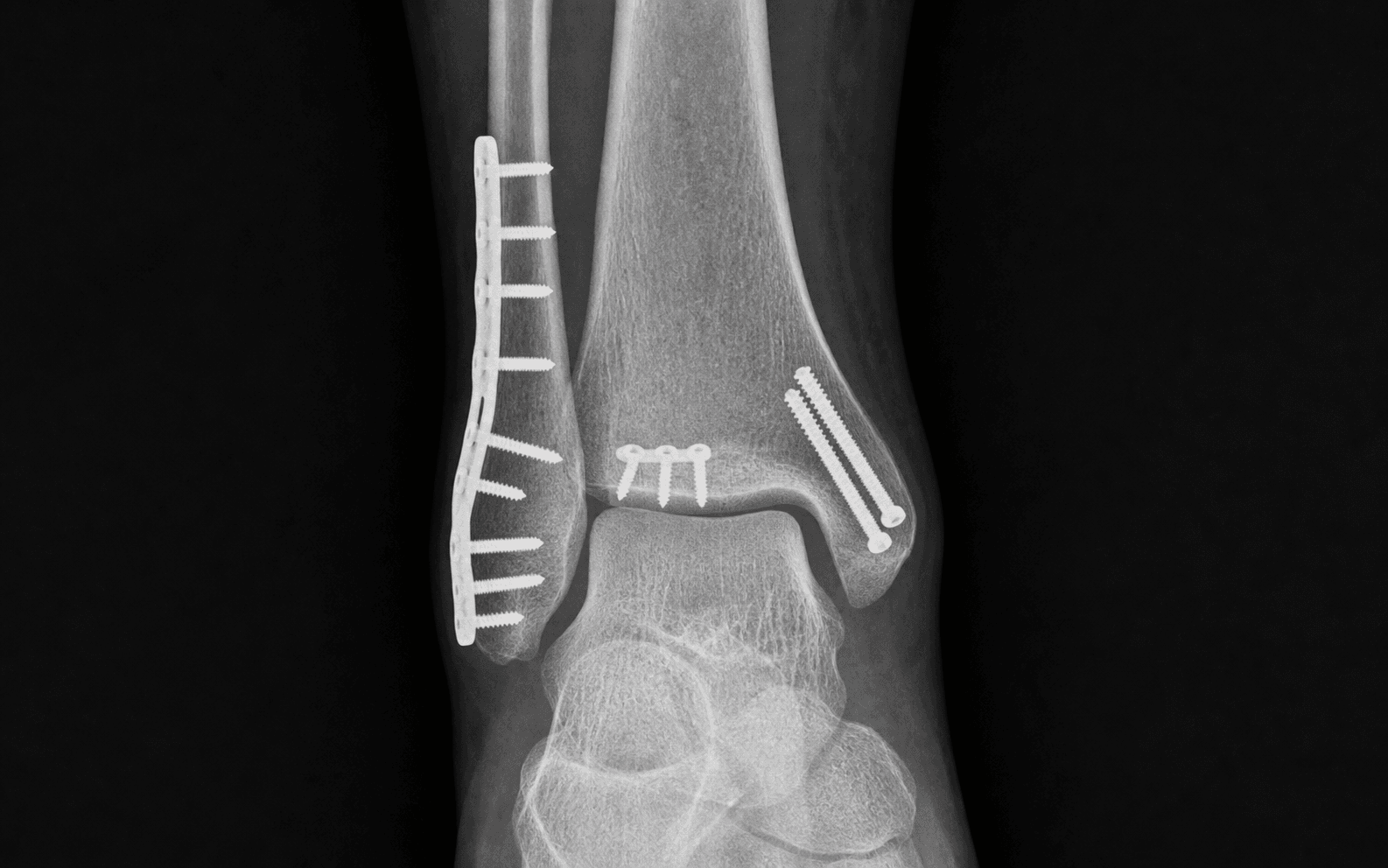
- Lateral longitudinal incision over the fibula (8-10cm), centred on the fracture.
- Anteromedial curved incision just anterior to the medial malleolar apex (5-7cm).
- Posterolateral incision between the fibula and Achilles (6-8cm) — only if the posterior malleolus needs open reduction.
- Lateral longitudinal incision over the distal third of the fibula for a Weber B; incise the fascia and identify and protect the superficial peroneal nerve if it lies anteriorly.
- Identify the peroneal tendons posteriorly and retract them.
- Expose the fracture with a periosteal elevator using minimal stripping to preserve blood supply; clear haematoma and debris from the fracture line.
- Restore fibular length (12-15mm from tip to plafond) and rotation (posterior surface flat, anterior ridge sharp); use the contralateral ankle as a template if available.
- Hold the reduction with a pointed reduction clamp or K-wires before definitive fixation.
- Apply a one-third tubular or precontoured locking plate (3.5mm system) posterolaterally — not directly lateral — to reduce hardware prominence and protect the peroneals.
- For an oblique fracture, place a lag screw perpendicular to the fracture line first, then a neutralisation plate; for a transverse fracture, compress through the plate.
- Use a minimum of 6 cortices (3 screws) proximal and distal to the fracture, and avoid screws penetrating the syndesmosis proximally.
- Reassess on fluoro after fibula fixation. Fix the posterior malleolus when it is greater than 25 percent of the articular surface (traditional threshold), has greater than 2mm of step-off, leaves the syndesmosis unstable after fibula fixation, or is a large fragment preventing talar reduction.
- Incise between the lateral malleolus and the Achilles tendon (6-8cm); this is an internervous plane — peroneals (superficial peroneal nerve) laterally, FHL (tibial nerve) medially.
- Identify the sural nerve subcutaneously and protect it; identify the posterior malleolus fragment still attached to the PITFL, and protect the FHL running in the groove directly beneath it.
- Reduce the fragment anatomically under direct vision, elevating any impaction with a tamp (bone graft if needed), and hold with provisional K-wires.
- Anterior-to-posterior lag screws are the gold standard: start 1-2cm above the joint line on the anterior tibial metaphysis, direct posteriorly into the fragment, and use two parallel 3.5mm or 4.0mm cortical screws for rotational stability — this gives the best purchase (metaphyseal to cortical).
- Posterior-to-anterior percutaneous screws are easier but weaker (cortical purchase only), suited to smaller fragments; a buttress plate is used for large or comminuted fragments from the posterolateral approach.
- Anteromedial curved incision (5-7cm) just anterior to the medial malleolar apex to protect the saphenous nerve and great saphenous vein, which lie more anteriorly.
- Identify and protect the saphenous nerve and vein (variable anatomy); incise the periosteum, clear debris and reduce the fragment anatomically — the articular surface and medial plafond must be perfect.
- Oblique or transverse pattern: two parallel 4.0mm partially threaded cancellous screws perpendicular to the fracture for lag compression.
- Vertical (supination-adduction) pattern: a buttress plate is mandatory — screws alone fail as deltoid tension pulls them superiorly out of the fragment.
- Comminuted: bridge plate with screws in intact bone only; small fragment: tension band wiring (2 K-wires and a figure-of-8 wire). Countersink the screw heads.
- Cotton test: grasp the fibula with a bone clamp and apply lateral stress — translation should be less than 2mm side to side.
- External-rotation stress under fluoro, looking for widening of the tibiofibular clear space; if a posterolateral approach was used, palpate the anterior and posterior tibiofibular ligaments directly.
- On the mortise view the tibiofibular clear space should be less than 6mm (1cm above the plafond) with overlap greater than 6mm on AP and greater than 1mm on mortise.
- Compress the fibula into the incisura with a clamp, confirm it is correctly seated (not anterior, which restricts dorsiflexion, nor posterior, which restricts plantarflexion), and hold the foot in neutral dorsiflexion — not forced dorsiflexion, which overtightens the syndesmosis.
- Place a 3.5mm fully-threaded cortical screw 2-4cm above the joint line, directed posterolateral-to-anteromedial at about 30 degrees and parallel to the joint line, engaging 3 cortices (tricortical) or 4 (quadricortical); one screw usually suffices, two for severe instability.
- Alternative: a suture button (TightRope), which allows physiologic micromotion and has a lower removal rate.
- Mortise view (15-20 degrees internal rotation): medial clear space equals superior clear space (both about 4mm), symmetric talar dome, tibiofibular clear space less than 6mm, tibiofibular overlap greater than 6mm.
- AP view: no talar shift, fibular length restored (12-15mm to plafond), no step at the posterior malleolus.
- Lateral view: no anterior or posterior talar subluxation, posterior malleolus reduced (less than 2mm step), congruent tibiotalar joint.
- Irrigate all wounds thoroughly; layered closure — periosteum and fascia with absorbable sutures, then subcutaneous layer, then skin with interrupted nylon or subcuticular (a small drain if the posterolateral dissection was significant).
- Apply a bulky padded dressing with a plaster backslab in neutral (90 degrees dorsiflexion, hindfoot neutral), elevate the limb, and start DVT prophylaxis (LMWH 40mg daily or aspirin per protocol).
The leg must be internally rotated 15-20 degrees to see the mortise en face. Without a true mortise view you cannot judge reduction quality, and a malreduced mortise is the dominant modifiable driver of a poor outcome.
Fibular length and rotation determine mortise congruity. A fibula left too short causes lateral talar shift and mortise widening; a malrotated fibula alters joint biomechanics. Restore length (12-15mm tip-to-plafond) and rotation (posterior surface flat) and confirm against the contralateral ankle before final tightening.
Plain X-rays underestimate posterior malleolus size. Measure the fragment on sagittal CT as a percentage of the tibial plafond. The posterior malleolus is the attachment of the PITFL, so fixing it restores syndesmotic stability — and anterior-to-posterior lag screws give better bone purchase than posterior-to-anterior screws.
Vertical medial malleolar fractures (the supination-adduction pattern) must have plate fixation. Screws alone fail because deltoid tension pulls them superiorly out of the fragment.
Seat the fibula correctly in the incisura before drilling — confirmed by direct vision or CT if uncertain. A fibula left too anterior restricts dorsiflexion; too posterior restricts plantarflexion. Foot in neutral, never forced dorsiflexion.
Accepting residual displacement leads to post-traumatic arthritis. One millimetre of lateral talar shift reduces tibiotalar contact area by about 42 percent — perfect reduction determines outcome.
Aftercare & Complications
Rehabilitation timeline | Timeframe | Activity | Goals | |-----------|----------|-------| | Day 1-14 | Non-weight-bearing in a backslab, elevation, ankle pumps | Wound healing, swelling control | | 2 weeks | Suture removal, transition to a CAM boot | Begin gentle range-of-motion exercises | | 2-6 weeks | Non-weight-bearing in a CAM boot, active ROM | Regain dorsiflexion and plantarflexion | | 6-8 weeks | Progressive weight-bearing if healing | Confirm radiographic union | | 8-12 weeks | Full weight-bearing, intensive physiotherapy | Strengthening and proprioception | | 3-4 months | Return to driving and light activities | Functional recovery | | 6-12 months | Return to sport or heavy labour | Full recovery | Syndesmotic screw management. A quadricortical screw is considered for removal at 3-4 months if symptomatic or before high-demand activity; a tricortical screw is often left in situ and may break with weight-bearing (acceptable). A suture button needs no removal. Modern evidence does not mandate routine screw removal if the patient is asymptomatic. Follow-up. 2 weeks (wound check, suture removal, X-ray), 6 weeks (clinical and radiographic assessment, advance weight-bearing), 3 months (assess union, consider screw removal), 6 months (functional assessment), 1 year (long-term assessment, screen for arthritis).
- Recognition
- Progressive ankle pain and stiffness with joint-space narrowing (10-30 percent)
- Prevention
- Anatomic reduction of every component; medial clear space equal to superior clear space, less than 2mm posterior step, stable fixation for early motion
- Management
- Activity modification, AFO, NSAIDs, injections; debridement, supramalleolar osteotomy if malaligned, ankle fusion (gold standard) or replacement in selected cases
- Recognition
- Chronic lateral ankle pain and instability, widened mortise on stress views (10-20 percent)
- Prevention
- Cotton test after all fixation; correct screw technique (2-4cm above the joint, 25-30 degrees, fibula seated in the incisura)
- Management
- Acute: revision reduction and fixation. Chronic: syndesmotic reconstruction with ligament graft, possibly combined procedures
- Recognition
- Lateral pain and instability, talar shift, valgus hindfoot (5-15 percent)
- Prevention
- Meticulous fibular reduction — restore length (12-15mm to plafond) and rotation; use the contralateral X-ray as a template
- Management
- Corrective osteotomy: fibular lengthening and derotation, supramalleolar osteotomy if the tibia is involved
- Recognition
- Dehiscence, erythema, drainage, exposed hardware (5-10 percent)
- Prevention
- Wait for the wrinkle sign (7-14 days), careful tissue handling, well-placed incisions, layered closure
- Management
- Superficial: local care and oral antibiotics. Deep: IV antibiotics, surgical debridement, possible hardware removal, flap cover if needed
- Recognition
- Painful prominence over screws or plate, especially medially (20-40 percent)
- Prevention
- Countersink screws, low-profile plates, posterolateral fibular plate position; warn the patient preoperatively
- Management
- Hardware removal after union (12-18 months for fracture fixation, 3-4 months for a symptomatic syndesmotic screw)
- Recognition
- Numbness or dysaesthesia in the nerve distribution, Tinel sign at the injury site (5-15 percent)
- Prevention
- Know the anatomy, careful dissection, identify nerves before retraction, protect the saphenous vein and nerve medially
- Management
- Observation — most are neuropraxia and recover; persistent neuroma: desensitisation, blocks, or excision; complete division: repair
- Recognition
- Persistent pain, motion at the fracture, radiographic lucency with no callus at 3-4 months (less than 5 percent)
- Prevention
- Anatomic reduction, stable fixation, avoid NSAIDs perioperatively, smoking cessation, optimise nutrition and diabetes
- Management
- Revision ORIF with bone graft, consider a bone stimulator; a medial malleolus nonunion may be asymptomatic
- Recognition
- Calf pain and swelling; PE gives dyspnoea, chest pain, tachycardia (2-10 percent)
- Prevention
- Pharmacological prophylaxis (LMWH or aspirin), early ankle pumps, elevation, hydration, early mobilisation when safe
- Management
- Ultrasound for DVT and anticoagulation; PE: CT pulmonary angiography, anticoagulation, ICU if massive
Viva & Exam Focus
FPMSFPMS — order of fixation
MORTISEMORTISE — reduction checklist
Clinical Decision Scenarios
Practise clinical reasoning and management decisions out loud
“A 45-year-old man presents with a displaced trimalleolar ankle fracture. The CT shows a posterior malleolus fragment of 35 percent of the articular surface. Walk me through your surgical planning and fixation sequence.”
“You have reduced and plated the fibula in an ankle fracture. On the Cotton test there is 4mm of lateral translation of the fibula compared with the opposite side. How do you manage this syndesmotic instability?”
“A patient returns to clinic 3 weeks after trimalleolar ORIF with wound dehiscence over the lateral incision and visible plate. There is purulent discharge. How do you manage this?”
Fixation sequence (FPMS)
- Fibula FIRST — restores length (12-15mm to plafond) and rotation
- Posterior malleolus second — buttress and PITFL restoration for syndesmotic stability
- Medial malleolus third
- Syndesmosis last — Cotton test after all fractures are fixed
Syndesmosis fixation
- Screw 2-4cm above the joint, 25-30 degrees, parallel to the joint
- Compress the fibula into the incisura, foot in NEUTRAL (not dorsiflexion)
- 3 cortices (tricortical) or 4 cortices (quadricortical)
- Alternative: suture button (TightRope) — allows micromotion
Posterior malleolus
- Greater than 25 percent of the articular surface on CT (traditional threshold)
- Greater than 2mm step-off (increasingly used threshold)
- Syndesmotic instability after fibula fixation
- Anterior-to-posterior lag screws are the gold standard
Mortise view (15-20 degrees IR)
- Medial clear space equals superior clear space (both about 4mm)
- Tibiofibular clear space less than 6mm
- Tibiofibular overlap greater than 6mm (AP), greater than 1mm (mortise)
- Symmetric talar dome in the mortise
Classifications
- Weber A/B/C — fibula level relative to the syndesmosis
- Lauge-Hansen — mechanism-based (SER is commonest)
- A vertical medial malleolus (SAD) needs a plate, not just screws
- Cotton translation greater than 2mm means syndesmotic instability
Background & Evidence
Epidemiology. Ankle fractures are among the most common operatively treated fractures, and their incidence in adults is rising — Court-Brown and colleagues documented a sustained increase through the 1990s, driven largely by low-energy falls in an ageing population. Trimalleolar injuries sit at the unstable, rotationally displaced end of that spectrum and almost always warrant ORIF; supination-external rotation is the commonest underlying mechanism. Two classifications frame the injury. The Weber (AO) system is defined by the level of the fibula fracture relative to the syndesmosis and is the one that drives the operative decision:
- Fibula level
- Below the syndesmosis
- Stability
- Stable — syndesmosis intact
- Typical management
- Usually non-operative
- Fibula level
- At the level of the syndesmosis
- Stability
- Variable — about half have syndesmotic injury
- Typical management
- ORIF if displaced or mortise disrupted
- Fibula level
- Above the syndesmosis
- Stability
- Unstable — syndesmosis disrupted
- Typical management
- ORIF; assess and usually fix the syndesmosis
- Mechanism
- The commonest pattern (40-75 percent)
- Weber equivalent
- Weber B
- Notes
- Stage IV adds a posterior malleolus or posterior tibial margin
- Mechanism
- Pronation plus external rotation
- Weber equivalent
- Weber C
- Notes
- High fibula with syndesmotic injury
- Mechanism
- Pronation plus abduction
- Weber equivalent
- Weber C
- Notes
- Bending-wedge fibula fragment
- Mechanism
- Supination plus adduction
- Weber equivalent
- Weber A
- Notes
- Vertical medial malleolus — needs a buttress plate
Critical radiographic measurements. Plain X-rays underestimate posterior malleolus size, so a CT is essential to measure the fragment as a percentage of the tibial plafond and to assess impaction, comminution and the syndesmosis.
- Normal
- Less than 6mm
- Suggests injury
- Greater than 6mm = syndesmotic injury
- Normal
- Greater than 6mm on AP, greater than 1mm on mortise
- Suggests injury
- Loss of overlap = diastasis
- Normal
- Equals superior clear space (about 4mm)
- Suggests injury
- Widening greater than 4mm = deltoid injury
- Normal
- 12-15mm
- Suggests injury
- Shortening = mortise widening
- Normal
- Anatomic on sagittal CT
- Suggests injury
- Greater than 25 percent or greater than 2mm step = fix
References
- Court-Brown CM, McBirnie J, Wilson G. Adult ankle fractures — an increasing problem? Acta Orthop Scand. 1998;69(1):43-47. 2. Tornetta P, Axelrad TW, Sibai TA, Creevy WR. Treatment of the stress positive ligamentous SE4 ankle fracture: incidence of syndesmotic injury and clinical decision making. J Orthop Trauma. 2012;26(11):659-661. PMID: 23100079. 3. Gardner MJ, Demetrakopoulos D, Briggs SM, Helfet DL, Lorich DG. The ability of the Lauge-Hansen classification to predict ligament injury and mechanism in ankle fractures: an MRI study. J Orthop Trauma. 2006;20(4):267-272. 4. Miller AN, Carroll EA, Parker RJ, Borber SP, Mauricio L, Helfet DL. Direct visualization for syndesmotic stabilization of ankle fractures. Foot Ankle Int. 2009;30(5):419-426. 5. Sagi HC, Shah AR, Sanders RW. The functional consequence of syndesmotic joint malreduction at a minimum 2-year follow-up. J Orthop Trauma. 2012;26(7):439-443. PMID: 22357084. 6. Ramsey PL, Hamilton W. Changes in tibiotalar area of contact caused by lateral talar shift. J Bone Joint Surg Am. 1976;58(3):356-357. (Replicated by Lloyd J, Elsayed S, Hariharan K, Tanaka H. Revisiting the concept of talar shift in ankle fractures. Foot Ankle Int. 2006;27(10):793-796. PMID: 17054879.) 7. Gardner MJ, Brodsky A, Briggs SM, Nielson JH, Lorich DG. Fixation of posterior malleolar fractures provides greater syndesmotic stability. Clin Orthop Relat Res. 2006;447:165-171. PMID: 16467626. 8. Clanton TO, Paul P. Syndesmosis injuries in athletes. Foot Ankle Clin. 2002;7(3):529-549. 9. Norkus SA, Floyd RT. The anatomy and mechanisms of syndesmotic ankle sprains. J Athl Train. 2001;36(1):68-73. 10. Andersen MR, Frihagen F, Hellund JC, Madsen JE, Figved W. Randomized trial comparing suture button with single syndesmotic screw for syndesmosis injury. J Bone Joint Surg Am. 2018;100(1):2-12. PMID: 29298255.
Fixation of posterior malleolar fractures provides greater syndesmotic stability
- Cadaveric pronation-external rotation model (n=10): fixing the posterior malleolus restored syndesmotic stiffness to 70% of intact
- Trans-syndesmotic screw fixation alone restored stiffness to only 40% of intact
- In 15 clinical PER-4 cases, no complete PITFL tears occurred - the ligament stays attached to the posterior fragment
Treatment of the stress positive ligamentous SE4 ankle fracture: incidence of syndesmotic injury and clinical decision making
- 114 stress-positive supination-external rotation injuries; 60 operated, 27 (45%) showed syndesmotic instability after fibular fixation
- Operative cases had significantly greater stress medial clear space widening (6.9mm vs 4.8mm, p less than 0.001)
- No patient healed with talar subluxation regardless of operative or cast treatment
The functional consequence of syndesmotic joint malreduction at a minimum 2-year follow-up
- Bilateral CT in 68 patients: 27 (39%) syndesmoses were malreduced versus the uninjured side
- Closed reduction was malreduced in 44% versus only 15% with open reduction
- Malreduced syndesmoses had significantly worse SMFA and Olerud-Molander scores at minimum 2 years (p less than 0.05)
Randomized trial comparing suture button with single syndesmotic screw for syndesmosis injury
- RCT of 97 patients: suture button versus a single quadricortical syndesmotic screw
- At 2 years suture button had higher AOFAS (96 vs 86) and Olerud-Molander (100 vs 90) scores and less walking pain
- Residual tibiofibular widening of at least 2mm occurred in 8/40 (button) versus 20/40 (screw); 7 screw patients developed recurrent diastasis versus none with button
Revisiting the concept of talar shift in ankle fractures
- Cadaveric replication of the classic Ramsey and Hamilton experiment under a 70-kg load
- 1mm of lateral talar shift produced a 40% loss of tibiotalar contact area (versus 42% originally reported)
- Confirmed the original finding while noting large variance with joint morphology