Portal placement for minimally invasive diagnostic and therapeutic access to all knee compartments
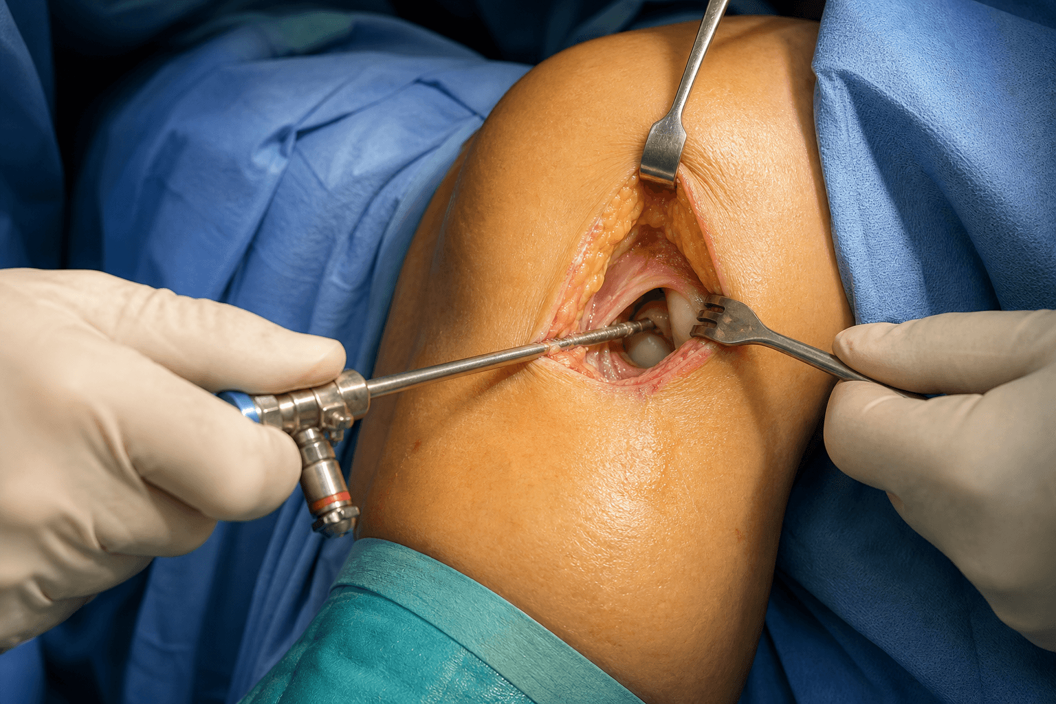
- Anterolateral (AL) portal is the primary VIEWING portal, placed 1cm lateral to the patellar tendon at joint-line level, and is established first.
- Anteromedial (AM) portal is the primary WORKING portal, placed 1cm medial to the patellar tendon, created UNDER DIRECT VISION — never blind.
- The infrapatellar branch of the saphenous nerve (IPBSN) crosses the field 3-5mm below the portals and is injured in 10-30 percent of cases, causing anterolateral knee numbness.
- Triangulation is the fundamental skill — working instruments through the AM portal while viewing through the AL portal.
- Posterior portals place the popliteal neurovascular bundle (within 5-15mm of the posterior capsule) at risk and demand transillumination and spinal-needle localisation.
When & Why
What it exposes. Knee arthroscopy gives minimally invasive diagnostic and therapeutic access to every intra-articular compartment of the knee — the suprapatellar pouch, medial and lateral gutters, patellofemoral joint, both tibiofemoral compartments and the intercondylar notch (ACL and PCL). It is among the most frequently performed orthopaedic procedures worldwide. Primary indications:
- Meniscal pathology — tears managed by repair or partial meniscectomy
- ACL and PCL reconstruction
- Cartilage procedures — microfracture, debridement, OATS
- Loose body removal
- Synovectomy for inflammatory arthritis or PVNS
- Lateral release for selected patellofemoral disorders
- Diagnostic arthroscopy when imaging is inconclusive Advantages: small incisions, direct visualisation of intra-articular structures, same-day surgery in most cases, rapid rehabilitation versus open procedures, and the ability to combine diagnosis with treatment in one sitting. Disadvantages: requires specialised equipment and training, fluid extravasation with compartment-syndrome risk, limited access to some structures without accessory portals, and a learning curve for complex procedures. Position & landmarks. Supine on the operating table with the leg hanging free off the side of the table or in a leg holder maintaining 70-90 degrees of flexion. A thigh tourniquet (about 300mmHg after exsanguination) is optional; a lateral post at thigh level allows valgus stress for medial-compartment access. The knee is palpated with the joint at 90 degrees to mark the patella (poles and borders), the patellar tendon (inferior patella to tibial tubercle), the joint line (the soft spot adjacent to the tendon) and the tibial tubercle.
Advantages: gravity assists flexion, easy to apply varus/valgus stress, simple set-up. Disadvantages: the surgeon must stabilise the leg, which can be awkward in prolonged cases.
Advantages: hands-free stabilisation, consistent positioning, easier for long cases such as ACL reconstruction. Disadvantages: cost, set-up time, may limit some positioning options.
Recent randomised evidence (the FIDELITY and METEOR trials) has questioned the role of arthroscopic partial meniscectomy in degenerative meniscal tears. For exams, know that APM is NOT superior to sham surgery for degenerative tears, but remains indicated for mechanical symptoms from acute traumatic tears and for meniscal repair in young patients.
The Exposure
The exposure is the standard two-portal technique: establish the anterolateral (viewing) portal first, then create the anteromedial (working) portal under direct vision, triangulate the instruments, and only then add accessory portals if the pathology demands. There is no true internervous plane — arthroscopy uses small stab incisions through skin and capsule directly into the joint.
Standard two-portal exposure
- Supine with the leg hanging or in a leg holder at 70-90 degrees of flexion; thigh tourniquet (about 300mmHg) optional; lateral post for valgus stress.
- Palpate the patella, patellar tendon, joint-line soft spot and tibial tubercle with the knee flexed to 90 degrees and mark the planned portals.
- Optional: insert a superolateral spinal needle into the suprapatellar pouch and inject 30-60mL saline to distend the joint (alternatively distend after the AL portal using the pump).
- Palpate the soft spot 1cm lateral to the patellar tendon at joint-line level (level with the inferior pole of the patella), knee flexed 70-90 degrees.
- Localise with an 18-gauge spinal needle aimed at the intercondylar notch; confirm intra-articular position by aspirating fluid if the joint was distended.
- Make a horizontal 5-10mm stab incision with an 11-blade centred on the needle — horizontal is parallel to the infrapatellar nerve and protects it.
- Insert the blunt trocar with sheath aiming toward the intercondylar notch; feel the capsule give way. Do NOT force.
- Remove the trocar, insert the 4mm 30-degree arthroscope, connect camera, light source and inflow, and confirm an intra-articular position.
- From the AL scope, look toward the medial compartment and identify the medial femoral condyle, the anterior horn of the medial meniscus and the fat pad.
- Insert a spinal needle under direct vision 1cm medial to the patellar tendon at joint-line level, watching the needle enter the joint on the monitor.
- Optimise the needle so it enters above the anterior horn of the medial meniscus and avoids the fat pad, adjusting the entry point before any skin incision.
- Make a horizontal stab over the needle, remove the needle, and pass a blunt trocar (or working cannula) through the portal under direct visualisation.
- Triangulate the working instruments through the AM portal while viewing through the AL scope.
- Examine in sequence: suprapatellar pouch (synovium, loose bodies, plica) → medial gutter → patellofemoral joint (tracking in extension) → medial compartment under valgus stress (MFC, medial plateau, medial meniscus) → intercondylar notch (ACL AM/PL bundles, PCL) → lateral compartment under varus stress (LFC, lateral plateau, lateral meniscus, popliteus hiatus) → lateral gutter.
- Document meniscal tears by zone (red-red, red-white, white-white) and clock position, and cartilage lesions by ICRS or Outerbridge grade.
- Superolateral: 2-3cm above the joint line, lateral — outflow and patellofemoral visualisation.
- Posteromedial: 1cm posterior to the MCL at the joint line — posterior-horn medial meniscus repair and PCL work. Saphenous nerve and vein lie anterior, popliteal vessels posterior.
- Posterolateral: 1cm posterior to the LCL at the joint line — popliteus and posterolateral corner. The common peroneal nerve is at risk if the portal is placed too posterior.
- Far AM / trans-tendon: for anatomic ACL femoral tunnel drilling; a far AM portal is now often preferred over a trans-patellar-tendon portal to avoid tendonitis.
- Remove all instruments, confirm haemostasis, and irrigate the joint thoroughly to remove debris.
- Express fluid by flexing the knee, then close skin only — the capsule is not closed.
- Use 3-0 or 4-0 nylon/Prolene, Steri-strips or dermabond, and apply a sterile compression dressing.
The AM portal must be created under direct arthroscopic vision. Blind AM placement risks injury to the anterior horn of the medial meniscus, the articular cartilage and the fat pad. Always localise with a spinal needle first and watch it enter the joint before making the skin incision.
The concept of an internervous plane does not apply to arthroscopic portals. These are small stab incisions (5-10mm) made directly through skin and capsule into the joint, not an interval between muscles of different nerve supply. The main nerve at risk is the infrapatellar branch of the saphenous nerve, which runs horizontally across the anterior knee about 3-5mm below the portals; it is protected by staying at joint-line level and using horizontal skin incisions. Injury causes anterolateral numbness that is usually well tolerated.
Dangers & Extensions
Neurovascular structures at risk, by portal
- Structure at risk
- Infrapatellar branch of saphenous nerve
- Distance from portal
- 3-5mm below
- Consequence of injury
- Lateral knee numbness (common)
- Structure at risk
- Infrapatellar branch of saphenous nerve
- Distance from portal
- 5-10mm below
- Consequence of injury
- Medial/anterior knee numbness
- Structure at risk
- Saphenous vein and nerve
- Distance from portal
- 10-15mm anterior
- Consequence of injury
- Numbness, bleeding, thrombosis
- Structure at risk
- Common peroneal nerve
- Distance from portal
- 25-30mm posterolateral
- Consequence of injury
- Foot drop (rare but devastating)
- Structure at risk
- Popliteal artery and vein
- Distance from portal
- 5-15mm (variable)
- Consequence of injury
- Haemorrhage, pseudoaneurysm
The popliteal artery lies within 5-15mm of the posterior capsule. When creating any posterior portal: (1) transilluminate from the anterior scope to identify vasculature, (2) insert the spinal needle under direct vision, (3) make only a skin incision with the blade then use a blunt trocar, and (4) stay superficial to the capsule until the position is confirmed arthroscopically. Establish posterior portals with the knee flexed to 90 degrees, which maximises the distance from the saphenous and common peroneal nerves.
The infrapatellar branch of the saphenous nerve (IPBSN). The IPBSN emerges from beneath sartorius and runs horizontally across the anterior knee about 3-5mm below the standard portal sites. It is the most commonly injured structure — injury occurs in 10-30 percent of cases and produces anterolateral or anteromedial knee numbness, usually well tolerated. Prevention uses horizontal skin incisions parallel to the nerve, staying at joint-line level, and avoiding overtight sutures. Complications
- Incidence
- 10-30%
- Prevention
- Horizontal incisions, stay at joint line
- Management
- Counselling; typically well tolerated
- Incidence
- Less than 1%
- Prevention
- Sterile technique, prophylactic antibiotics
- Management
- Arthroscopic washout, IV antibiotics
- Incidence
- Less than 1%
- Prevention
- Early mobilisation, chemical prophylaxis if high risk
- Management
- Anticoagulation, IVC filter if indicated
- Incidence
- 2-5%
- Prevention
- Meticulous haemostasis, drain suprapatellar pouch
- Management
- Aspiration, compression, rarely re-arthroscopy
- Incidence
- 1-2%
- Prevention
- Early ROM, avoid excessive synovitis
- Management
- Physiotherapy, MUA, arthroscopic lysis
- Incidence
- Rare
- Prevention
- Monitor pump pressure, recognise extravasation
- Management
- Immediate fasciotomies
- Incidence
- Rare
- Prevention
- Correct portal technique, avoid posterior structures
- Management
- Vascular surgery consult, nerve exploration
Compartment syndrome — recognise extravasation early. Fluid extravasation through a capsular defect into the calf or thigh is the mechanism. Risk factors are prolonged surgery, high pump pressures and capsular tears. Watch for increasing leg swelling, pain out of proportion and tense compartments; prevention means monitoring pump pressure (keep less than 60mmHg), periodically palpating the calf and thigh, limiting operative time, and recognising capsular tears. Established compartment syndrome is an orthopaedic emergency treated by immediate four-compartment fasciotomy.
During arthroscopy, periodically palpate the calf and thigh for swelling. If significant extravasation is detected, lower the pump pressure, consider switching to gravity inflow, and expedite the procedure. Postoperatively, monitor for compartment-syndrome symptoms.
Instrument breakage. Shavers, graspers and other instruments can break intra-articularly; perform an instrument count before and after the case, retrieve broken fragments immediately (a loose metal body damages cartilage), and add portals or convert to a mini-open approach if needed. Extensile options. When the two-portal set-up is insufficient: add far medial/lateral portals for ACL femoral tunnel drilling and better triangulation; use posteromedial and posterolateral portals with transillumination; convert to inside-out or outside-in meniscal-repair techniques; or extend a portal to a mini-open arthrotomy (for example a mini-open posterior-horn meniscal repair). Pump pressure can be raised (up to 60mmHg) for bleeding control, but only with awareness of the extravasation risk.
- Viewing portal
- Anterolateral
- Working portal
- Anteromedial
- Accessory portals
- Posteromedial (suture retrieval)
- Viewing portal
- Anterolateral
- Working portal
- Far AM (accessory)
- Accessory portals
- Anteromedial, sometimes superolateral
- Viewing portal
- Anterolateral
- Working portal
- Anteromedial
- Accessory portals
- Posteromedial essential
- Viewing portal
- Anterolateral
- Working portal
- Posteromedial
- Accessory portals
- Posterolateral may be needed
Procedures Through This Approach
- ACL reconstruction — patellar tendon (BTB) autograft and ACL reconstruction — hamstring autograft — the principal ligament reconstructions performed through these portals.
- ACL reconstruction technique and revision ACL reconstruction — including the far AM portal for anatomic femoral tunnel drilling.
- PCL reconstruction — requires a posteromedial portal.
- Arthroscopic partial meniscectomy and meniscal repair — all-inside and inside-out.
- MPFL reconstruction — for patellofemoral instability.
- Cartilage procedures (microfracture, debridement, OATS), loose body removal, synovectomy, lateral release, and diagnostic arthroscopy.
Viva & Exam Focus
PORTALSPORTALS — standard knee arthroscopy portals
SAFETYSAFETY — structures at risk
The anterolateral portal is always established first as the viewing portal. The anteromedial portal is then created under direct arthroscopic vision. Never create the AM portal blind.
The infrapatellar branch of the saphenous nerve (IPBSN) is injured in 10-30 percent of cases, causing anterolateral knee numbness. Prevention is horizontal skin incisions and staying at joint-line level.
Exam viva scenarios
Practise clinical reasoning and management decisions out loud
“Describe your technique for establishing portals for knee arthroscopy.”
“A patient develops severe calf pain and swelling 4 hours after knee arthroscopy, with a tense calf. What is your concern and how do you manage it?”
“You are performing an ACL reconstruction. What portals do you need and why?”
“A 52-year-old has a degenerative medial meniscal tear on MRI and asks whether arthroscopy will help. How do you counsel them?”
Portal positions
- Anterolateral: 1cm lateral to patellar tendon at joint line — VIEWING
- Anteromedial: 1cm medial to patellar tendon at joint line — WORKING
- AL portal FIRST; AM portal UNDER DIRECT VISION
- Spinal-needle localisation before ALL portal incisions
- Horizontal incisions parallel to the IPBSN
Structures at risk
- IPBSN: 10-30% injury rate, causes anterolateral numbness
- Anterior horn of meniscus: with improper AM placement
- Popliteal vessels: with posterior portals (5-15mm from capsule)
- Common peroneal nerve: with posterolateral portal
- Fat pad: if portals are too high, causing bleeding
Systematic examination
- Suprapatellar pouch — synovium, loose bodies
- Medial gutter — plica, medial condyle surface
- Patellofemoral joint — tracking, articular surfaces
- Medial compartment (valgus stress) — MFC, meniscus
- Intercondylar notch — ACL, PCL
- Lateral compartment (varus stress) — LFC, lateral meniscus
Accessory portals
- Superolateral: outflow, patellofemoral visualisation
- Posteromedial: posterior horn medial meniscus, PCL surgery
- Posterolateral: popliteus, PLC surgery
- All posterior portals with TRANSILLUMINATION
- Far AM: ACL femoral tunnel drilling access
Complications
- IPBSN injury: 10-30%, counselling, well tolerated
- Infection: less than 1%, washout plus antibiotics
- Compartment syndrome: monitor pump pressure and leg swelling
- DVT: rare, early mobilisation, prophylaxis if high risk
- Stiffness: early ROM, physiotherapy, MUA if severe
Evidence base
- FIDELITY: APM no better than sham for degenerative tears
- METEOR: PT as effective as surgery for degenerative tears
- First-line for degenerative tears: NON-OPERATIVE
- Reserve surgery for mechanical symptoms, PT failure, acute tears
- Meniscal repair carries roughly a one-in-four long-term failure rate
References
FIDELITY Trial — Arthroscopic Partial Meniscectomy vs Sham Surgery
- Multicentre double-blind sham-controlled RCT, 146 patients aged 35-65
- Degenerative medial meniscus tear WITHOUT radiographic knee osteoarthritis
- 12-month Lysholm and WOMET scores: no significant between-group difference
- Knee pain after exercise: between-group difference only -0.1 (95% CI -0.9 to 0.7)
METEOR Trial — Surgery vs Physical Therapy for Meniscal Tear with OA
- Multicentre RCT, 351 patients aged 45 or older
- Meniscal tear PLUS mild-to-moderate osteoarthritis on imaging
- WOMAC physical-function change at 6 months: mean difference 2.4 (95% CI -1.8 to 6.5), not significant
- 30% of the physical-therapy group crossed over to surgery by 6 months
Meniscal Repair Outcomes at Greater Than Five Years — Meta-analysis
- Systematic review and meta-analysis of 13 studies (566 repairs), minimum 5-year follow-up
- Pooled meniscal-repair failure rate (reoperation or clinical failure) 23.1% (131 of 566)
- Failure rate similar for medial vs lateral meniscus
- Failure rate similar for intact vs reconstructed ACL
- Long-term data for modern all-inside devices were not yet available
Neural Structures and Posterior Portals by Knee Position
- Cadaveric study (10 knees) of nerve proximity to posteromedial and posterolateral portals
- Sartorial branch of saphenous nerve and infrapatellar branches measured from the posteromedial portal
- At 90 degrees flexion the posteromedial portal was a mean 26.1mm from the sartorial branch vs 14mm in extension
- At 90 degrees flexion the posterolateral portal was a mean 25.4mm from the common peroneal nerve vs 20.1mm in extension
Experimental Risk of Compartment Syndrome During Knee Arthroscopy
- Controlled porcine model with deliberate capsulotomies allowing free fluid extravasation
- Intra-articular pressures tested at 100, 150 and 200 mmHg over 30 to 90 minutes
- Maximum compartment pressures averaged 78.75 mmHg during infusion
- Elevated compartment pressures resolved rapidly (mean 25.5 min) once infusion stopped, with normal nerve conduction and no myonecrosis
Guidelines, registries and global practice. Volumes for degenerative meniscal pathology have fallen substantially since the FIDELITY and METEOR trials. The global consensus across major societies now favours non-operative management first for degenerative tears and meniscal preservation (repair) over resection in younger patients with traumatic, repairable tears.
- Position on arthroscopy for a degenerative meniscal tear
- Arthroscopic lavage and debridement NOT recommended for knee OA; meniscal surgery reserved for true mechanical locking
- Position on arthroscopy for a degenerative meniscal tear
- 2016 consensus: APM is not a first-line treatment for degenerative meniscus lesions; trial structured exercise first
- Position on arthroscopy for a degenerative meniscal tear
- Strong evidence against arthroscopy with lavage/debridement for symptomatic OA; meniscal surgery for selected mechanical symptoms
- Position on arthroscopy for a degenerative meniscal tear
- Strong recommendation against arthroscopy for nearly all patients with degenerative knee disease
Practice points with global validity:
- Document that non-operative management (structured physiotherapy, activity modification, weight optimisation) was offered before arthroscopy for any degenerative tear.
- Reserve arthroscopy for true mechanical symptoms (locking blocking full extension), a failed conservative trial, or an acute traumatic tear pattern.
- Counsel that surgery does not slow the natural history of osteoarthritis progression.
- Favour repair over resection in young patients with peripheral, vertical longitudinal tears, especially with concurrent ACL reconstruction.
For the Operative Surgery station (advanced orthopaedic practice, DNB/MS, MRCS, SICOT), be able to describe portal placement in detail: positioning, the AL portal first (landmarks and technique), the AM portal under direct vision, the systematic joint-examination sequence, and common complications. Know the FIDELITY/METEOR evidence and the major-society guidance on APM for degenerative tears, and be prepared to discuss non-operative alternatives.