Marrow stimulation · Fibrocartilage repair · First-line for small full-thickness cartilage lesions
- Fibrocartilage forms, not hyaline — mechanically inferior but functional
- Marrow stimulation — holes penetrate the subchondral plate to release mesenchymal stem cells
- 3-4mm spacing between holes preserves subchondral bone bridges
- Protected weight-bearing for 6-8 weeks is essential for healing
- Deterioration after 2-5 years is common — the benefit is time-limited
When & Why
Microfracture is a single-stage marrow-stimulation technique that remains a common first-line treatment for small focal cartilage lesions because it is simple, low-cost and entirely arthroscopic. Its central limitation — that the repair tissue is fibrocartilage, mechanically inferior to native hyaline cartilage, with results that deteriorate over time — is the single most important concept to hold for the exam. Indications and contraindications
- Full-thickness (ICRS Grade 4) cartilage defect - Lesion under 2cm² - Intact subchondral bone - Contained lesion with stable shoulders - Failed conservative management
- Lesion over 4cm² - Degenerative OA (diffuse disease) - Bipolar (kissing) lesions untreated - Uncorrected malalignment - Inflammatory arthropathy - Subchondral bone disease
The size threshold governs everything. Under 2cm² is the magic number — outcomes are significantly better below it. Between 2 and 4cm² the results are acceptable but reduced, and over 4cm² microfracture should generally be avoided in favour of OATS, ACI/MACI or osteochondral allograft.
- First-line treatment
- Microfracture
- Alternative
- OATS (single plug)
- Avoid
- Overtreating a small lesion
- First-line treatment
- OATS or ACI/MACI
- Alternative
- Microfracture if contained
- Avoid
- Microfracture as first choice
- First-line treatment
- ACI/MACI or osteochondral allograft (OCA)
- Alternative
- OATS mosaicplasty
- Avoid
- Microfracture (poor outcomes)
- First-line treatment
- Address alignment first
- Alternative
- Combined procedure
- Avoid
- Isolated cartilage surgery
Assess the whole knee, not just the defect. Before committing to microfracture, take a focused history and examine for the factors that decide success or failure:
- Mechanism — traumatic versus degenerative onset - Symptoms — mechanical (catching, locking) versus pain - Duration — acute versus chronic - Activity level and sporting demands and expectations - Previous treatment — conservative measures tried
- Effusion — suggests synovitis or cartilage damage - Joint line tenderness over the affected compartment - Range of motion — usually preserved unless severe - Alignment — varus or valgus assessment - Stability — ligamentous integrity
Malalignment is a major cause of microfracture failure. Always assess alignment clinically and on full-length films. If there is significant varus (a medial lesion) or valgus (a lateral lesion), consider an osteotomy before or together with the microfracture.
Investigations
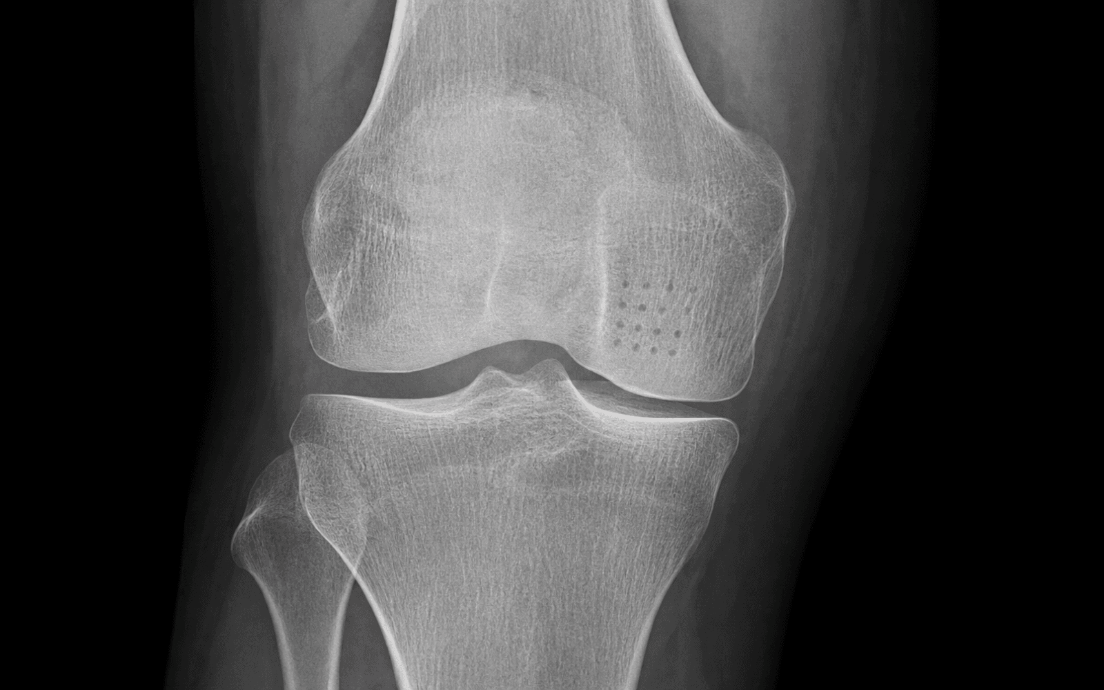
Weight-bearing AP, lateral, Rosenberg (45 degree PA) and skyline. Assess joint-space narrowing, alignment and OA grade. Normal X-rays do not exclude cartilage damage.
Cartilage-specific sequences are essential. Map lesion location, size, depth and containment; assess subchondral bone for oedema or cysts; identify associated meniscal or ligamentous pathology.
Full-length standing alignment films when malalignment is a clinical concern, to calculate mechanical axis deviation.
Look for the defect location and size, subchondral oedema (which may indicate ongoing damage), cyst formation (a relative contraindication), bone-marrow lesions, and any associated meniscal or ligamentous pathology that needs addressing at the same sitting.
Predicting outcome. These factors separate a lesion that will do well from one that will fail:
- Lesion under 2cm² - Age under 40 - Acute traumatic defect - Single lesion - Normal alignment - Compliant with rehabilitation
- Lesion over 4cm² - Age over 40 - Multiple lesions - Degenerative (versus traumatic) - Malalignment - Early weight-bearing - Subchondral bone disease
Consent. Counsel the patient explicitly that microfracture produces fibrocartilage (Type I collagen), not native hyaline cartilage (Type II), that the benefit is time-limited with deterioration common at 2-5 years, and that further surgery may be needed. Critically, warn that violating the subchondral plate can roughly triple the failure rate of any future ACI (Minas 2009) — so in a larger lesion that may later need cell-based repair, marrow stimulation should be chosen judiciously.
The Operation
The goal is an all-arthroscopic marrow-stimulation procedure: expose and assess the defect, debride it to stable vertical shoulders, remove the calcified cartilage layer, then perforate the subchondral plate at a defined spacing and depth to release marrow elements that form a clot and mature into a fibrocartilage repair. The exposure and the perforation technique are the heart of the operation.
Operative sequence
- Supine on the operating table with a lateral post or thigh holder; thigh tourniquet.
- Standard knee arthroscopy setup with controlled pump pressure.
- Establish the standard anterolateral (viewing) and anteromedial (working) portals; a supplementary portal directly above the lesion may be added so the awl can enter perpendicular to the articular surface.
- Complete any planned procedure (for example a meniscectomy) first.
- Inspect all compartments, locate and probe the chondral defect.
- Confirm it is full-thickness with exposed subchondral bone (ICRS Grade 4), measure the size precisely, and confirm containment with stable surrounding cartilage shoulders.
- Use a shaver and ring curette to remove loose, unstable cartilage flaps.
- Create stable vertical walls of healthy cartilage around the defect — a perpendicular rim that will hold the clot. Tapered, sloping shoulders will not contain the repair.
- Curette away the calcified cartilage layer overlying the subchondral bone to expose the bone beneath, but preserve the integrity of the subchondral plate itself — do not scrape deeply or gouge.
- With an arthroscopic awl or microfracture pick, make holes perpendicular to the articular surface.
- Space the holes 3-4mm apart, starting at the periphery and working toward the centre, preserving the bone bridges between them so the subchondral plate does not collapse.
- Penetrate 2-4mm into the subchondral bone to reach the marrow.
- Reduce or turn off the arthroscopic pump and watch for marrow bleeding and fat droplets rising from each hole — confirmation that the subchondral plate has been breached and MSCs can reach the defect.
- Remove the instruments, release the tourniquet and confirm haemostasis, and close the portal sites.
Microfracture produces fibrocartilage (Type I collagen), mechanically inferior to native hyaline cartilage (Type II) — lower compressive stiffness (roughly 50-80 percent), less water content and poorer wear resistance. This is why the benefit is time-limited (deterioration at 2-5 years) and why the technique is reserved for small lesions. Breaching the subchondral plate also roughly triples the failure rate of any future ACI (Minas 2009) — choose it deliberately.
The awl must enter perpendicular to the curved condylar surface — add a supplementary portal directly above the lesion if needed. Always confirm bleeding with the pump pressure reduced or off: fat droplets and marrow blood rising from every hole is the intra-operative proof of an adequate breach.
- Subchondral penetration releases marrow elements - Mesenchymal stem cells populate the defect - A blood clot forms as a scaffold for healing - CPM stimulates cartilage-like differentiation - Fibrocartilage fills the defect over 6-12 months
- Fibrocartilage is mechanically inferior - Subchondral changes develop over time - Cyclic loading degrades the repair tissue - Large lesions cannot fill adequately - Type I collagen lacks the resilience of Type II
Aftercare & Complications
Rehabilitation The rehabilitation protocol protects the forming fibrocartilage while using mechanical stimulus (CPM) to drive repair-tissue maturation. | Phase | Timing | Weight-bearing | Therapy | |-------|--------|----------------|---------| | 1 | 0-6 weeks | Strict non-weight-bearing (touch-down only) | CPM started immediately, 6-8 hours per day; active ROM of the unaffected joints | | 2 | 6-8 weeks | Progressive partial to full weight-bearing | Continued CPM and ROM; quadriceps activation and closed-chain strengthening introduced | | 3 | 8-16 weeks | Full weight-bearing as tolerated | Functional rehabilitation, proprioception and progressive strengthening | | 4 | 4-9 months | Full | Sport-specific training; return to sport at 9-12 months | Premature weight-bearing is the most common cause of failure — strict non-weight-bearing for 6-8 weeks is essential. CPM (or aggressive early ROM) promotes fibrocartilage maturation toward a more hyaline-like phenotype and prevents adhesions. Outcomes. Around 85 percent report good results at 2 years, but deterioration occurs after 2-5 years in the majority as the fibrocartilage degrades — microfracture may therefore serve as a bridge to definitive treatment in some patients. Complications
- Recognition
- Recurrent pain and effusion after early loading
- Prevention
- Strict non-weight-bearing for 6-8 weeks; clear patient counselling
- Management
- Repeat imaging; revision to ACI/MACI or OATS if symptomatic
- Recognition
- Pain and swelling at 1-2 years; MRI shows incomplete fill with subchondral oedema
- Prevention
- Correct patient selection (under 2cm², young, aligned)
- Management
- Escalate to ACI/MACI (preferred), OATS or allograft; address subchondral bone
- Recognition
- MRI subchondral oedema, cyst formation or bone overgrowth at the defect
- Prevention
- Avoid over-penetrating or debriding the subchondral plate; preserve bone bridges
- Management
- Bone grafting at revision if cystic; definitive cartilage procedure
- Recognition
- Higher failure rate if cell-based repair is attempted later
- Prevention
- Use marrow stimulation judiciously in lesions that may need ACI
- Management
- Counsel pre-operatively; factor prior microfracture into ACI planning
- Recognition
- Loss of flexion or extension during the restricted phase
- Prevention
- Early CPM and active ROM
- Management
- Aggressive physiotherapy; rare manipulation under anaesthesia
- Recognition
- Calf pain and swelling (risk from restricted weight-bearing)
- Prevention
- Chemical and mechanical prophylaxis as indicated
- Management
- Anticoagulation per protocol
- Recognition
- Increasing pain, effusion and fever
- Prevention
- Aseptic portal technique
- Management
- Urgent washout and antibiotics
- Recognition
- Worsening joint-space narrowing over years
- Prevention
- Treat only focal defects; address alignment
- Management
- Manage as OA; consider realignment or arthroplasty if advanced
Viva & Exam Focus
MICROMICRO — key principles
HOLESHOLES — the surgical technique
High-yield question points
Type I collagen — fibrocartilage. Microfracture does NOT regenerate native hyaline cartilage (Type II collagen). The fibrocartilage is mechanically inferior, which is the basis of its time-limited benefit.
Under 2cm². Lesions of 2-4cm² have acceptable but reduced outcomes; over 4cm² should be treated with OATS, ACI/MACI or allograft.
3-4mm apart (to preserve bone bridges and prevent subchondral collapse) and 2-4mm deep into the subchondral bone.
Strict non-weight-bearing for 6-8 weeks. Premature loading is the most common cause of failure.
Around 2-5 years. Short-term results are good (roughly 85 percent at 2 years) but decline as the fibrocartilage degrades under cyclic loading.
CPM is thought to promote fibrocartilage differentiation toward a more hyaline-like phenotype, while preventing adhesions and maintaining range of motion. The mechanical stimulus during early healing influences tissue quality.
Exam viva scenarios
Practise clinical reasoning and management decisions out loud
“A 28-year-old recreational footballer has a 1.5cm² full-thickness cartilage defect on the medial femoral condyle found incidentally during meniscectomy. How would you manage this?”
“A 35-year-old woman presents 18 months after microfracture for a 2.5cm² medial femoral condyle lesion with recurrent pain and swelling. MRI shows incomplete fill with subchondral oedema. How do you approach this?”
“A 42-year-old presents with a 5cm² cartilage defect on the lateral femoral condyle. The referring surgeon suggests microfracture. What are your thoughts?”
Definition
- Marrow-stimulation technique for cartilage repair
- Penetrate the subchondral plate to release MSCs
- Produces fibrocartilage (Type I collagen)
- First-line for small full-thickness defects
Key numbers
- Under 2cm² — optimal lesion size
- 3-4mm — hole spacing
- 2-4mm — hole depth into subchondral bone
- 6-8 weeks — non-weight-bearing
- Around 85 percent — good results at 2 years
- 2-5 years — typical time to deterioration
Ideal patient
- Young (under 40)
- Single lesion
- Under 2cm²
- Normal alignment
- Good subchondral bone
Technique (HOLES)
- Healthy borders (stable shoulders)
- Offset 3-4mm between holes
- Leave the subchondral plate intact
- Enter perpendicular with the awl
- See fat droplets (confirm bleeding)
Fibrocartilage limitations
- Type I collagen, not Type II hyaline
- Lower compressive stiffness (50-80 percent)
- Less water content than hyaline
- Poorer wear resistance
- Deteriorates over 2-5 years
When to avoid microfracture
- Lesion over 4cm²
- Age over 40 (relative)
- Diffuse OA (not focal)
- Uncorrected malalignment
- Bipolar lesions untreated
- Subchondral bone disease
Background & Evidence
The technique. Microfracture, popularised by Steadman, is a marrow-stimulation technique in the family of cartilage-repair procedures that breach the subchondral plate to recruit mesenchymal stem cells. It is the least technically demanding of these options, which accounts for its place as a common first-line treatment worldwide for small focal defects. Cartilage lesion grading (ICRS). Microfracture is primarily indicated for full-thickness (ICRS Grade 4) lesions.
- Description
- Normal cartilage
- Depth
- Intact
- Microfracture suitability
- No treatment needed
- Description
- Softening or superficial fissures
- Depth
- Superficial
- Microfracture suitability
- Conservative management
- Description
- Lesion depth under 50 percent
- Depth
- Partial thickness
- Microfracture suitability
- Usually conservative
- Description
- Lesion depth over 50 percent
- Depth
- Near full-thickness
- Microfracture suitability
- Consider microfracture if symptomatic
- Description
- Full-thickness, subchondral bone exposed
- Depth
- Full-thickness
- Microfracture suitability
- Microfracture indicated
Repair tissue properties. The biomechanical gap between the repair fibrocartilage and native hyaline cartilage explains the time-limited benefit:
- Hyaline cartilage
- Type II
- Fibrocartilage (microfracture)
- Type I (inferior)
- Hyaline cartilage
- High
- Fibrocartilage (microfracture)
- Lower (50-80 percent)
- Hyaline cartilage
- 65-80 percent
- Fibrocartilage (microfracture)
- Lower
- Hyaline cartilage
- High aggrecan
- Fibrocartilage (microfracture)
- Less organised
- Hyaline cartilage
- Excellent
- Fibrocartilage (microfracture)
- Inferior
- Hyaline cartilage
- Decades
- Fibrocartilage (microfracture)
- 2-5 years before degradation
Guidelines, registries & global practice
- Microfracture remains a common single-stage first-line option worldwide for small focal defects - Growing use of scaffold-augmented marrow stimulation (AMIC) for medium lesions - ICRS and ESSKA cartilage consensus favour cell-based or osteochondral repair over microfracture for defects over 2-3cm² - Fresh osteochondral allograft availability varies markedly by region (well established in North America, limited elsewhere by tissue-bank supply) - MACI/ACI availability and reimbursement differ by health system; access is the main practical constraint, not the technique itself
- Document lesion size precisely at arthroscopy - Record the alignment assessment - Document the technical parameters (spacing, depth) - Record that marrow bleeding was confirmed - Consent for the time-limited benefit
Key evidence. The systematic review by Mithoefer (2009) showed reliable functional improvement in the first 24 months but conflicting durability thereafter, with better outcomes in small lesions and younger patients. Steadman's long-term series (2003, mean 11 years) demonstrated durable gains in carefully selected young patients with isolated traumatic defects. The SUMMIT trial (Brittberg, 2018) showed MACI durably superior to microfracture for defects of 3cm² or larger at 5 years. The AMIC trial (Volz, 2017) showed a collagen scaffold protected against the mid-term decline seen with microfracture alone. Critically, Minas (2009) showed that prior marrow stimulation roughly triples the failure rate of subsequent ACI — marrow stimulation is not a consequence-free first step.
References
Clinical Efficacy of Microfracture - Systematic Review
- 28 studies, 3122 patients; mean follow-up 41 months (only 5 studies reached 5 years or more)
- Microfracture improved knee function in all studies during the first 24 months
- Durability of the initial improvement was conflicting - functional deterioration reported after 2 years in several series
- MRI defect fill was highly variable and correlated with functional outcome; repair tissue was limited hyaline-like fibrocartilage
Long-Term Microfracture for Traumatic Chondral Defects (Average 11 Years)
- 75 knees, isolated traumatic full-thickness defects, age 45 or younger, mean follow-up 11.3 years (range 7-17)
- Lysholm improved from 59 to 89 and Tegner from 3 to 6 (both significant)
- 80% of patients rated themselves improved at 7 years
- Younger age was an independent predictor of functional improvement
SUMMIT Trial - MACI versus Microfracture (5-Year RCT)
- 144 patients randomised, defects 3cm² or larger; 5-year data on 128 (89%)
- At 2 years MACI was already significantly superior to microfracture for the co-primary KOOS pain and function endpoint (P =.001)
- At 5 years the MACI advantage in KOOS pain and function was maintained and remained significant (P =.022)
- MRI defect fill improved with both treatments with no significant between-group difference
AMIC versus Microfracture - Sustained Benefit at 5 Years (RCT)
- 47 patients randomised, mean defect 3.6cm², to microfracture or AMIC (type I/III collagen membrane, sutured or glued)
- All groups improved for the first 2 years
- From 2 to 5 years the microfracture group showed progressive significant score deterioration while AMIC scores stayed stable
- MRI defect fill was more complete in the AMIC groups at 2 and 5 years
Prior Marrow Stimulation Increases Subsequent ACI Failure
- 321 patients (522 defects) treated with ACI; grouped by whether the subchondral bone had been violated by prior marrow stimulation
- Defects with prior marrow stimulation failed at roughly 3 times the rate of untreated defects (26% versus 8%)
- Failure rates were similar across drilling (28%), abrasion (27%) and microfracture (20%)
- Penetrating the subchondral bone has a strong negative effect on later cell-based repair