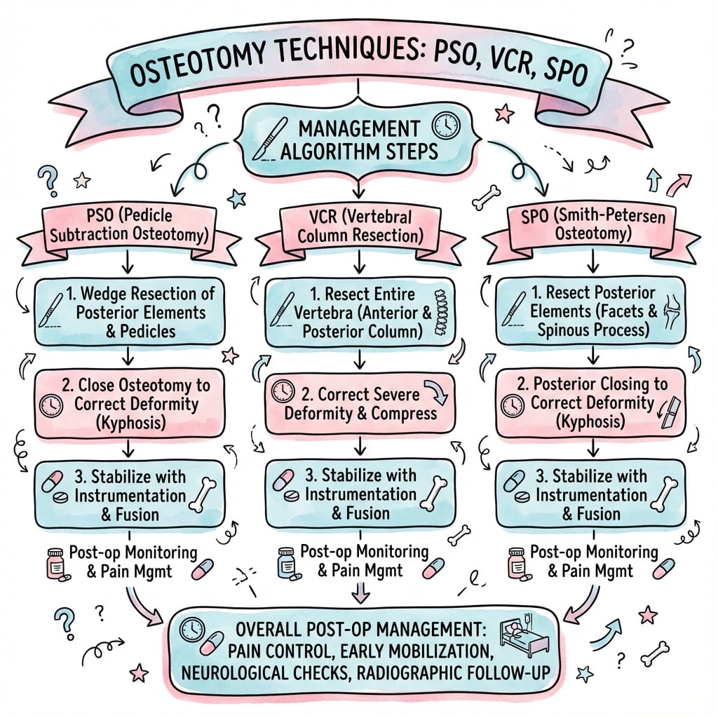
PSO, VCR, and SPO for fixed deformity correction
- SPO (Smith-Petersen) is a posterior-column-only osteotomy: it opens posteriorly and hinges on an intact anterior column (the ALL), giving about 10 degrees per level with low neurological risk (1–3 percent).
- PSO (pedicle subtraction) resects all three columns through a posterior approach as a closing wedge of the vertebral body, achieving 30–40 degrees at a single level (usually L3) with moderate neurological risk (5–11 percent).
- VCR (vertebral column resection) removes an entire vertebra for 50–70 degrees of correction but carries the highest neurological risk (10–25 percent) and the highest blood loss.
- Match the osteotomy to the deformity: SVA less than 100mm and flexible favours multiple SPOs; SVA 100–150mm and rigid favours a single PSO; SVA greater than 150mm or a sharp angular deformity favours a PSO plus SPOs or a VCR.
- Multimodal neuromonitoring (SSEP plus transcranial MEP) is mandatory for any three-column osteotomy; maintain MAP greater than 85 mmHg during closure and follow the structured rescue protocol the instant a signal changes.
When & Why
Indication. A spinal osteotomy is offered for a fixed spinal deformity that produces sagittal and/or coronal imbalance — typically inability to stand upright, forward-leaning posture, fatigue and pain — that has failed at least six months of conservative management and is not correctable by positioning alone. The choice of osteotomy is driven by the magnitude, rigidity and shape of the deformity, not by surgeon preference in isolation. The single decision that matters — match the osteotomy to the deformity. The correction available rises, and so does the risk, as you move from posterior column to three-column resection:
Posterior column only, about 10 degrees per level. Needs a mobile anterior column to act as the hinge. Flexible deformity, ankylosing spondylitis with mild-moderate kyphosis, iatrogenic flatback. Low neurological risk (1–3 percent).
All three columns through a posterior approach, 30–40 degrees at one level. Rigid flatback, fixed sagittal imbalance, PI-LL mismatch greater than 20 degrees, failed fusion with kyphosis. Moderate neurological risk (5–11 percent).
Complete vertebrectomy, 50–70 degrees. Severe rigid angular kyphosis, kyphoscoliosis, congenital deformity, tumour. Reserved for the most extreme deformities. High neurological risk (10–25 percent) and large blood loss.
Osteotomy selection algorithm. Decide from the standing films, not the supine films: 1. Deformity magnitude — SVA less than 100mm with a flexible deformity: multiple SPOs. SVA 100–150mm with a rigid deformity: a single PSO at L3. SVA greater than 150mm or a sharp angular deformity: a PSO plus SPOs, or a VCR. 2. Rigidity — greater than 20 degrees of correction on flexibility (supine hyperextension or traction) films: SPOs may suffice. Less than 10 degrees of correction: a bony osteotomy (PSO or VCR) is required. 3. Correction available — SPO about 10 degrees per level (posterior column); PSO 30–40 degrees per level (three columns); VCR 50–70 degrees per level (complete vertebrectomy).
The ideal candidate for a PSO has a fixed sagittal imbalance (SVA greater than 50mm with a PI-LL mismatch greater than 20 degrees), has failed conservative care, has a rigid deformity not correctable by positioning, has good bone quality (T-score greater than -2.0), is medically fit for major surgery, and holds realistic expectations. Contraindications. Absolute: active infection at the osteotomy site; severe untreated osteoporosis (T-score less than -3.5); prohibitive medical comorbidity (severe cardiac or pulmonary disease); unrealistic expectations. Relative: age greater than 75 years; moderate osteoporosis (T-score -2.5 to -3.0); prior radiation to the site; severe obesity (BMI greater than 40); revision at the same level. Deformity assessment. Standing full-length AP and lateral films are mandatory. Normal sagittal parameters: SVA less than 50mm; PI-LL mismatch within 10 degrees; pelvic tilt less than 20 degrees; thoracic kyphosis 20–50 degrees. Supine hyperextension and traction films assess flexibility and distinguish a flexible deformity (greater than 20 degrees correction, consider SPOs) from a rigid one (less than 10 degrees correction, a bony osteotomy is needed). Preoperative optimisation. Cardiac and pulmonary clearance (PFTs if restrictive lung disease); renal function and a type and screen (anticipate a 2–4 unit transfusion); a documented baseline neurological exam and MRI to assess the cord; and a DEXA — osteoporosis must be optimised (vitamin D, calcium, teriparatide) and cement augmentation considered when the T-score is less than -2.5.
The Operation
The goal of every spinal osteotomy is to restore sagittal and coronal balance by removing a controlled wedge of bone and closing the defect under instrumentation, with the neural elements protected and perfused throughout. All three osteotomies share the same posterior midline exposure, which simply scales with the magnitude of the resection — smaller for an SPO, wide (3–4 segments above and below) for a PSO, and circumferential (5–6 segments above and below) for a VCR. The pedicle subtraction osteotomy is the workhorse three-column procedure, so it is taught here in full as the reference technique; the SPO and VCR are then presented as its simpler and more extreme variants.
- Columns
- Posterior only
- Correction / level
- About 10°
- Axis of rotation
- Anterior to the ALL (hinge)
- Spinal shortening
- Minimal
- Neuro risk
- Low (1–3%)
- Columns
- All three
- Correction / level
- 30–40°
- Axis of rotation
- Middle of the vertebral body
- Spinal shortening
- 10–15mm
- Neuro risk
- Moderate (5–11%)
- Columns
- All three (complete)
- Correction / level
- 50–70°
- Axis of rotation
- Surgeon-controlled
- Spinal shortening
- 20–40mm
- Neuro risk
- High (10–25%)
Pedicle subtraction osteotomy (PSO) — operative sequence
- Prone on a relton-hall or jackson frame with all pressure points padded; neuromonitoring baseline (SSEP and MEP) recorded before incision.
- Midline incision carried three to four levels above and below the planned osteotomy; subperiosteal exposure of the laminae, facets and transverse processes out to the apophyseal joints.
- Confirm the osteotomy level (usually L3) with intraoperative fluoroscopy and a marker radiograph — never rely on surface counting.
- Place all pedicle screws first, including at the osteotomy level, before any bony resection.
- Mount temporary rods bilaterally to stabilise the construct during the osteotomy and prevent catastrophic translation while the posterior elements are removed.
- Complete laminectomy at L3 and bilateral facetectomy (L2–L3 and L3–L4); resect the spinous processes of L2, L3 and L4.
- Expose both pedicles circumferentially and identify and protect the nerve roots — the exiting L3 root in the foramen is the structure most at risk during pedicle work.
- Remove both pedicles with osteotomes or a high-speed burr, working from lateral to medial to keep instruments away from the theca.
- Create a cortical shell of the posterior vertebral body wall and expose the lateral walls of the body.
- With curettes, osteotomes and a high-speed burr create a closing wedge — wider posteriorly, angled about 30–40 degrees from the horizontal.
- Preserve a 1–2mm shell of anterior cortex to act as a controlled hinge during closure; copious irrigation prevents thermal injury.
- Keep the temporary rods under moderate compression while completing the second side to prevent premature or uncontrolled closure.
- Maintain a decompressed canal and keep the neural elements under direct vision.
- Gradually compress the bilateral rods; the posterior wedge closes and the preserved anterior cortex fractures as a controlled hinge.
- Monitor neuromonitoring continuously — stop and run the rescue protocol for any significant signal change.
- Aim for 10–15mm of shortening, which relaxes the neural elements; maintain MAP greater than 85 mmHg for cord perfusion.
- Replace the temporary rods with permanent cobalt-chromium rods (6.0–6.35mm); use dual rods for high-stress cases.
- Add cross-links every three to four levels for rotational stability and lay bone graft along the entire construct.
The SPO resects the posterior column only and hinges on an intact anterior column, giving about 10 degrees per level. Steps: (1) midline exposure over the planned levels with subperiosteal dissection of the laminae and facets and radiographic level confirmation; (2) remove the inferior facet completely (leave the superior facet intact), the ligamentum flavum, the spinous process and the interspinous ligament, thinning the superior lamina to a hinge point to create a V-shaped gap; (3) place pedicle screws two to three levels above and below each osteotomy with temporary rods; (4) close by gradual rod compression — the anterior column acts as the hinge, so it must be intact and mobile; (5) final rods, cross-links and bone graft. Pearls: multiple levels are better than one (they distribute stress), site the osteotomy at the apex of the kyphosis, and never violate the anterior longitudinal ligament. Pitfalls: fracture through the anterior column (loss of the hinge and instability), excessive correction at a single level (neurological injury), and inadequate facet resection (limited correction and rod stress).
Vertebral column resection (VCR) — the extreme variant
- Wide exposure five to six levels above and below with circumferential mobilisation of the vertebra to be resected.
- Place all pedicle screws except at the VCR level; mount temporary rods.
- Complete laminectomy at the VCR level and adjacent levels, with bilateral facetectomies above and below.
- Identify and protect the nerve roots; the dura is fully exposed.
- Resect both pedicles with a burr or osteotome and remove the posterior wall of the vertebral body to create a working channel to the anterior column.
- Blunt dissection laterally to protect the great vessels; ligate segmental arteries only as necessary.
- Resect the vertebral body completely, removing the anterior cortex last, and elevate and excise the discs above and below.
- Maintain temporary rods throughout; the dura is completely exposed with a 360-degree decompression and the sac of neural elements is free-floating.
- Slowly compress the rods to close the gap under continuous neuromonitoring.
- Translate the spine for coronal correction and rotate it for rotational correction; aim for 20–40mm of shortening.
- Place a structural graft (cage, mesh or allograft) for anterior column support, bone ingrowth and fusion, and to prevent recurrence of kyphosis.
- Robust fixation with dual rods (a four-rod construct) of cobalt-chromium (6.35mm), multiple cross-links, and fixation extended four to five levels beyond the VCR.
A posterior-only (single-stage) VCR is preferred for most cases when the surgeon is experienced — one surgery, less morbidity, shorter recovery — at the cost of limited anterior access, higher blood loss and greater technical demand. A staged anterior-posterior approach gives better anterior visualisation and controlled resection but means two surgeries with greater morbidity and longer stay; it is reserved for tumour resection, extensive anterior pathology or vascular anomalies.
The artery of Adamkiewicz (the major segmental feeder to the anterior spinal artery) typically arises between T9 and L2 on the left in about 75 percent of people. Injury causes anterior cord ischaemia and devastating paraplegia with no effective treatment once it occurs. Prevention is everything: maintain MAP greater than 85 mmHg (90–100 mmHg if a signal change occurs), avoid excessive hypotensive anaesthesia during closure, and respect the lateral vertebral body wall during resection. Vascular surgery standby is wise for thoracic VCR.
A MEP amplitude drop greater than 50 percent, or an SSEP amplitude drop greater than 50 percent or latency increase greater than 10 percent, is a significant alert. Respond in order: (1) notify the team and stop all compression immediately; (2) augment the blood pressure to MAP 90–100 mmHg for cord perfusion; (3) release the recent manipulation — reduce the correction by 5–10 degrees to relieve neural tension; (4) reassess after 10–15 minutes; (5) if signals do not return, perform a wake-up test; (6) accept less correction if needed — neurological preservation always outranks an ideal radiograph.
Use a four-rod technique (temporary rods during resection, permanent rods for closure) to control the osteotomy. Run a cell saver to reduce allogeneic transfusion in these high-blood-loss cases. Hypotensive anaesthesia (MAP 65–70 mmHg) reduces blood loss during the resection, but raise the MAP to greater than 85 mmHg during closure for cord perfusion. Close in small increments (5–10 degrees at a time), waiting two to three minutes between compressions, and keep a wake-up test ready.
Aftercare & Complications
Immediate postoperative care (ICU, first 24–48 hours). Frequent neurological checks every one to two hours (all myotomes, sensation and reflexes), with early waking to assess for deficit and an urgent MRI if any new deficit appears (rule out epidural haematoma, hardware malposition or cord ischaemia). Maintain MAP greater than 85 mmHg for the first 48 hours (vasopressors — phenylephrine or norepinephrine — as needed), target haemoglobin greater than 8 g/dL and urine output greater than 0.5 mL/kg/hour. Use multimodal analgesia (acetaminophen, NSAIDs if renal function allows, gabapentin, PCA opioids) and avoid excessive opioids. Remove closed suction drains when output falls below 50 mL per eight hours (typically day 2–3); drainage greater than 500 mL in eight hours prompts consideration of re-exploration. Early mobilisation and bracing. Sit at the edge of bed on day 1 if neurologically intact, stand and walk with assistance by day 2–3, aiming for independent walking by discharge. A TLSO brace is worn for 12 weeks after a PSO or VCR (not required for an SPO alone unless multiple levels or poor bone stock). Early physiotherapy reduces DVT, pneumonia and ileus — gentle core isometrics with no trunk rotation for 12 weeks, plus gait and ADL training. Neurological deficit after surgery. An immediate deficit is an emergency: the most common cause is an epidural haematoma, and decompression within eight hours gives the best recovery; other causes are hardware malposition (revise to remove the offending screw/rod) and cord ischaemia (augment MAP to 90–100 mmHg). A delayed deficit (day 1–7) is investigated identically with urgent MRI. Incomplete deficits recover substantially in 50–70 percent; complete deficits carry a poor prognosis (less than 20 percent recovery); nerve-root injuries fare better than cord injuries.
- Overall neuro risk
- 1–3%
- Motor / sensory
- 0.5–1% / 1–2%
- Mechanisms
- Nerve-root traction from excessive correction at one level
- Recovery
- 70–80% significant recovery
- Overall neuro risk
- 5–11%
- Motor / sensory
- 3–6% / 4–8%
- Mechanisms
- Root injury during pedicle resection, cord ischaemia from correction, epidural haematoma
- Recovery
- 50–60% significant recovery
- Overall neuro risk
- 10–25%
- Motor / sensory
- 8–15% / 10–18%
- Mechanisms
- Cord manipulation, vascular injury (Adamkiewicz), excessive shortening or translation
- Recovery
- 40–50% significant recovery
Other complications. Vascular — great-vessel or iliac injury is rare (less than 1 percent) but life-threatening; blunt lateral dissection, vascular standby for thoracic VCR, and avoiding anterior-cortex violation until final closure protect the vessels. Mechanical — rod fracture 5–10 percent at two years (prevent with dual 6.35mm cobalt-chromium rods and solid fusion); screw loosening 3–5 percent (higher in osteoporosis — cement-augment when T-score less than -2.5); proximal junctional kyphosis 20–40 percent (prophylactic vertebroplasty at the UIV, gradual lordosis transition); pseudarthrosis 10–20 percent (smoking cessation, autograft plus BMP, teriparatide). Wound — haematoma/seroma, superficial or deep infection (irrigation and debridement, retain hardware if early). Medical — DVT/PE (mechanical prophylaxis day 0, chemical day 1 after drain removal), ileus, pneumonia and delirium (especially over age 65).
- Achieve planned correction
- 90–95%
- Significant pain relief
- 70–80%
- Fusion rate
- 90–95%
- Overall complications
- 15–25% (mostly minor)
- Satisfaction
- 75–85%
- Achieve planned correction
- 80–90%
- Significant pain relief
- 65–75%
- Fusion rate
- 85–90%
- Overall complications
- 40–50% (major 10–15%)
- Satisfaction
- 65–75%
- Achieve planned correction
- 75–85%
- Significant pain relief
- 60–70%
- Fusion rate
- 75–85%
- Overall complications
- 50–70% (major 20–30%)
- Satisfaction
- 55–65%
Surveillance. Two weeks (wound check, staple removal, neurological exam), six weeks (standing radiographs, advance therapy), 12 weeks (radiographs, discontinue brace), six months (radiographs, CT if fusion in doubt, return to light activity), 12 months (radiographs and CT to confirm fusion — greater than 50 percent bridging bone — and return to full activity), and 24 months (final films, assess adjacent-segment degeneration). Track ODI, SRS-22, VAS pain and walking distance. At five years, 75–85 percent maintain SVA less than 50mm; reoperation runs 15–25 percent (PJK most common, then pseudarthrosis, rod fracture and infection). Predictors. Favourable: appropriate osteotomy selection, solid fusion, SVA restored to less than 50mm with PI-LL within 10 degrees, good bone quality, age less than 65, non-smoker, a single (non-revision) osteotomy. Unfavourable: severe osteoporosis (T-score less than -3.0), active smoking, multiple comorbidities (Charlson index greater than 3), revision at the same level, chronic preoperative opioid use, poor nutrition (albumin less than 3.5), and unrealistic expectations.
Viva & Exam Focus
Spinal osteotomies deliver powerful deformity correction at the price of rising complexity and risk. SPO is a posterior-column-only procedure giving about 10 degrees per level through a posterior-opening wedge that hinges on an intact anterior column. PSO resects all three columns via a posterior approach for 30–40 degrees at one level with 10–15mm of shortening. VCR removes an entire vertebra for 50–70 degrees but carries the highest risk. Neurological risk climbs with complexity — SPO 1–3 percent, PSO 5–11 percent, VCR 10–25 percent — so match the osteotomy to the deformity and treat multimodal neuromonitoring as non-negotiable.
An SPO/Ponte osteotomy (Schwab grade 1–2) gives about 10 degrees per level (posterior column only). A PSO (grade 3) gives about 30–40 degrees at a single level (all three columns; published means cluster near 30 degrees). A VCR (grade 4–6) gives greater than 40 degrees, with near-unlimited correction. Choose by the correction needed and the deformity type.
An SPO resects the posterior elements only; correction occurs through the disc space opening anteriorly and hinges on the ALL, so it needs a mobile anterior column. A PSO resects the pedicles and a wedge of vertebral body; closure is bone-on-bone with no disc involvement. A PSO is therefore preferred when the anterior column is fused or osteoporotic.
PSO blood loss averages 1.5–3 litres with a neurological risk of about 5–10 percent; VCR blood loss averages 3–5 litres with a neurological risk of about 15–20 percent. A VCR requires circumferential decompression with temporary spinal-cord instability. Both need neuromonitoring; the VCR is reserved for severe rigid deformity, tumour or failed prior surgery.
Grade 1 partial facetectomy; grade 2 complete facetectomy (Ponte); grade 3 PSO through a single vertebra; grade 4 VCR of a single vertebra; grade 5 VCR of two adjacent vertebrae; grade 6 VCR of three or more. Higher grades allow more correction but carry greater morbidity and guide surgical planning.
Fixed (rigid) sagittal imbalance not correctable by positioning or a posterior-column osteotomy — typically SVA greater than 5cm with a PI-LL mismatch greater than 20 degrees (often much larger); an ankylosed spine (ankylosing spondylitis, DISH); a failed prior fusion with kyphosis; and iatrogenic flatback syndrome. It is performed at L3 for lumbar deformity (L2–L4 acceptable) or at the apex of a thoracic/thoracolumbar deformity, with the goal of restoring SVA to within about 5cm of the posterosuperior corner of S1 and matching lumbar lordosis to pelvic incidence.
OSTEOTOMYOSTEOTOMY — patient selection criteria
TECHNIQUETECHNIQUE — stepwise PSO execution
Clinical Decision Scenarios
Practise clinical reasoning and management decisions out loud
“You are performing an L3 PSO for flatback deformity in a 62-year-old woman. After completing the vertebral body resection you begin gradual closure; at 20 degrees of correction neurophysiology reports bilateral MEP amplitudes have dropped by 70 percent from baseline. SSEPs are stable. The patient is haemodynamically stable with a MAP of 75 mmHg. How do you manage this?”
“A 58-year-old man with a previous L4–S1 fusion presents with progressive inability to stand upright. Standing films show SVA 135mm, PI-LL mismatch 38 degrees and pelvic tilt 35 degrees; a supine hyperextension film shows minimal change (SVA 125mm). He has moderate osteoporosis (T-score -2.3), controlled diabetes (HbA1c 6.9 percent) and BMI 32, and has failed 12 months of conservative care. What is your plan?”
Match the osteotomy to the deformity
- SPO: posterior column only, about 10 degrees per level, flexible deformity, intact anterior column as hinge, low risk (1–3% neuro)
- PSO: all three columns, 30–40 degrees per level, rigid flatback or severe sagittal imbalance, moderate risk (5–11% neuro)
- VCR: complete vertebrectomy, 50–70 degrees, severe rigid angular deformity, high risk (10–25% neuro)
- Selection: SVA less than 100mm flexible then SPOs; SVA 100–150mm rigid then PSO; SVA greater than 150mm or angular then PSO plus SPOs or VCR
PSO level selection
- L3 most common — below the conus (T12–L2), cauda equina tolerates manipulation, good bone stock
- L2 acceptable if L3 anatomy is unfavourable
- Avoid L4 (too close to L5–S1) and the thoracic spine (cord present)
- Avoid T12–L2 (conus medullaris)
- Flexibility films distinguish rigid (less than 10 degrees correction) from flexible (greater than 20 degrees)
PSO technique — TECHNIQUE mnemonic
- T — Temporary rods after all instrumentation
- E — Expose and remove posterior elements (laminectomy L3, facetectomy L2–L3 and L3–L4)
- C — Cut pedicles carefully (preserve the L3 roots)
- H — Hollow the vertebral body (30–40 degree wedge, preserve 1–2mm anterior cortex)
- N — Neuromonitoring active throughout (SSEP, MEP)
- I — Instrument with permanent rods (gradual compression)
- Q — Quality check fluoroscopy; U — Utilise cross-links
- E — Elevate blood pressure (MAP greater than 85) during closure
Neuromonitoring and the response to change
- Maintain MAP greater than 85 mmHg for cord perfusion
- Observe SSEP and MEP continuously
- Notify the surgeon of ANY signal change immediately
- Close incrementally (gradual, never sudden)
- Wake-up test if signals are lost
- Optimise BP to 90–100 mmHg if a change occurs
- Release correction if signals do not recover (safety first)
- Alert criteria: MEP amplitude drop greater than 50%; SSEP amplitude drop greater than 50% or latency increase greater than 10%
Patient selection — OSTEOTOMY mnemonic
- Objectives clear; Sagittal imbalance severe; Trial of conservative care failed
- Expectations realistic; Osteoporosis optimised; Type matches deformity
- Other comorbidities controlled; Medical fitness confirmed; Young enough (under 75 preferred)
- Absolute contraindications: active infection, untreated severe osteoporosis, prohibitive medical risk
Complications by type
- SPO: 1–3% neuro, 15–25% overall (mostly minor), 90–95% fusion
- PSO: 5–11% neuro, 40–50% overall (major 10–15%), 85–90% fusion, 5–10% rod fracture at 2 years
- VCR: 10–25% neuro, 50–70% overall (major 20–30%), 75–85% fusion, blood loss 2–5 litres
- All types: DVT/PE 3–5%, infection 3–8%, PJK 20–40%
Intraoperative pearls
- Four-rod technique: temporary rods during resection, permanent rods for closure
- Cell saver to reduce allogeneic transfusion (important in PSO/VCR)
- Hypotensive anaesthesia (MAP 65–70) during resection to reduce blood loss
- MAP greater than 85 during closure for cord perfusion
- Gradual closure: 5–10 degrees at a time, monitor signals, wait two to three minutes between compressions
- Wake-up test ready if MEPs are lost and do not recover
Expected outcomes
- SPO: 90–95% achieve correction, 70–80% pain relief, 75–85% satisfaction, 90–95% fusion
- PSO: 80–90% achieve correction (SVA less than 50mm in 85–90%), 65–75% pain relief, 85–90% fusion
- VCR: 75–85% achieve correction, 60–70% pain relief, 55–65% satisfaction, 75–85% fusion
- Favourable: appropriate selection, solid fusion, SVA less than 50mm, age less than 65, non-smoker
- Unfavourable: severe osteoporosis, smoking, multiple comorbidities, revision at same level
Postoperative management
- ICU for 24–48 hours: neuro checks every 1–2 hours, MAP greater than 85 mmHg
- Early mobilisation: sit day 1, stand/walk day 2, reduces DVT/pneumonia/ileus
- TLSO brace for 12 weeks after PSO/VCR
- DVT prophylaxis: mechanical day 0, chemical day 1 after drain removal
- Follow-up: 6-week radiographs, 12-week brace off, 12-month CT to confirm fusion
- Urgent MRI for any deficit — rule out haematoma, hardware malposition, cord ischaemia
Background & Evidence
Epidemiology and service configuration. Three-column osteotomies (PSO and VCR) are concentrated at high-volume tertiary deformity centres with neuromonitoring, cell salvage, blood-bank support and a dedicated neuro-spinal ICU. Surgeon and centre volume correlate with lower complication and reoperation rates, and most units restrict PSO and VCR to fellowship-trained deformity surgeons within multidisciplinary clinics (surgery, anaesthesia, neurophysiology, rehabilitation). Named-society guidance and registries. The Scoliosis Research Society (SRS) and AOSpine define the Schwab–SRS adult-deformity and osteotomy classifications, the morbidity-and-mortality reporting standards, and the deformity-fellowship curricula used internationally. NASS and EUROSPINE publish perioperative neuromonitoring statements; multimodal SSEP plus transcranial MEP is regarded as the standard of care for deformity correction. The AO Foundation / AOSpine teaches the posterior-only VCR and three-column osteotomy techniques. Large prospective multicentre adult-deformity databases (the International Spine Study Group and European cohorts) drive the evidence on three-column osteotomy complications, proximal junctional failure and the protective effect of supplemental rods. The main genuine global variation is in rod metallurgy and multi-rod use (cobalt-chromium versus titanium, two-rod versus four-rod constructs) and in biologic augmentation (iliac-crest autograft, allograft, BMP — used off-label in the spine in many jurisdictions). Denis three-column concept. The spine is divided into an anterior column (ALL, anterior half of the vertebral body and disc), a middle column (posterior half of the body and disc, and the PLL) and a posterior column (pedicles, laminae, facets, ligamentum flavum and the interspinous and supraspinous ligaments). An SPO affects the posterior column only; a PSO affects all three columns through a closing wedge of the body; a VCR removes all three columns completely. Biomechanics. The axis of rotation differs: for an SPO it lies anterior to the ALL (the posterior gap opens and the anterior column closes, so the anterior column must be intact and mobile); for a PSO it lies through the middle of the vertebral body (symmetrical closure, no anterior hinge needed); for a VCR it is surgeon-controlled via rod position (maximum flexibility, allowing simultaneous sagittal and coronal correction but requiring temporary instability during closure). Correction potential is about 10 degrees per level for an SPO (range 5–15), 30–40 degrees for a PSO (range 25–45) and 50–70 degrees for a VCR (range 40–80). Spinal shortening is minimal for an SPO, 10–15mm for a PSO and 20–40mm for a VCR — shortening relaxes the neural elements but may affect visceral structures. Vascular anatomy. Lumbar segmental arteries exit at the mid-vertebral body and are at risk during a PSO or VCR; their sacrifice is usually tolerated through collaterals but bleeding can be brisk. The artery of Adamkiewicz typically arises at T9–L2 on the left in about 75 percent of people and is the major feeder to the anterior spinal artery; its injury causes paraplegia (anterior cord syndrome) and is prevented by maintaining MAP greater than 85 mmHg and respecting the lateral body wall. Neural anatomy. The conus medullaris lies at T12–L2 (higher myelopathy risk if the osteotomy is here, which is why L3–L4 is preferred for a PSO). Below L2 the cauda equina is more tolerant of manipulation and individual roots can be mobilised. Distinguish the exiting root in the neural foramen (vulnerable to pedicle work) from the traversing root medial to the pedicle (vulnerable to canal work). Cervical osteotomies carry the highest neurological risk (cord injury equals quadriplegia) and are usually SPO or opening-wedge procedures; thoracic osteotomies (T1–T10) place the cord at risk in a narrow canal; thoracolumbar (T11–L2) is the conus level and best avoided; L3–L4 is preferred for a PSO; L5 is avoided (proximity to the sacrum and large vessels). Anatomical-region and deformity-type considerations. By region, cervical osteotomies are the highest risk (typically SPO/opening-wedge; PSO/VCR rarely performed); thoracic osteotomies carry cord/myelopathy risk; thoracolumbar T11–L2 is the conus level to avoid; lumbar L3–L4 is preferred for a PSO; L5 is avoided. By deformity type: a flatback is corrected by a PSO; kyphosis by a PSO or VCR depending on magnitude and rigidity; chin-on-chest by multiple SPOs or a cervicothoracic VCR; scoliosis or coronal imbalance by an asymmetric VCR; and kyphoscoliosis or a 3D deformity by a VCR or multiple osteotomies.
- Technique
- Partial facetectomy
- Structures resected
- Inferior facet only
- Columns
- Posterior (partial)
- Correction / level
- Less than 10°
- Neuro risk
- Very low
- Technique
- SPO (Smith-Petersen)
- Structures resected
- Facets, ligamentum flavum, spinous process
- Columns
- Posterior (complete)
- Correction / level
- About 10°
- Neuro risk
- Low (1–3%)
- Technique
- PSO (pedicle subtraction)
- Structures resected
- Posterior elements, pedicles, posterior body wedge
- Columns
- All three
- Correction / level
- 30–40°
- Neuro risk
- Moderate (5–11%)
- Technique
- VCR (posterior only)
- Structures resected
- Entire vertebra from a posterior approach
- Columns
- All three (complete)
- Correction / level
- 50–60°
- Neuro risk
- High (10–15%)
- Technique
- VCR (staged posterior-anterior)
- Structures resected
- Complete vertebrectomy via two approaches
- Columns
- All three, wide exposure
- Correction / level
- 60–70°
- Neuro risk
- Very high (15–20%)
- Technique
- VCR (multi-level / asymmetric)
- Structures resected
- Multiple vertebrae or asymmetric resection
- Columns
- Extended multi-level
- Correction / level
- Greater than 70°
- Neuro risk
- Very high (20–25%)
Key evidence in brief. Cho and Bridwell showed that multiple SPOs give angular correction similar to a single PSO but a PSO restores true sagittal balance far more (11.2cm versus 5.5cm improvement in the C7 plumb), at the cost of higher blood loss. Auerbach and colleagues reported a roughly one-in-three major-complication rate across 240 three-column osteotomies, with preoperative sagittal imbalance of 40mm or greater, age 60 or older and three or more comorbidities as the key risk factors — yet a major complication did not reduce final SRS scores at two years. Thirumala's meta-analysis established combined SSEP plus transcranial MEP monitoring as a high-specificity early-warning system (pooled specificity 94.4 percent) and the standard of care. Lenke demonstrated that single-stage posterior-only VCR can achieve dramatic correction with no permanent neurological deterioration when evoked-potential loss is acted on promptly. Passias showed that across a decade of practice, supplemental (accessory) rods and PJF prophylaxis have reduced rod fracture, junctional failure and reoperation while improving patient-reported outcomes — robust multi-rod fixation is now expected around PSO and VCR levels. Related topics. Adult deformity surgery (the primary indication); revision deformity surgery (often needs a PSO or VCR); proximal junctional kyphosis (a common complication); spinal cord monitoring (essential for safe execution); flatback deformity (the classic PSO indication); ankylosing spondylitis (the classic SPO indication); sagittal balance (the parameters that guide selection); and rod fractures (the characteristic mechanical failure).
References
Smith-Petersen osteotomy versus pedicle subtraction osteotomy for fixed sagittal imbalance
- 30 SPO patients (14 with three or more SPOs) compared with 41 single-level PSO patients at one institution.
- Mean correction per SPO segment was 10.7 degrees; three or more SPOs gave total kyphosis correction (33.0 degrees) nearly identical to a single PSO (31.7 degrees).
- A single PSO restored sagittal balance significantly more than three or more SPOs (11.2cm versus 5.5cm improvement in the C7 plumb, p less than 0.01).
- Three or more SPOs decompensated patients toward the concavity more often than a single PSO (p less than 0.02).
- Estimated blood loss was substantially higher with PSO (2617mL versus 1398mL, p less than 0.001).
Major complications of three-column osteotomies (PSO versus VCR)
- 240 consecutive three-column osteotomies (156 PSO, 84 VCR); 105 patients with complete 2-year data analysed.
- Major complications occurred in 35 percent overall, at similar rates for PSO (38 percent) and VCR (22 percent).
- Independent risk factors for a major complication: preoperative sagittal imbalance of 40mm or greater, age 60 years or older, and three or more medical comorbidities.
- Mean estimated blood loss was higher in PSO (1867mL) than VCR (1278mL); PSO patients were older (53 versus 29 years).
- A major complication did not reduce ultimate Scoliosis Research Society outcome scores at 2 years.
Diagnostic accuracy of combined SSEP plus transcranial MEP monitoring in deformity surgery
- Systematic review and meta-analysis of 7 studies, 2052 idiopathic scoliosis correction patients.
- Incidence of a new neurological deficit was 0.93 percent.
- Combined SSEP plus transcranial MEP pooled sensitivity 82.6 percent and specificity 94.4 percent (area under the curve 0.928).
- Patients with a new deficit were 106 times more likely to have shown an intraoperative SSEP and/or MEP change (diagnostic odds ratio 106).
- Combined multimodality monitoring outperformed either modality alone.
All-posterior vertebral column resection for severe spinal deformity
- 43 patients with severe scoliosis, global or angular kyphosis, or kyphoscoliosis treated by single-stage posterior-only VCR with pedicle screws and anterior cages.
- 40 of 43 procedures (93 percent) were performed at L1 or cephalad, within the spinal-cord territory.
- 7 patients (18 percent) lost intraoperative evoked-potential data during correction; signals returned to baseline after prompt surgical intervention in every case.
- No patient had permanent neurological deterioration; 2 transient nerve-root palsies resolved spontaneously.
- Single-stage posterior VCR obviates the need for a circumferential (anterior plus posterior) approach.
Three-column osteotomy trends: supplemental rods reduce junctional and hardware failure
- 752 adult spinal deformity patients with 2-year data; 138 underwent a three-column osteotomy.
- Over 2008–2018 the rate of three-column osteotomy use fell (31 percent to 21 percent) as anterior and lateral interbody fusion use rose.
- More recent cases used supplemental (accessory) rods far more often (odds ratio 21.8).
- The supplemental-rod era showed lower proximal junctional failure (odds ratio 0.23) and fewer overall hardware complications at 2 years (odds ratio 0.28).
- The recent cohort had better 2-year ODI and SRS-22 outcomes and more often reached the best clinical-outcome threshold.