Physeal-respecting ORIF of displaced Salter-Harris III-IV, Tillaux and triplane fractures of the distal tibia · trauma · advanced
- Displaced Salter-Harris (SH) Type II-IV fractures of the distal tibia or fibula with articular displacement greater than 2 mm or physeal displacement greater than 3 mm require operative fixation
- Tillaux fractures are SH III injuries of the anterolateral distal tibia occurring at age 12-14 years when the medial physis has closed but the lateral remains open - the AITFL avulses the anterolateral fragment
- Triplane fractures are complex three-dimensional injuries with sagittal, coronal and axial components that require CT for surgical planning and often a combination of approaches for reduction
- Physeal-respecting fixation is critical - NEVER place screws perpendicular to an open physis; use K-wires parallel to the physis, screws crossing obliquely, or epiphyseal screws to prevent growth arrest
When & Why
The operation is open reduction and physeal-respecting internal fixation of a displaced physeal fracture of the paediatric ankle. The threshold is unforgiving: an articular step greater than 2 mm on any view (SH III, SH IV, Tillaux, triplane) or a physeal displacement greater than 3 mm that is irreducible or unstable after a proper closed reduction attempt. Everything else can be managed in a cast.
Intra-articular displacement greater than 2 mm (SH III/IV, Tillaux, triplane); physeal displacement greater than 3 mm irreducible or unstable after closed reduction; open physeal fractures needing washout; compartment syndrome with an unstable fracture; polytrauma needing stabilisation to mobilise.
SH II fractures with 2-3 mm displacement that reduce but are unstable; a syndesmotic injury preventing mortise reduction; a distal fibula fracture preventing mortise reduction (fibula too long); delayed presentation (more than 7-10 days) healing in malposition; a family unable to comply with non-operative cast management.
Undisplaced or minimally displaced fractures (less than 2 mm articular, less than 3 mm physeal); fractures that achieve and hold anatomic reduction closed; SH V crush injuries (no surgical role - already maximally injured); severe soft-tissue compromise (defer until recovered); active infection at the surgical site.
Plan from imaging first. Plain radiographs (AP, lateral and a mortise view - the single most critical film for assessing reduction) are mandatory; image the contralateral ankle to compare physeal width and identify normal variants. A CT is mandatory for triplane fractures (and for any borderline Tillaux) - plain films systematically under-represent the three-dimensional displacement. Use gentle stress views under fluoroscopy only if stability is uncertain after closed reduction. Age and growth remaining drive implant choice. Less than 10 years (maximal growth remaining) - be most conservative, prefer smooth K-wires and avoid crossing the physis. Ten to 14 years (transitional age, Tillaux begins to appear) - individualise on bone age and physeal status. Greater than 14 years (minimal growth remaining) - screws used more liberally, but still respect an open physis. Check a bone-age radiograph if skeletal maturity is uncertain. Setup and positioning. Supine on a radiolucent table. Add a bump under the ipsilateral hip for medial malleolar fractures (rotates the ankle medially, bringing the medial malleolus to the apex); use no bump for lateral/Tillaux approaches (neutral or slight external rotation suits the anterolateral exposure). Prep the entire lower leg circumferentially from mid-thigh to toes so rotation can be assessed and compared with the other side. Thigh tourniquet (never the calf - it blocks ankle access), inflated to 200-250 mmHg (50-75 mmHg above systolic). Use it judiciously - paediatric skin is pressure-sensitive; keep tourniquet time under 60 minutes where possible and consider omitting it for straightforward cases. Position the C-arm for AP, lateral and mortise views and test all three before draping (the mortise requires 20 degrees of internal rotation of the whole leg); the C-arm usually works best from the contralateral side. Mark the incision and palpate the displaced fragment before prepping - it guides placement. Consent specifically for physeal arrest and the need for long-term surveillance (3-5 percent for SH I/II, 15-20 percent for SH III/IV, and possible even with a perfect operation), post-traumatic arthritis if an intra-articular fracture is malreduced, superficial peroneal nerve numbness (anterolateral approach), hardware prominence and a likely second procedure for K-wire or screw removal, and ankle stiffness from immobilisation.
The Operation
The goal is an anatomic articular reduction (zero step for SH III/IV), a physeal-respecting fixation, and a symmetric mortise - achieved through an approach chosen by the fracture pattern. The three exposures are laid out in full below as the first operative steps; complex triplane fractures often need a combination of them.
Operative sequence
- Supine, radiolucent table. Bump under the ipsilateral hip for medial malleolar fractures (rotates the leg medially); no bump for lateral/Tillaux approaches.
- Thigh tourniquet (never the calf), 200-250 mmHg, used judiciously - paediatric skin is pressure-sensitive, keep time under 60 minutes.
- Prep the whole lower leg circumferentially from mid-thigh to toes; flex the knee 20-30 degrees.
- C-arm for AP, lateral and MORTISE views, tested before draping. The mortise (20 degrees internal rotation of the whole leg) is the most critical view - it shows a symmetric joint and reveals talar shift.
- Anterolateral - Tillaux and the lateral component of a triplane (between tibialis anterior and EHL).
- Anteromedial - the medial malleolus (anterior to the malleolar tip).
- Posteromedial - a large posterior metaphyseal fragment of a triplane (between Achilles and FDL).
- Combination approaches for complex three-part triplane fractures.
- Palpate and mark the displaced fragment before prepping - it guides incision placement. Paediatric skin is mobile; make the incision slightly longer than the anticipated bony exposure.
- A 5-7 cm longitudinal incision over the anterolateral ankle, centred over the (usually palpable) fragment.
- Identify and protect the superficial peroneal nerve in the subcutaneous fat - it emerges from the anterior compartment 8-10 cm proximal to the joint line and crosses the field; it is the structure most often injured (1-2 percent incidence). Loop it with a vessel loop, retract gently, and avoid electrocautery near it.
- Incise the deep fascia longitudinally and develop the interval between tibialis anterior (medially) and extensor hallucis longus (laterally) - an anatomic rather than a true internervous interval (both are deep peroneal nerve).
- Retract EHL laterally, carrying the deep peroneal nerve and anterior tibial artery with it (they lie on the medial side of EHL). Avoid aggressive medial retraction.
- Incise the periosteum longitudinally, expose the fracture with minimal stripping, and preserve the AITFL insertion on the anterolateral fragment. Use small Hohmann retractors placed on bone, not soft tissue.
- A 5-7 cm curvilinear incision starting anterior and superior to the medial malleolus, curving posteriorly and distally if a posterior metaphyseal spike needs access.
- The incision lies anterior to the malleolar tip. Identify and protect the saphenous vein and nerve (anterior/superior) and retract them anteriorly with a vessel loop.
- Incise the periosteum directly over the fracture and expose with minimal stripping. For a SH II with a large posterior spike the dissection may extend posteriorly but stays anterior to the posterior tibial neurovascular bundle.
- Preserve the deltoid ligament fibres behind the malleolus unless they block reduction.
- A 5-6 cm incision posterior to the medial malleolus, between the Achilles tendon and the posterior tibial tendon.
- Incise the deep fascia, identify the flexor retinaculum, and retract FDL and the posterior tibial tendon anteriorly - this protects the posterior tibial neurovascular bundle, which lies between FDL and FHL.
- Expose the posterior tibia with subperiosteal dissection; dorsiflex the ankle to improve visualisation. Protect the saphenous vein and nerve if the incision extends anteriorly.
- Every displaced fracture deserves a closed reduction attempt under adequate sedation or anaesthesia - never in an awake, distressed child. Even if ORIF is planned this reduces soft-tissue trauma and occasionally makes surgery unnecessary.
- Reverse the injury mechanism (most are supination-external rotation): gentle longitudinal traction to unlock the fragments, then direct pressure on the displaced fragment with the ankle positioned to close the gap.
- Medial malleolus: direct lateral pressure, pronate the foot, slight eversion (avoid excessive force - it can create an iatrogenic SH V crush). Tillaux: direct medial-to-lateral pressure, internal rotation, slight plantarflexion to relax the AITFL - the fragment often locks in place. Triplane: longitudinal traction, correct rotation (the lateral fragment is usually externally rotated), then direct pressure. Fibula: direct medial pressure, slight internal rotation.
- Accept anatomic reduction (articular step less than 2 mm, physeal gap less than 3 mm) that is stable on fluoroscopy and cast. If non-anatomic or unstable, proceed to open reduction.
- Evacuate the haematoma with irrigation and suction to see the surfaces and any interposed tissue.
- Remove interposed periosteum (the commonest block to reduction) with a small elevator or dental pick; divide deltoid fibres partially only if they block a medial malleolus reduction.
- Manipulate fragments gently with pointed reduction forceps, dental picks or small elevators - do not crush soft physeal tissue.
- Hold provisionally with 1.6 or 2.0 mm smooth K-wires placed parallel to the physis, positioned so they will not clash with definitive screws. Confirm on fluoroscopy before definitive fixation.
- Demand an anatomic articular surface (zero step) by direct vision, with physeal surfaces apposed.
- Smooth K-wires parallel to the physis: 1.6 or 2.0 mm, two to three wires, safest for young children (less than 10 years) with maximal growth remaining; bury or bend and remove at 4-6 weeks.
- Epiphyseal screws (safest): a screw placed entirely within the epiphysis that does not cross the physis at all - for example a medial malleolus SH IV screw from the malleolar epiphysis into the talar body; zero arrest risk.
- Screws parallel to the physis or crossing obliquely (greater than 45 degrees): acceptable in older children (greater than 12 years) with limited growth; partially threaded 3.5 or 4.0 mm cannulated screws for compression; plan removal at 3-6 months if growth remains.
- NEVER a screw perpendicular to an open physis: it creates a physeal bar, focal arrest and progressive angular deformity - acceptable only if closure is imminent (bone age greater than 14 in girls, greater than 16 in boys).
- Technique: guidewire first under fluoroscopy (AP, lateral, mortise), measure (subtract 5 mm to avoid far-cortex prominence), drill over the wire, insert the partially threaded screw for compression, countersink the head, and confirm reduction and hardware on all three views.
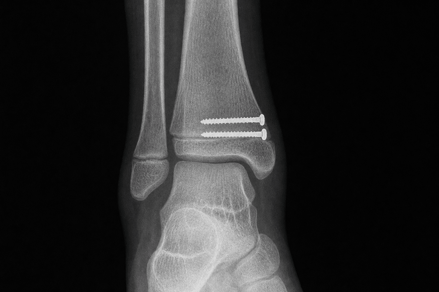
- Tillaux: a single 3.5 or 4.0 mm cannulated screw, anterolateral to posteromedial, parallel to the physis or entirely epiphyseal; partially threaded for compression; confirm an articular step less than 2 mm on the mortise view.
- Medial malleolus SH II: young children - two smooth K-wires parallel to the physis; older children - a single 3.5 mm screw parallel or oblique to the physis; never perpendicular.
- Medial malleolus SH IV: preferred - an epiphyseal screw from the malleolar epiphysis into the talar body (safest, and anatomic for this intra-articular fracture); alternative - a screw parallel to the physis in the metaphysis.
- Triplane: usually two screws - a lateral epiphyseal screw from the lateral malleolus across the lateral fragment toward the medial epiphysis, plus an anterior-to-posterior screw parallel to the physis for the metaphyseal component; add a medial screw for three-part patterns.
- Distal fibula: if the ankle mortise is anatomic after tibial fixation and the fibula is undisplaced, leave the fibula alone - it heals reliably. Fix the fibula only if it is too long and prevents mortise reduction; in children under 12 use K-wires parallel to the fibular physis, in older adolescents fix as in adults.
- Final fluoroscopy in three views before closing. AP: medial clear space less than 4 mm and equal to the superior clear space (both 2-4 mm).
- Mortise (most critical, whole leg internally rotated 20 degrees): the medial, superior and lateral (tibiofibular) clear spaces must all be equal (1-4 mm). Any asymmetry means talar shift from malreduction, syndesmotic injury or fibular length - revise.
- Lateral: no anterior or posterior talar subluxation; check the posterior malleolus and the hardware.
- Acceptable: articular step less than 2 mm (anatomic for SH III/IV), physeal gap less than 3 mm, symmetric mortise. Unacceptable and requiring revision: articular step greater than 2 mm, medial clear space wider than superior (talar shift), talar subluxation, or a screw perpendicular to the physis.
- Irrigate thoroughly (3 litres; pulsatile lavage for high-energy fractures), remove debris and devitalised tissue.
- Close in layers with absorbable sutures - periosteum (3-0 Vicryl), subcutaneous tissue (3-0/4-0 Vicryl), skin with a 4-0/5-0 Monocryl subcuticular (preferred in children - avoids traumatic suture removal) or 4-0 nylon if healing is in doubt.
- Apply a well-padded long-leg posterior splint or bivalved long-leg cast with the ankle in slight equinus (10-15 degrees, to relax the Achilles and protect the physis - avoid more than 20 degrees, which causes stiffness) and the knee flexed 20-30 degrees.
- Strict non-weight-bearing for 4-6 weeks. AP, lateral and mortise radiographs in the splint to document maintained reduction.
Never place a screw perpendicular to an open physis: it creates a physeal bar, focal growth arrest and progressive angular deformity. Use the physeal-respecting ladder - smooth K-wires parallel to the physis, an epiphyseal screw that does not cross the physis, or a screw crossing obliquely at greater than 45 degrees - and remove any implant crossing the physis at 3-6 months if significant growth remains. This applies even in the transitional-age patient near skeletal maturity, because open physeal segments can persist.
Before you close, the mortise view (whole leg internally rotated 20 degrees) must show three equal clear spaces - medial, superior and lateral tibiofibular - each 1-4 mm. A medial clear space wider than the superior space means talar shift from residual displacement, a missed syndesmotic injury or a malreduced fibula, and mandates revision. Obtain a true mortise by rotating the entire leg, not just the foot.
It is the most commonly injured structure in the anterolateral approach (1-2 percent). It emerges from the anterior compartment 8-10 cm proximal to the joint line and crosses the field in the subcutaneous tissue. Identify it before any deep dissection, loop it with a vessel loop, retract gently, keep the incision slightly more anterior if its course is uncertain, and avoid electrocautery near it. If transected, repair primarily under loupe magnification; a contused nerve usually recovers over 3-6 months.
In the viva, state the ladder explicitly: young child (less than 10 years) - smooth K-wires parallel to the physis; SH IV medial malleolus - an epiphyseal screw from malleolus into talus that never crosses the physis (safest); older child needing rigidity - a screw parallel to or crossing the physis obliquely at greater than 45 degrees, removed at 3-6 months. End with "I never place a screw perpendicular to an open physis" - it shows you understand why growth arrest happens.
Even when ORIF is planned, attempt a gentle closed reduction under adequate sedation or anaesthesia first - many physeal fractures reduce anatomically and stay stable in a cast, avoiding surgery entirely, and closed reduction decreases soft-tissue trauma in the rest. Reverse the injury mechanism (usually supination-external rotation), use gentle traction, and assess stability on fluoroscopy. Repeated forceful attempts carry a high odds ratio for physeal arrest - if one gentle attempt fails, proceed to open reduction rather than remanipulating.
Aftercare & Complications
Rehabilitation and surveillance protocol. A physeal ankle fracture needs supervised, phased rehabilitation and - critically - long-term surveillance for growth arrest, because arrest develops months later and is unrelated to whether the skin has healed.
- Clinical
- Well-padded long-leg posterior splint in slight equinus (10-15 degrees); elevate above heart; neurovascular checks every 2-4 hours for 24 hours; scheduled acetaminophen and ibuprofen with opioids for 48-72 hours
- Radiographs
- AP, lateral and mortise in the splint to document maintained reduction and hardware; compare with final intra-operative fluoroscopy
- Activity
- Strict non-weight-bearing with crutches; toe-touch for balance only; no impact or sports
- Clinical
- Wound check; if healing and swelling improved, convert to a bivalved or circumferential long-leg cast in slight equinus, knee flexed 20-30 degrees; wean opioids
- Radiographs
- AP, lateral and mortise to confirm maintained reduction (rarely lost, but must document); revise if lost
- Activity
- Continue strict non-weight-bearing; may return to school, no physical education
- Clinical
- Remove cast; expect stiffness. Begin physiotherapy for ROM (dorsiflexion/plantarflexion, inversion/eversion), Achilles stretching, gentle strengthening; transition to a CAM boot or short-leg walking cast for comfort if needed
- Radiographs
- AP, lateral and mortise to assess healing (callus, regular physeal line, hardware position); progress weight-bearing if healed, otherwise continue protection 2-4 more weeks
- Activity
- Progressive weight-bearing over 2-3 weeks; no running, jumping or sports; stationary bike and swimming once healed
- Clinical
- Examine ROM, strength and gait; address persistent stiffness; plan K-wire removal (should already be out) and screw removal if crossing the physis in a young child
- Radiographs
- Bilateral AP, lateral and mortise to compare; assess union, physeal symmetry, angular alignment and leg length; look for early asymmetric physeal line or sclerosis
- Activity
- Low-impact sports (swimming, cycling, light jogging) if full ROM and strength; no contact sports
- Clinical
- ROM, strength, single-leg balance, sport-specific testing; measure leg lengths; plan removal of any screw crossing the physis not yet out
- Radiographs
- Bilateral AP, lateral and mortise comparing physeal width; look for a sclerotic physeal bar; add CT or MRI if arrest suspected
- Activity
- Full sports including contact and high-impact if ROM, strength and testing are normal; a protective ankle brace may help confidence initially
- Clinical
- Examine ROM, gait, leg length and angular alignment; discharge routine SH I/II if no concerns; continue surveillance for SH III/IV and triplane
- Radiographs
- Bilateral AP, lateral and mortise - critical comparison for asymmetric growth or bar; continue annual films for high-risk fractures with growth remaining
- Activity
- Full unrestricted activity; modify only if arrest is progressing
- Clinical
- Final assessment for high-risk fractures (SH III/IV, triplane) with growth remaining; discharge if approaching skeletal maturity with no arrest; educate the family on late signs to watch for
- Radiographs
- Final surveillance films; standing alignment films if any angular deformity; if a bar is present, quantify its extent and estimate remaining growth to plan intervention
- Activity
- Full activity if no arrest; restrict extreme high-impact activity if a bar is established and correction is pending
Growth arrest - the long-term threat. All physeal ankle fractures need long-term follow-up; explain this to the family before surgery. Risk factors are SH III/IV intra-articular fractures (15-20 percent arrest risk), initial displacement greater than 3 mm, a crush component suggesting SH V, screws crossing the physis perpendicularly, and high-energy mechanisms. Counsel that arrest can occur even with a perfect operation - it reflects the injury energy. Watch for progressive angular deformity (varus more common than valgus from medial arrest), leg-length discrepancy (usually less than 1 cm from the ankle and well tolerated), and a physeal bar seen as a sclerotic bridge with asymmetric growth. Management depends on the bar and the growth remaining: physeal bar resection with fat or cement interposition if less than 50 percent of the physis is involved and significant growth remains; completion epiphysiodesis if the bar is greater than 50 percent or little growth remains; corrective osteotomy for established deformity after growth is complete; and contralateral epiphysiodesis for leg-length discrepancy greater than 2 cm. Hardware removal. K-wires MUST come out at 4-6 weeks without exception (migration, infection, breakage if left) - usually in clinic under local anaesthetic, or with brief sedation in an uncooperative child. Screws crossing the physis are controversial; preferentially remove them at 3-6 months if more than two years of growth remains, to minimise any tether effect. Epiphyseal screws not crossing the physis, and screws in older adolescents near maturity, can stay unless symptomatic. After removal, protect in a CAM boot or short-leg cast for 2-3 weeks and avoid impact sports for 4-6 weeks (refracture risk through the screw tracts, which temporarily weaken bone - highest in the first 4-6 weeks; lower in paediatric bone, which heals faster than adult). Complications.
- Recognition
- Progressive angular deformity (varus common), leg-length discrepancy (usually less than 1 cm), a sclerotic physeal bar on radiographs at 6-12 months, asymmetric growth on serial films
- Prevention
- Anatomic reduction with physeal gap less than 3 mm, atraumatic technique, never a perpendicular screw, smooth K-wires or oblique screws in young children, remove crossing implants at 3-6 months
- Management
- Serial radiographs every 3-6 months. Bar resection with fat interposition if less than 50 percent physis and growth remains; completion epiphysiodesis if greater than 50 percent or minimal growth; corrective osteotomy after growth completes; contralateral epiphysiodesis for LLD greater than 2 cm
- Recognition
- Activity-related ankle pain years later, loss of dorsiflexion/plantarflexion, joint-space narrowing, sclerosis and osteophytes on radiographs
- Prevention
- Anatomic reduction of the articular surface (less than 2 mm step mandatory for SH III/IV), perfect mortise alignment, stable fixation, early mobilisation when stable
- Management
- Non-operative first (activity modification, NSAIDs, therapy, bracing); arthroscopy and debridement in the young; supramalleolar osteotomy for malalignment; ankle arthrodesis for end-stage disease
- Recognition
- Displacement on the 2-week film, progressive deformity, an asymmetric mortise, pain and swelling after initial improvement
- Prevention
- Adequate fixation for the pattern (two to three K-wires or appropriately sized screws), protected weight-bearing for 4-6 weeks, a long-leg cast to control rotation, a film in the splint immediately and again at 2 weeks
- Management
- Early (less than 2 weeks) and unacceptable - return to theatre for re-reduction and revision fixation; late but acceptable - accept and monitor; late and unacceptable - osteotomy after healing
- Recognition
- Clinical internal/external rotation versus the other side, abnormal gait and foot progression angle, CT with contralateral comparison if uncertain
- Prevention
- Assess rotation during reduction (clinical and fluoroscopic), compare with the other side intra-operatively, stable fixation, a long-leg cast to control rotation
- Management
- Minor (less than 10 degrees) - observe (hip and knee compensate); significant (greater than 15 degrees) - supramalleolar rotational osteotomy after healing and growth complete; angular malunion - corrective osteotomy if symptomatic
- Recognition
- Numbness or dysaesthesia over the dorsum and first web space, a positive Tinel over the nerve at the anterolateral ankle
- Prevention
- Careful anterolateral dissection, identify the nerve in the subcutaneous tissue before deeper work, gentle vessel-loop retraction, avoid electrocautery near it
- Management
- If transected - primary repair under loupes with 8-0 nylon and sensory re-education; if contused - observe, usually recovers in 3-6 months; persistent beyond 6 months - exploration and neurolysis or graft
- Recognition
- Skin irritation or prominence over a screw head (medial malleolus commonest), K-wire migration on radiographs or clinically, screw breakage, pin-site infection
- Prevention
- Countersink screw heads flush, measure length carefully to avoid far-cortex penetration, bury or adequately bend K-wires, protected weight-bearing, plan K-wire removal at 4-6 weeks
- Management
- Prominent screw - remove once healed (3-6 months) or earlier if skin compromised; migrated K-wire - remove urgently if near neurovascular structures; broken screw - leave if healed and asymptomatic, remove if symptomatic or crossing the physis
- Recognition
- Reduced ROM versus the other side, difficulty with stairs, running and squatting, capsular contracture in chronic cases
- Prevention
- Minimise immobilisation (4-6 weeks typical), early ROM when stable, aggressive therapy, avoid more than 10-15 degrees equinus in the cast
- Management
- Therapy (progressive ROM, Achilles stretching, joint mobilisation, proprioception); night dorsiflexion splint for residual equinus; persistent beyond 6 months - arthroscopy and manipulation under anaesthesia; severe equinus - gastrocnemius recession or Achilles lengthening
- Recognition
- Wound erythema and drainage (superficial); systemic upset and deep wound breakdown (deep)
- Prevention
- Perioperative antibiotics, meticulous sterile technique, minimise soft-tissue trauma, early treatment of wound problems
- Management
- Superficial - oral antibiotics and local wound care; deep - return to theatre for irrigation and debridement, IV antibiotics for 4-6 weeks, retain hardware if unhealed
- Recognition
- Pain out of proportion, pain with passive stretch, tense compartments
- Prevention
- High index of suspicion with high-energy mechanisms or prolonged surgery; minimise tourniquet time
- Management
- Measure pressures (greater than 30 mmHg or within 30 mmHg of diastolic); emergency four-compartment fasciotomy
- Recognition
- Medial clear space greater than superior clear space on the mortise view, from inadequate reduction or a missed syndesmotic injury
- Prevention
- Perfect mortise reduction confirmed on final fluoroscopy; assess the syndesmosis
- Management
- Return to theatre for revision reduction; a syndesmotic screw if unstable
- Recognition
- New injury in the first 4-6 weeks after removal, through the weakened screw tracts
- Prevention
- Protected weight-bearing for 2-3 weeks after removal; avoid impact sports for 4-6 weeks
- Management
- Lower risk in paediatric bone (heals faster); treat the new fracture on its merits
Viva & Exam Focus
SALTERSALTER - Salter-Harris classification of physeal fractures
PHYSISPHYSIS - physeal-respecting fixation strategy
Five danger zones. Five anatomical structures account for almost all the avoidable harm in this operation - know the location and the protection for each.
Location: horizontal growth plate 1-2 cm proximal to the ankle joint, thickest anteromedially, closing predictably central then medial then anterolateral from age 12 to 16. Protection: never a perpendicular screw; use K-wires parallel to the physis, screws crossing obliquely at greater than 45 degrees, or epiphyseal screws that stay within the epiphysis.
Location: 1 cm proximal to the tibial physis, closing later (15-17 years); commonly injured with ankle fractures but often undisplaced. Protection: in children under 12 with significant growth, avoid crossing it with screws; use smooth K-wires parallel to the physis, or accept fibular displacement if the ankle mortise is anatomic.
Location: emerges from the anterior compartment 8-10 cm proximal to the joint line and crosses the anterolateral ankle toward the first web space - directly in the Tillaux/triplane incision. Protection: keep the incision slightly more anterior, identify the nerve in the subcutaneous tissue before deeper dissection, retract gently with a vessel loop, avoid electrocautery near it.
Location: travel together between EHL (lateral) and tibialis anterior (medial), on the anterior tibia deep to the extensor retinaculum; at risk in every anterior ankle approach. Protection: develop the interval between tibialis anterior and EHL, retract EHL laterally with the whole neurovascular bundle, avoid aggressive medial retraction, place Hohmann retractors on bone.
Location: anterior and superior to the medial malleolus; the nerve supplies sensation to the medial ankle and foot and the vein is often prominent in children; both are vulnerable in the medial malleolar approach. Protection: make the anteromedial incision anterior to the malleolar tip, identify and protect both in the subcutaneous layer, retract gently, avoid electrocautery near the nerve.
Clinical Decision Scenarios
Practise clinical reasoning and management decisions out loud
“A 13-year-old presents with an ankle injury after a football tackle. X-rays show a displaced fracture of the anterolateral distal tibia. How would you classify this injury and what is your management approach?”
“Describe the Salter-Harris classification and explain why it is important when treating paediatric ankle fractures. Which types require operative fixation and why?”
“You are fixing a Salter-Harris Type IV medial malleolar fracture in a 12-year-old. What fixation options do you have, and how do you decide? Explain your technique for protecting the physis.”
Indications
- Intra-articular displacement greater than 2 mm (SH III/IV, Tillaux, triplane) - zero tolerance for an articular step
- Physeal displacement greater than 3 mm irreducible or unstable after a closed reduction attempt
- Tillaux fractures (SH III anterolateral distal tibia, age 12-14, AITFL avulsion) if displaced greater than 2 mm
- Triplane fractures (complex three-part with sagittal, coronal and axial components) if articular displacement greater than 2 mm
- Open physeal fractures needing washout; compartment syndrome; polytrauma needing stabilisation
- A distal fibula fracture preventing mortise reduction (fibula too long, causing talar shift)
Key anatomy
- Distal tibial physis: horizontal growth plate 1-2 cm proximal to the joint, closing predictably central (12-14y) then medial (14-15y) then anterolateral (15-16y)
- Superficial peroneal nerve: emerges 8-10 cm proximal to the ankle, crosses the anterolateral ankle, at risk in the Tillaux/triplane approach
- Deep peroneal nerve and anterior tibial artery: between tibialis anterior (medial) and EHL (lateral), vulnerable in every anterior approach
- Saphenous vein and nerve: anterior and superior to the medial malleolus, at risk in the medial malleolar approach
- AITFL: inserts on the anterolateral distal tibial epiphysis and avulses it in Tillaux fractures during supination-external rotation
- Ankle mortise: symmetric joint with medial, superior and lateral clear spaces all equal (2-4 mm), assessed on the mortise view with 20-degree internal rotation of the whole leg
Critical steps
- Classify by Salter-Harris (I-V) - it determines prognosis, arrest risk and treatment urgency; SH III/IV are intra-articular and demand anatomic reduction
- Always attempt closed reduction first under adequate sedation/anesthesia - many reduce anatomically avoiding surgery, and it decreases soft-tissue trauma even if ORIF is needed
- CT is mandatory for triplane - it shows the three-dimensional anatomy; plain films underestimate complexity
- Anterolateral approach for Tillaux/triplane: between tibialis anterior and EHL, protect the superficial peroneal nerve in the subcutaneous tissue
- Anteromedial approach for the medial malleolus: anterior to the malleolar tip, protect the saphenous vein and nerve, preserve the deltoid
- Achieve anatomic articular reduction to less than 2 mm under direct vision and fluoroscopy; physeal-respecting fixation; final mortise view must show symmetric clear spaces
Danger zones
- Distal tibial physis - a perpendicular screw causes a physeal bar and arrest with angular deformity; use K-wires parallel, epiphyseal screws, or oblique crossing only
- Distal fibular physis - avoid crossing with screws in children under 12; use smooth K-wires parallel or accept displacement if the mortise is anatomic
- Superficial peroneal nerve - crosses the anterolateral ankle; identify in the subcutaneous tissue and protect with a vessel loop before deep dissection
- Deep peroneal nerve and anterior tibial artery - between tibialis anterior and EHL; retract EHL laterally with the whole bundle, avoid aggressive medial retraction
- Saphenous vein and nerve - anterior to the medial malleolus; incise anterior to the tip, identify and retract anteriorly, avoid electrocautery near the nerve
Technique pearls
- The mortise view (20-degree internal rotation of the whole leg) is the most critical radiograph - it shows a symmetric joint and detects talar shift; the medial clear space must equal the superior
- Tillaux occurs at age 12-14 when the medial physis is closed but the lateral is open - the AITFL avulses the anterolateral fragment; a transitional fracture during predictable closure
- Triplane has three fracture planes - sagittal through the lateral metaphysis, coronal through the physis, axial through the epiphysis; requires CT for planning
- Epiphyseal screws (medial malleolus to talus) are the safest fixation for SH IV - entirely in the epiphysis, zero arrest risk, excellent stability
- Distal fibula: if the mortise is anatomic after tibial fixation and the fibula is undisplaced, leave it alone - it heals without fixation; fix only if it prevents mortise reduction
- Bump under the ipsilateral hip for the medial malleolus; no bump for lateral/Tillaux; long-leg cast in slight equinus (10-15 degrees); K-wires out at 4-6 weeks, crossing screws out at 3-6 months if growth remains
Complications
- Physeal arrest (5-10 percent overall, 15-20 percent SH III/IV) - progressive angular deformity (varus common), leg-length discrepancy, physeal bar; needs long-term surveillance
- Post-traumatic arthritis (5-15 percent intra-articular) - from articular incongruity greater than 2 mm; prevent with anatomic reduction
- Loss of reduction (5-8 percent) - from inadequate fixation or premature weight-bearing; prevent with adequate fixation and protected weight-bearing for 4-6 weeks
- Rotational malunion - affects gait; prevent by assessing rotation during reduction, stable fixation, and a long-leg cast to control rotation
- Superficial peroneal nerve injury (1-2 percent) - sensory deficit over the dorsum; prevent with careful anterolateral dissection identifying the nerve first
- Hardware complications (5-10 percent) and ankle stiffness (10-15 percent) - countersink heads, bury or bend K-wires, minimise cast time, early ROM at 6 weeks
Post-op protocol
- Immediate: long-leg posterior splint in slight equinus (10-15 degrees), non-weight-bearing, elevate, ice; films in the splint to document maintained reduction
- 2 weeks: wound check, convert to a long-leg cast if swelling improved; film to confirm maintained reduction
- 4-6 weeks: cast removal, assess healing, initiate ROM and progressive weight-bearing over 2-3 weeks with therapy
- 3 months: bilateral films comparing for early arrest; plan hardware removal if K-wires remain or screws cross the physis in a young child
- 6-12 months: serial films monitoring for arrest (bar, asymmetric growth, angular deformity); SH III/IV and triplane need longer surveillance
- Return to sport: low-impact at 3 months if healed; full contact at 6-9 months if ROM and strength are normal and there are no arrest concerns
Exam tips
- Always classify by Salter-Harris first - shows a systematic approach; emphasise that SH III/IV are intra-articular demanding anatomic reduction, while SH I/II can accept some displacement
- State 'I always attempt closed reduction first' even if planning ORIF - many reduce anatomically avoiding surgery, and it decreases soft-tissue trauma
- Emphasise 'never perpendicular to an open physis' when discussing fixation - describe the physeal-respecting alternatives (K-wires, epiphyseal screws, oblique crossing)
- The mortise view is the most critical radiograph - the medial clear space must equal the superior; asymmetry means talar shift requiring revision
- Long-term follow-up is mandatory for ALL physeal fractures - counsel the family on arrest risk (3-5 percent SH I/II, 15-20 percent SH III/IV) and serial films at 3, 6 and 12 months minimum
- Distinguish Tillaux (SH III anterolateral, simple two-part) from triplane (complex three-part needing CT) - a common examiner question on transitional fractures
- If asked about complications, lead with physeal arrest - discuss recognition, prevention (physeal-respecting fixation) and management (bar resection versus epiphysiodesis)
Background & Evidence
Epidemiology. Salter-Harris Type II is the most common paediatric ankle fracture (60-70 percent). The transitional fractures cluster between ages 12 and 15 years and are a direct consequence of the asymmetric distal tibial physeal closure pattern: the physis closes central first (age 12-14), then medial (14-15), then anterolateral (15-16). A Tillaux fracture (SH III) arises when the medial and central physis are closed but the anterolateral segment is still open; a triplane fracture arises in the same window but with a more complex three-dimensional geometry. The distal fibular physis sits 1 cm proximal to the tibial physis and closes later (15-17 years), so it is frequently injured alongside tibial fractures (30-40 percent of paediatric ankle fractures) but is often undisplaced. Salter-Harris classification. The classification is demoted here deliberately - in the viva you state it first to frame prognosis and treatment, but it is background, not the operation.
- Pattern
- Through the physis only; rare in the ankle
- Arrest risk
- About 3 percent
- Management implication
- Extra-articular; some displacement acceptable; usually non-operative
- Pattern
- Through the physis with a metaphyseal (Thurston-Holland) fragment; the commonest
- Arrest risk
- About 5 percent
- Management implication
- Extra-articular; up to 3 mm physeal gap acceptable; ORIF only if unstable after closed reduction
- Pattern
- Through the physis with an epiphyseal fragment into the joint (e.g. Tillaux)
- Arrest risk
- About 15 percent
- Management implication
- Intra-articular; demand anatomic reduction to less than 2 mm; ORIF if displaced
- Pattern
- Through metaphysis-physis-epiphysis (e.g. medial malleolus with articular involvement)
- Arrest risk
- About 20-25 percent
- Management implication
- Intra-articular; demand anatomic reduction to less than 2 mm; ORIF if displaced
- Pattern
- Crush injury to the physis
- Arrest risk
- Near 100 percent
- Management implication
- No surgical role - already maximally damaged; surveillance for arrest complications
Triplane geometry. A triplane fracture has three components in orthogonal planes: a sagittal fracture through the lateral metaphysis, a coronal fracture through the physis (central to lateral), and an axial fracture through the epiphysis (an anterolateral articular fragment). Two-part fractures (about 70 percent) have a single medial fragment (epiphysis plus medial metaphysis fused) and a lateral fragment (anterolateral epiphysis plus lateral metaphysis); three-part fractures (about 30 percent) have separate medial epiphyseal, lateral epiphyseal and posterior metaphyseal fragments. Pre-operative CT is mandatory - plain films significantly underestimate the complexity and cannot show the three-dimensional anatomy that determines the approach. Key evidence. The pooled meta-analysis of physeal distal tibial fractures (Jalkanen, 2021) found an overall premature physeal closure rate of 13 percent, highest in SH IV (20 percent), and showed that residual displacement after reduction was the single strongest predictor of arrest, while more than two repeated reduction attempts carried an odds ratio of 8.5 - and open reduction was associated with a lower arrest risk in displaced fractures. The message: the quality and completeness of reduction, not the act of surgery, drives outcome - avoid repeated forceful closed manipulations, and proceed to a gentle open reduction if one gentle attempt fails. For displaced SH II fractures (Park, 2017), surgery did not reliably abolish the risk of arrest - injury energy and mechanism (pronation-abduction / pronation-external rotation, younger age) mattered more than the treatment method, so families must be counselled that arrest can occur despite optimal fixation. For triplane fractures, the original description (Cooperman, 1978) established that tomography was essential to define the three-dimensional fragment anatomy and that anatomic reduction was the goal. For Tillaux fractures (Horn, 2001), a cadaveric study showed CT is more sensitive than plain films for detecting displacement greater than 2 mm - the operative threshold - so obtain CT before deciding between casting and ORIF when the plain films are borderline.
References
Physeal Fractures of Distal Tibia: A Systematic Review and Meta-analysis
- Pooled analysis of 12 studies and 1997 patients - overall premature physeal closure (PPC) rate 13 percent, ranging 0.2 percent to 42 percent across studies
- Salter-Harris IV fractures carried the highest PPC risk (20 percent), followed by SH II (12 percent) - confirming intra-articular and through-and-through patterns are highest risk
- Residual displacement after reduction was the single most significant predictor of PPC; repeated (more than 2) reduction maneuvers carried a pooled odds ratio of 8.5 (95 percent CI 6.3-12.2)
- Open reduction was associated with LOWER risk of PPC in displaced fractures (OR 0.63, 95 percent CI 0.38-0.91)
What is the best treatment for displaced Salter-Harris II physeal fractures of the distal tibia?
- 95 displaced SH II distal tibial fractures with greater than 3 mm residual gap after closed reduction; 25 treated non-operatively, 70 operatively
- PPC incidence was 13/52 (non-operative) versus 24/70 (operative) - no statistically significant difference (p=0.1)
- Independent risk factors for PPC were younger age at injury and injury mechanism (pronation-abduction / pronation-external rotation), NOT method of treatment or residual displacement in this cohort
- Implant type, sex, and presence of a fibular fracture were not predictive of PPC
Tibial fractures involving the ankle in children: the so-called triplane epiphyseal fracture
- Original description of the triplane fracture in 15 children (mean age 13 years), representing 6 percent of 237 consecutive paediatric ankle physeal fractures
- Tomography (the era's CT equivalent) was essential to define the three-dimensional fragment anatomy - medial malleolar/anteromedial epiphysis versus the lateral epiphysis-plus-posterior-metaphysis fragment
- External rotation deformity (5-10 degrees) and articular incongruity occurred with inadequate reduction, establishing anatomic reduction as the treatment goal
- Defined the two-fragment versus three-fragment patterns that still guide surgical planning today
Fractures of the distal tibial epiphysis in adolescence
- Characterised the transitional fractures (juvenile Tillaux SH III and triplane) and their shared external-rotation mechanism in 17 adolescents
- Demonstrated that the pattern of asymmetric distal tibial physeal closure (central to medial to anterolateral) dictates which transitional fracture occurs
- Three-fragment triplane fractures occurred in younger patients and more often required operative treatment than two-fragment patterns
- Linked fracture morphology to skeletal age, explaining why Tillaux and triplane fractures cluster at ages 12 to 15 years
Radiologic evaluation of juvenile Tillaux fractures of the distal tibia
- Cadaveric simulated Tillaux fractures displaced at 0, 1, 2, 3 and 5 mm imaged with plain radiographs and CT
- Plain radiographs and CT were accurate within 1 mm only about 50 percent of the time, but CT was MORE sensitive than plain films for detecting displacement greater than 2 mm
- Because greater than 2 mm displacement is the operative threshold, CT is the preferred modality for assessing juvenile Tillaux fractures
- Confirms plain films systematically under-call clinically important Tillaux displacement
Further reading 1. Caterini R, Farsetti P, Ippolito E. Long-term followup of physeal injury to the ankle. Foot & Ankle. 1991;11(6):372-383. PMID: 1894231. DOI: 10.1177/107110079101100607 - 68 distal tibial/fibular physeal injuries followed a mean of 27 years; Salter-Harris type, initial displacement and quality of reduction determined outcome, with radiographic osteoarthritis (11.8 percent) occurring almost exclusively in SH III/IV lesions. 2. Spiegel PG, Cooperman DR, Laros GS. Epiphyseal fractures of the distal ends of the tibia and fibula: a retrospective study of two hundred and thirty-seven cases in children. Journal of Bone and Joint Surgery (American). 1978;60(8):1046-1050. PMID: 721852 - classic study of 237 consecutive paediatric distal tibia/fibula physeal fractures correlating Salter-Harris type and displacement with complication risk, underpinning the operative displacement thresholds. 3. Ertl JP, Barrack RL, Alexander AH, VanBuecken K. Triplane fracture of the distal tibial epiphysis: long-term follow-up. Journal of Bone and Joint Surgery (American). 1988;70(7):967-976. PMID: 3403587 - long-term outcomes of 23 triplane fractures: residual displacement of 2 mm or more within the weight-bearing zone was associated with suboptimal results, and plain films alone did not demonstrate fracture configuration. 4. Rapariz JM, Ocete G, González-Herranz P, López-Mondejar JA, Domenech J, Burgos J. Distal tibial triplane fractures: long-term follow-up. Journal of Pediatric Orthopaedics. 1996;16(1):113-118. PMID: 8747367. DOI: 10.1097/00004694-199601000-00023 - 35 triplane fractures: prognosis was good provided reduction achieved less than 2 mm displacement; CT is required to define the pattern and closed reduction should be attempted first. 5. Shin AY, Moran ME, Wenger DR. Intramalleolar triplane fractures of the distal tibial epiphysis. Journal of Pediatric Orthopaedics. 1997;17(3):352-355. PMID: 9150025 - describes the intramalleolar triplane variant and a CT-based classification (intra-articular within/outside the weight-bearing zone, and extra-articular) guiding individualised treatment. 6. Kleiger B, Mankin HJ. Fracture of the lateral portion of the distal tibial epiphysis. Journal of Bone and Joint Surgery (American). 1964;46:25-32. PMID: 14104312 - original description of the juvenile Tillaux fracture mechanism and its relationship to the physeal closure pattern. 7. Kärrholm J, Hansson LI, Laurin S. Pronation injuries of the ankle in children: retrospective study of radiographical classification and treatment. Acta Orthopaedica Scandinavica. 1983;54(1):1-17. PMID: 6402887. DOI: 10.3109/17453678308992863 - classification of 457 paediatric ankle fractures showing pronation-mechanism injuries (18 percent) have distinct patterns and are more often displaced and difficult to reduce closed than supination injuries.