Midline approach | Quadrilateral surface | Suprapubic window
- Intrapelvic approach — works inside the pelvis through the space of Retzius, the retropubic extraperitoneal plane between the bladder and the pubic symphysis.
- Superior access to the quadrilateral surface — better direct visualization than the medial window of the ilioinguinal approach.
- Bladder mobilization is the key step — mobilize the bladder posterolaterally off the quadrilateral surface to expose bone; a Foley catheter is mandatory.
- Corona mortis is encountered crossing the superior pubic ramus and must be identified and ligated before proceeding laterally.
- Cannot access the iliac wing or high anterior column — limited to medial structures; combine with another approach for superior pathology.
When & Why
What it exposes. The modified Stoppa (anterior intrapelvic) approach gives direct intrapelvic access to the quadrilateral surface of the acetabulum, the medial wall, the superior pubic ramus and the low anterior column. Working from inside the pelvis through the space of Retzius, it allows buttress plating of the quadrilateral plate under direct vision to prevent medial subluxation of the femoral head. Why intrapelvic (and not ilioinguinal). The medial window of the ilioinguinal approach gives only limited inferior and posterior access to the quadrilateral surface. The Stoppa approach approaches the same bone from the intrapelvic side, giving superior visualization and plate application, with a lower nerve-injury profile (no lateral femoral cutaneous nerve at risk), and a more cosmetic Pfannenstiel incision. Its trade-off is that it cannot reach the iliac wing or the high anterior column. Historical context. The approach was adapted from the Stoppa hernia repair and popularised for acetabular fractures in the 1990s: - Original Stoppa — developed for inguinal/groin hernia repair using the retropubic (preperitoneal) space.
- Hirvensalo and colleagues (1990s) — described the intrapelvic/ilioinguinal modification for pelvic and acetabular surgery.
- Cole and Bolhofner (1994) — popularised the modified Stoppa approach for acetabular fracture surgery (Clin Orthop Relat Res).
- Sagi, Collinge and others — coined the broader term anterior intrapelvic (AIP) approach and refined its indications.
- Keel et al (2012) — described the Pararectus variant, entering extraperitoneally along the lateral border of rectus abdominis to create five working windows.
- Current — intrapelvic approaches are increasingly preferred for quadrilateral surface and anterior column fractures, especially in the elderly.
- Stoppa role
- Primary approach
- Combined approach
- Stoppa alone or + lateral window
- Rationale
- Direct quadrilateral access
- Stoppa role
- Cannot use Stoppa
- Combined approach
- Ilioinguinal required
- Rationale
- No access to iliac wing
- Stoppa role
- Anterior component
- Combined approach
- Stoppa + Kocher-Langenbeck
- Rationale
- Stoppa for stem, K-L for transverse
- Stoppa role
- Anterior fixation
- Combined approach
- Stoppa + K-L
- Rationale
- Excellent medial buttress access
- Stoppa role
- Rarely indicated
- Combined approach
- Kocher-Langenbeck preferred
- Rationale
- Posterior approach addresses both
Contraindications and when not to use it. - Absolute: previous lower midline laparotomy with extensive adhesions; active pelvic infection.
- Relative: previous pelvic surgery; extensive scarring in the retropubic space; confirmed bladder injury requiring repair (may still proceed with urology input).
- When NOT to use Stoppa: high anterior column fractures (no access to the iliac wing); anterior wall without quadrilateral involvement (ilioinguinal is better); SI joint pathology (needs the ilioinguinal lateral window).
If there is blood at the urethral meatus or gross hematuria, obtain a retrograde urethrogram (if urethral injury is suspected) and a CT cystogram before surgery. A known bladder injury is not an absolute contraindication, but it requires urology consultation and careful surgical planning.
Position and landmarks. - Supine on a radiolucent table (essential for fluoroscopy).
- Arms tucked (preferred) to allow the surgeon to move around.
- A bump under the ipsilateral hip is usually NOT recommended — it makes the quadrilateral surface more vertical and harder to visualize; a flat pelvis is preferred.
- Foley catheter is MANDATORY, placed before draping to decompress the bladder.
- Wide preparation from nipples to knees. The C-arm must be able to swing for Inlet, Outlet and Judet views.
- The surgeon stands on the CONTRA-lateral side, looking across the pelvis at the fracture; the assistant stands on the IPSI-lateral side to retract the bladder.
- Best approach
- Modified Stoppa
- Rationale
- Direct visualization, better plate placement
- Alternative
- Ilioinguinal medial window (inferior access)
- Best approach
- Ilioinguinal
- Rationale
- Need lateral and middle windows
- Alternative
- Cannot access with Stoppa alone
- Best approach
- Combined: Stoppa + lateral ilioinguinal
- Rationale
- Stoppa for quadrilateral, lateral window for column
- Alternative
- Full ilioinguinal but less quadrilateral access
- Best approach
- Stoppa + Kocher-Langenbeck
- Rationale
- Stoppa for anterior, K-L for posterior
- Alternative
- Ilioinguinal + K-L (traditional)
The modified Stoppa provides superior access to the quadrilateral surface compared with the medial window of the ilioinguinal approach. Working intrapelvically allows direct visualization and buttress plate application to prevent medial subluxation. However, Stoppa cannot access the iliac wing or high anterior column — those still require ilioinguinal lateral and middle windows.
The modified Stoppa is NOT a replacement for the ilioinguinal approach — it is an alternative for specific patterns involving the quadrilateral surface. High anterior column and iliac wing fractures still require the ilioinguinal lateral and middle windows.
The Exposure
Work down the midline to the pubis, develop the space of Retzius bluntly, mobilise the bladder off the quadrilateral surface, ligate the corona mortis on the superior pubic ramus, and complete a subperiosteal exposure from the superior ramus to the ischium — ready for buttress plating.
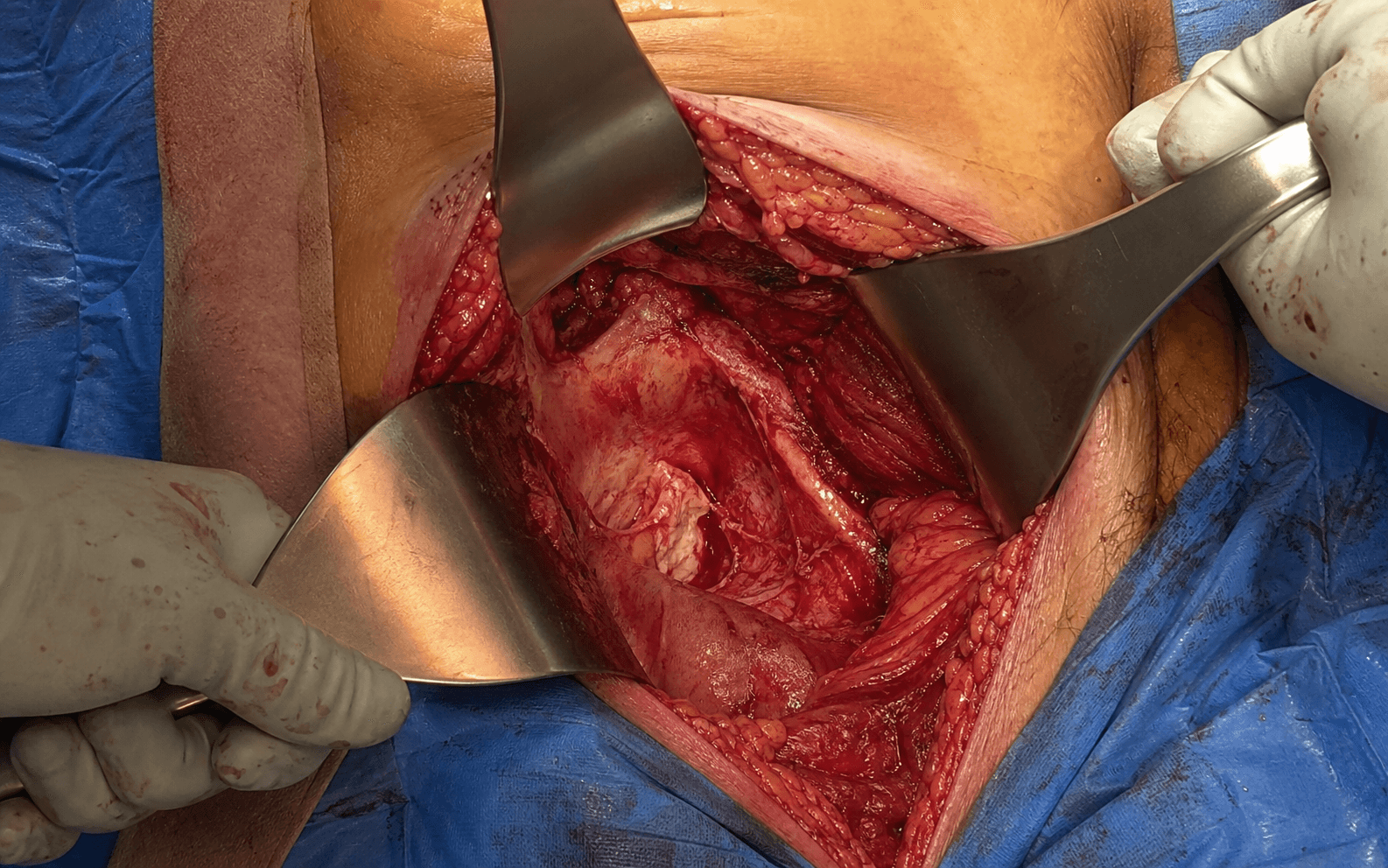
The space of Retzius is the key corridor — a potential extraperitoneal space between the bladder and the posterior surface of the pubic symphysis. - Anterior: posterior surface of the pubic symphysis and superior rami.
- Posterior: anterior wall of the bladder and peritoneum.
- Superior: peritoneal reflection.
- Inferior: pelvic floor (levator ani, obturator internus).
- Lateral: pelvic sidewall and external iliac vessels.
- Location
- Posterior to pubis
- Relationship to approach
- Must mobilize posterolaterally
- Clinical significance
- Injury risk if not in correct plane — use Foley
- Location
- Covers bladder dome
- Relationship to approach
- Stay below the peritoneal reflection
- Clinical significance
- Entry causes bowel complications — repair if entered
- Location
- Lateral pelvic sidewall
- Relationship to approach
- Lateral boundary of the first window
- Clinical significance
- Retract laterally for extended exposure
- Location
- Obturator foramen, on obturator internus
- Relationship to approach
- Identify and protect during dissection
- Clinical significance
- Injury risk during lateral dissection
- Location
- Superior pubic ramus (40-96mm from symphysis)
- Relationship to approach
- Encountered during lateral dissection
- Clinical significance
- Present in ~80% anatomically; ligate before proceeding
Quadrilateral surface anatomy. The quadrilateral surface is the medial wall of the acetabulum on the inner table of the pelvis — four-sided, with the pelvic brim superiorly, the obturator foramen inferiorly, the superior ramus anteriorly and the ischium posteriorly. It resists medial subluxation of the femoral head; when fractured, the head can displace medially into the pelvis.
- Boundaries
- Bladder (medial), external iliac vessels (lateral)
- Exposes
- Quadrilateral surface, superior ramus
- Fixation
- Quadrilateral buttress plate
- Boundaries
- External iliac vessels (medial), psoas (lateral)
- Exposes
- Low anterior column, pelvic brim
- Fixation
- Infrapectineal plate if needed
Unlike the ilioinguinal approach, which works between nerve territories (femoral, gluteal, lateral femoral cutaneous), the Stoppa is an intrapelvic, extraperitoneal approach that avoids the major traversing nerves of the anterior thigh. The obturator nerve is the only major nerve at risk, lying on the pelvic sidewall on obturator internus.
Exposure sequence
- Pfannenstiel (preferred): a transverse suprapubic incision 2-3cm above the pubic symphysis, 10-15cm long — better cosmetic result, familiar to general surgeons and gynaecologists.
- Low midline (alternative): vertical midline from the umbilicus toward the symphysis — better extensibility but a higher wound-complication rate; reserved when Pfannenstiel is contraindicated.
- Incise the anterior rectus sheath transversely (Pfannenstiel) or vertically (midline).
- Identify the rectus abdominis muscles; separate them in the midline (linea alba for midline) or retract them laterally (Pfannenstiel).
- Identify the transversalis fascia and dissect gently down to the pubic symphysis.
- Feel the bladder dome (soft, mobile, Foley balloon palpable).
- Begin blunt dissection between the bladder and the symphysis.
- Use a sponge stick or finger dissection; sweep the bladder posteriorly and laterally off the pubis.
- Develop the space laterally to the pelvic sidewall.
- Identify the superior pubic rami bilaterally and confirm you are extraperitoneal (no bowel visible).
- Continue blunt dissection posterolaterally; mobilize the bladder off the quadrilateral surface toward the obturator foramen.
- Retract the bladder medially with a broad retractor (assistant on the ipsilateral side).
- Identify the external iliac artery and vein on the pelvic sidewall as the lateral boundary of the first window.
- Approach the superior pubic ramus carefully and identify any vessels crossing it (the corona mortis, 40-96mm from the symphysis).
- Clip or ligate with 2-0 silk; confirm hemostasis before proceeding laterally.
- Complete a subperiosteal dissection along the quadrilateral surface; identify the fracture lines.
- Expose the superior ramus anteriorly and the ischium posteriorly for plate application.
- Identify and protect the obturator neurovascular bundle throughout.
The correct plane is between the bladder wall and the bone — relatively avascular loose areolar tissue that sweeps easily with blunt dissection. If you see detrusor muscle you are in the bladder wall; if you see glistening peritoneum you are too superficial. Significant bleeding means you are likely in the bladder wall or have entered a vessel. Always place a Foley catheter to decompress the bladder before dissection, and use a sponge stick for gentle blunt sweeping.
The quadrilateral buttress plate acts as an internal shelf preventing medial subluxation of the femoral head. It spans from the superior pubic ramus anteriorly to the ischium posteriorly, creating a buttress that resists medial displacement. Screw fixation into intact bone (ramus and ischium) provides stability — biomechanically superior to trying to reduce and fix the quadrilateral fragment itself.
Reduction and buttress plating
- Pelvic clamp to compress a laterally displaced fracture.
- Ball-spike pusher to push the displaced quadrilateral fragment laterally.
- Pointed reduction forceps for temporary hold.
- Assess reduction with fluoroscopy — the inlet view shows the quadrilateral reduction.
- Pre-contoured quadrilateral plate (if available), or a 3.5mm reconstruction plate contoured to shape.
- The plate spans from the superior ramus (anteriorly) to the ischium (posteriorly).
- Place the plate on the quadrilateral surface (inner pelvic wall).
- Anterior screws into the superior pubic ramus; posterior screws into the ischium or retroacetabular surface.
- Central screw holes may be left empty so the plate acts as a buttress.
- Drill and measure carefully; 3.5mm cortical screws, typically 40-60mm in length.
- Avoid intra-articular penetration (check with fluoroscopy).
- The obturator oblique view shows the retroacetabular safe zone between the sciatic notches.
- AP, obturator oblique and inlet views.
- Confirm plate position and screw lengths; assess reduction of the quadrilateral surface.
- Check for any intra-articular hardware.
Posterior screws from the quadrilateral plate can be directed into the ischium or into the retroacetabular safe zone (between the sciatic notches). The obturator oblique view shows this zone well. Screws must avoid the hip joint — aim posteriorly and inferiorly, not laterally into the acetabulum.
Dangers & Extensions
- Incidence
- 1-3%
- Prevention / management
- Foley catheter, correct-plane dissection; repair primarily if injured, consult urology
- Incidence
- 5-10%
- Prevention / management
- Stay below the peritoneal reflection; repair if entered, no long-term consequence
- Incidence
- Less than 1%
- Prevention / management
- Gentle dissection if mobilizing for the second window; have vascular backup
- Incidence
- Anastomosis in ~80% anatomically; troublesome bleeding less common clinically
- Prevention / management
- Identify and ligate early; control with clips or ties if bleeding
- Incidence
- 10-20%
- Prevention / management
- Indomethacin 75mg daily for 6 weeks, or single-dose radiation
- Incidence
- 2-5%
- Prevention / management
- Prophylactic antibiotics, minimize dead space, drain placement
- Incidence
- Rare (less than 1%)
- Prevention / management
- Identify and protect the nerve during dissection
Bladder injury management. Recognize intraoperatively when possible (methylene blue test); perform a primary two-layer repair with 3-0 absorbable suture; leave the Foley catheter for 7-10 days; obtain a cystogram before catheter removal; consult urology for complex injuries near the trigone. Vascular injury management. Apply direct pressure for control; obtain vascular surgery consultation; perform primary repair for small injuries; use a graft for extensive injury.
The modified Stoppa has a significantly lower nerve injury risk than the ilioinguinal approach. There is no lateral femoral cutaneous nerve at risk (injured or sacrificed in 10-15% of ilioinguinal cases), and the femoral nerve is not dissected. Only the obturator nerve is at potential risk, and it is rarely injured with proper technique — a major advantage of Stoppa for isolated quadrilateral fractures.
Extensions and combining approaches. The modified Stoppa is rarely used in isolation — it typically combines with other techniques for complete fracture management. - Stoppa + Kocher-Langenbeck (most common): both-column with medial displacement and T-type fractures. Position sequentially (supine for Stoppa, then lateral for K-L), as a two-team lateral approach, or a floating position.
- Stoppa + lateral window of ilioinguinal: single supine position, via a combined or separate incision, for low anterior column extending beyond the quadrilateral.
- Stoppa + percutaneous screw: quadrilateral surface plus posterior column without wall — Stoppa for the anterior component, screw for the posterior, minimizing surgical trauma.
When combining Stoppa with Kocher-Langenbeck, consider operative time carefully — prolonged prone or lateral positioning increases DVT risk. Many surgeons prefer staged procedures 5-7 days apart for complex fractures to reduce physiological stress.
Closure. - Hemostasis: meticulous — there is a large dead space in the retropubic region; check for bladder injury (fill with methylene blue if suspicious) and ensure the corona mortis is ligated.
- Layers: posterior rectus sheath (if entered) with 0 or 1 absorbable continuous; anterior rectus sheath with 0 or 1 absorbable continuous; Scarpa's fascia with 2-0 absorbable interrupted; skin with staples or subcuticular suture.
- Drain: a retropubic drain (10mm flat or 15Fr round) through a separate stab incision, removed at 24-48h when output is less than 30mL/day.
- Post-closure: check the Foley is still draining (not kinked or clamped); final fluoroscopy (AP, obturator, inlet); document neurovascular status.
Recovery protocol
- Foley catheter remains for 24-48 hours; monitor drain output (remove when less than 30mL/24h).
- DVT prophylaxis (LMWH or rivaroxaban); early mobilization to chair with assistance.
- Toe-touch weight bearing (10-20kg); hip and knee ROM exercises.
- Avoid hip flexion greater than 90 degrees initially; continue DVT prophylaxis for 4-6 weeks.
- Partial weight bearing (50%); increase ROM and strengthening.
- X-rays at 6 weeks to assess healing; progress weight bearing based on healing.
- Full weight bearing when callus is visible; progressive strengthening.
- Return to activities as tolerated; monitor for post-traumatic arthritis.
- Modified Stoppa
- Excellent
- Ilioinguinal
- Good
- Significance
- Stoppa provides superior access
- Modified Stoppa
- 0%
- Ilioinguinal
- 10-15%
- Significance
- No LFCN in Stoppa field
- Modified Stoppa
- Less than 2%
- Ilioinguinal
- 5-10%
- Significance
- Less nerve dissection required
- Modified Stoppa
- 2-5%
- Ilioinguinal
- 5-8%
- Significance
- Smaller incision in Stoppa
- Modified Stoppa
- Shorter
- Ilioinguinal
- Longer
- Significance
- Less dissection needed
Long-term outcomes. Good-to-excellent results in 75-85% with anatomic reduction. Post-traumatic arthritis occurs in 20-30% at 10-20 years even with good reduction; heterotopic ossification in 15-25% (reduced with prophylaxis); conversion to total hip arthroplasty in 10-20% at 10 years; avascular necrosis of the femoral head in 5-10% (higher with initial dislocation).
Acetabular fracture outcomes are graded with Matta radiographic criteria: Excellent = anatomic reduction (0mm displacement); Good = less than 2mm; Fair = 2-3mm; Poor = greater than 3mm. Clinical outcomes correlate strongly with the quality of reduction — anatomic reduction gives 80% or more good/excellent results.
Procedures Through This Approach
- Acetabular fracture ORIF — anterior column — fixation of the low anterior column and quadrilateral surface through this intrapelvic corridor.
- Acetabular fracture ORIF — both-column — anterior (medial wall) component of associated both-column fractures.
- Quadrilateral buttress plating for medial-wall fractures with femoral head subluxation — the principal fixation this approach is built for.
- Anterior stem fixation in T-type fractures (combined with Kocher-Langenbeck for the transverse component).
- Low anterior column with quadrilateral involvement (Stoppa alone, or combined with the lateral window of the ilioinguinal for superior column extension).
Viva & Exam Focus
STOPPASTOPPA — key steps of the approach
RETZIUSRETZIUS — anatomical boundaries
The space of Retzius is the retropubic extraperitoneal space between the bladder and the posterior surface of the pubic symphysis. Boundaries: anterior = pubis; posterior = bladder and peritoneum; lateral = pelvic sidewall and external iliac vessels; inferior = pelvic floor.
Superior direct visualization and access to the quadrilateral surface working intrapelvically. The Stoppa approach allows buttress plate application under direct vision, whereas the ilioinguinal medial window has limited inferior and posterior access to the quadrilateral surface.
The plate acts as an internal shelf preventing medial subluxation of the femoral head into the pelvis. It spans from the superior pubic ramus anteriorly to the ischium posteriorly, creating a mechanical buttress that resists medial displacement forces.
The Stoppa does not dissect the lateral femoral cutaneous nerve (injured or sacrificed in 10-15% of ilioinguinal cases) and does not mobilize the femoral nerve. The only nerve at potential risk is the obturator nerve, which is rarely injured with proper technique.
Stoppa cannot access the iliac wing, sacroiliac joint, or high anterior column — it is limited to medial structures (quadrilateral surface, superior pubic ramus, low anterior column). These superior structures require the ilioinguinal lateral window or a separate lateral approach.
Exam viva scenarios
Practise clinical reasoning and management decisions out loud
“A 50-year-old female has an associated both-column acetabular fracture with 15mm medial displacement of the quadrilateral surface on CT, and the femoral head is subluxing medially into the pelvis. What approach would you use for the anterior component and why?”
“During development of the space of Retzius in a modified Stoppa approach, you notice pink fluid in the wound and the anesthesiologist reports the Foley catheter is no longer draining. What is your immediate assessment and management?”
“A 40-year-old male has a T-type acetabular fracture. The transverse component is minimally displaced posteriorly, but the anterior component (stem of the T) shows a low anterior column fracture with quadrilateral surface involvement and 10mm displacement. The iliac wing and high anterior column are intact. Which approach(es) would you use and why?”
Space of Retzius anatomy
- Retropubic extraperitoneal space between the bladder and pubis
- Anterior: posterior surface of pubic symphysis
- Posterior: bladder wall and peritoneum
- Lateral: pelvic sidewall and external iliac vessels
- Developed bluntly — avascular plane when in the correct layer
Key surgical steps
- 1. Pfannenstiel or low midline incision (Pfannenstiel preferred)
- 2. Develop the space of Retzius bluntly with a Foley catheter in place
- 3. Mobilize the bladder posterolaterally off the quadrilateral surface
- 4. Identify and ligate the corona mortis on the superior pubic ramus
- 5. Expose the quadrilateral surface from superior ramus to ischium
- 6. Apply a buttress plate from ramus to ischium
Indications
- Quadrilateral surface fractures (primary indication)
- Associated both-column fractures with medial displacement
- T-type fractures (anterior stem component)
- Low anterior column with quadrilateral involvement
Advantages over ilioinguinal
- Superior quadrilateral surface access (vs limited medial window)
- Lower nerve injury risk (no LFCN at risk, no femoral nerve dissection)
- Better cosmetic result (Pfannenstiel incision)
- Less operative time for isolated quadrilateral fractures
Limitations
- Cannot access the iliac wing or high anterior column
- Cannot access the SI joint
- Limited to medial structures only
- Requires combination with a lateral approach for extensive anterior column
Key complications and prevention
- Bladder injury (1-3%): Foley catheter, correct plane, gentle dissection
- Peritoneal entry (5-10%): stay below the reflection, repair if entered
- Corona mortis hemorrhage: identify and ligate early
- Obturator nerve injury: rare — identify and protect the nerve
The retropubic space between the bladder and the pubic symphysis. This avascular plane is the key to the approach — develop it bluntly to reach the medial pelvic wall without entering the peritoneum or damaging the bladder.
The modified Stoppa provides superior access to the quadrilateral surface compared with the ilioinguinal medial window. Direct visualization allows placement of buttress plates to prevent medial subluxation of the femoral head.
Mobilize the bladder posterolaterally off the quadrilateral surface — the key maneuver, with gentle blunt dissection in the correct plane. A Foley catheter is essential to decompress the bladder and reduce injury risk.
As in the ilioinguinal approach, the corona mortis crosses the superior pubic ramus, typically 40-96mm lateral to the symphysis. Anatomical prevalence is high (around 80%; large vessels over 3mm in roughly 60% of hemipelves), so anticipate, identify and ligate it before proceeding laterally — a venous variant is most common and harder to control once torn.
References
Globally consistent principles: displaced acetabular fractures are managed in specialist pelvic/acetabular units with fellowship-trained surgeons; inter-hospital transfer of complex patterns to high-volume centres improves reduction quality and outcomes. AO Foundation and BOA (BOAST: Pelvic and Acetabular Fracture Management) endorse early CT, multidisciplinary care and timely transfer. Training pathways (advanced orthopaedic practice Tr & Orth, advanced orthopaedic practice/AOA, advanced orthopaedic practice, DNB/MS) converge on the same competency; an AO Pelvis course and cadaveric simulation are widely recommended before independent intrapelvic surgery.
Reduction-driven outcomes (Matta): anatomic reduction (under 2mm) is the strongest modifiable predictor of good/excellent results across all series. VTE prophylaxis (society guidance differs): mechanical prophylaxis universally; chemical prophylaxis once haemostasis is secure. AAOS, NICE and regional consensus support extended prophylaxis after major pelvic/acetabular surgery, individualised to bleeding risk; no single global duration is mandated, commonly continued for several weeks.
Across advanced orthopaedic practice and DNB vivas, you must be able to justify the intrapelvic (modified Stoppa / AIP / Pararectus) approach versus the ilioinguinal, name the key structures (space of Retzius, corona mortis, obturator and external iliac vessels), and describe the technique step-by-step. Acetabular fracture management is a recurring viva topic testing decision-making and surgical planning.
Cole and Bolhofner — Modified Stoppa for Acetabular Fractures (Landmark Technique)
- Popularised the modified Stoppa approach for acetabular fractures, adapted from the Stoppa hernia technique
- Direct intrapelvic access to the quadrilateral surface and medial pelvic wall
- Allows buttress plating under direct vision to resist medial femoral head displacement
- Avoids dissection of the lateral femoral cutaneous and femoral nerves
Matta — Accuracy of Reduction and Clinical Results in Acetabular Fractures
- 262 displaced acetabular fractures fixed within 21 days, mean 6-year follow-up
- Anatomic reduction achieved in 71%; reduction quality fell with fracture complexity, older age and delay
- Excellent/good clinical result in 76% (40% excellent, 36% good); poor in 16%
- Clinical outcome correlated closely with quality of radiographic reduction
Keel et al — The Pararectus Approach: Anatomical Study and Clinical Evaluation
- Described the Pararectus intrapelvic approach using five extraperitoneal windows along the lateral border of rectus abdominis
- Cadaveric validation (10 hemipelves) confirmed safe dissection and multidirectional screw placement
- 20 patients: mean step-off improved 3.3mm to 0.1mm and mean gap 11.5mm to 0.8mm (both p less than 0.001)
- Access morbidity low: 2 peritoneal lesions and 2 minor vascular injuries
Bastian/Keel et al — Pararectus versus Modified Stoppa: Exposure and Instrumentation
- Paired cadaveric study (5 specimens) comparing both approaches through a 10cm incision
- Exposed inner hemipelvis bone: 42% with Pararectus versus 29% with modified Stoppa (p=0.011)
- Pararectus allowed posterior column screws 1.8-2.1x longer and more posteromedial trajectories
- Greater false-pelvis access and the option to extend without a new incision
Darmanis et al — Corona Mortis: Anatomical Study with Clinical Implications
- 80 hemipelves dissected: a retropubic anastomosis (corona mortis) present in 83%
- Large-calibre channel over 3mm behind the superior pubic ramus in 60% of specimens, 40-96mm from the symphysis
- In 492 clinical anterior approaches, only 5 problematic vessels and 2 troublesome bleeds occurred
- High anatomical prevalence but lower clinical hazard than feared — caution, not avoidance
Nadeem et al — Infrapectineal Buttress Plating of the Quadrilateral Surface via AIP
- 21 patients with quadrilateral-plate acetabular fractures fixed via anterior intrapelvic (modified Stoppa) approach
- Matta reduction anatomic in 14, imperfect in 5, poor in 2
- Functional outcome (modified Merle d'Aubigne) excellent 47.6%, good 42.9%, fair 9.5%
- Both fair outcomes had non-anatomical reduction; one required later total hip arthroplasty