Gold-standard treatment for chronic periprosthetic joint infection · Advanced
- Two-stage exchange is the gold standard for chronic periprosthetic joint infection (symptoms greater than 3–4 weeks) — the mature biofilm cannot be eradicated with the implant in situ.
- Confirm PJI with the MSIS/ICM criteria before surgery: serum CRP and ESR, synovial WBC and PMN, alpha-defensin and culture — a sinus tract or two positive cultures is definite PJI.
- Stage 1: through the previous incision remove ALL components and ALL cement, take 5–6 tissue cultures before any antibiotics, debride to healthy bleeding bone, and insert a high-dose antibiotic cement spacer.
- Antibiotic spacer cement: vancomycin 2–4 g plus tobramycin 2–4 g per 40 g PMMA — high-dose loading is acceptable because mechanical strength is sacrificed for elution.
- Interim: 6 weeks of IV organism-specific antibiotics, then a 2-week antibiotic holiday before reaspiration — the holiday is critical to avoid false-negative cultures.
- Stage 2 only when infection is eradicated: normalized CRP, negative aspiration, healed wound — reimplanting into an infected joint fails.
When & Why
Indication. Two-stage exchange is the gold-standard surgical treatment for chronic periprosthetic joint infection (PJI) of a total knee arthroplasty — infection present for greater than 3–4 weeks, or any infection in which the biofilm has matured and cannot be cleared with the implant retained. Typical scenarios are: - Chronic PJI with symptoms greater than 3–4 weeks
- Infection that has failed DAIR (debridement, antibiotics and implant retention)
- An unknown organism, or culture-negative disease that nevertheless meets the MSIS criteria
- Resistant or virulent organisms (MRSA, VRE, multi-drug-resistant gram-negatives)
- A sinus tract communicating with the joint
- A poor soft-tissue envelope that needs a staged reconstruction
- The severely immunocompromised patient The one decision that matters — acute versus chronic. The whole strategy turns on how long the infection has been present, because that determines whether the biofilm can be eradicated with the implant left in place. The biofilm takes 2–4 weeks to mature and then becomes resistant to antibiotic penetration and to the host immune response. - Acute PJI (less than 3–4 weeks from symptom onset, or within 4 weeks of the index operation) may be treated with DAIR if the implant is well-fixed, the soft tissues are viable, and the organism is susceptible.
- Chronic PJI (greater than 3–4 weeks) requires removal of all foreign material — hence two-stage exchange.
For acute PJI only — a susceptible organism, a stable implant and viable soft tissues. Debride openly, exchange the liner, and give organism-specific antibiotics. High failure rate (50–80 percent) if used for chronic infection.
For highly selected cases — a known susceptible organism that is not highly virulent, good soft tissues, no sinus tract, no major bone loss. Reported success of 85–90 percent in selected series (notably high-volume European centres).
The international default for chronic PJI. Remove all components and cement, place an antibiotic spacer, give interval IV antibiotics, then reimplant once infection is eradicated. Eradication in 85–90 percent.
Contraindications. A patient medically unfit for two staged operations, unable to comply with the antibiotic regimen, so severely immunosuppressed that suppression alone is preferable, or with a life-expectancy too short to complete both stages. When the limb cannot be salvaged, the alternatives are arthrodesis (a failed two-stage, severe bone loss, or extensor-mechanism loss) and, as a last resort, amputation (life-threatening sepsis or a non-reconstructable limb). Chronic oral antibiotic suppression is reserved for the patient unfit for any surgery. Before theatre — confirm the diagnosis and plan. - Serology: CRP and ESR, with D-dimer as an adjunct.
- Joint aspiration, stopping antibiotics 2 weeks beforehand if the patient is already on treatment — send for cell count and differential, alpha-defensin, and culture (aerobic, anaerobic, fungal if indicated).
- Nuclear imaging (a labelled-leucocyte scan) only if the diagnosis remains unclear.
- Imaging: AP and lateral knee radiographs for component position and bone loss, long-leg standing films if malalignment is suspected, and a CT if Stage-2 reconstruction needs bone-loss quantification.
- Organism identification is critical for antibiotic selection. Request sensitivities including to aminoglycosides for cement. Culture-negative disease (10–25 percent of cases) that still meets the MSIS criteria is managed with empiric broad-spectrum cover — vancomycin plus gram-negative cover — and an infectious-diseases consultation is essential throughout.
One-stage exchange can be considered only when all five apply: a known organism susceptible to available antibiotics; not highly virulent (exclude MRSA, VRE, resistant gram-negatives); a good soft-tissue envelope with no sinus tract or skin necrosis; no significant bone loss needing structural grafting; and a fit, motivated patient reliable for follow-up. If any criterion is missing, two-stage is safer. Contemporary randomised evidence for knee PJI shows broadly similar functional outcomes between single- and two-stage revision, so the choice is increasingly driven by host, organism and soft-tissue factors rather than dogma — but two-stage remains the international default for chronic disease.
- Attempting DAIR for chronic infection fails in 50–80 percent — use two-stage.
- Culture-negative disease is still PJI if the MSIS criteria are met — do not delay treatment.
- A sinus tract is definite PJI regardless of culture result.
- Pushing one-stage in an unsuitable patient (resistant organism, poor soft tissues) has a high failure rate.
- Underestimating frailty — two-stage carries significant morbidity, and the patient must be able to survive both operations.
The Operation
The goal is to render the joint free of all foreign material and biofilm, deliver a high local antibiotic concentration through a cement spacer, then — once the infection is eradicated — reimplant a durable revision arthroplasty. The exposure is laid out as the first operative steps because everything that follows depends on a safe, adequate field: the previous incision, full-thickness flaps, and a medial parapatellar arthrotomy that is extended with a quadriceps snip or tibial tubercle osteotomy when the knee is stiff or scarred.
Stage 1 — explantation, debridement and antibiotic spacer
- Supine on a radiolucent table, thigh tourniquet, leg holder or foot positioner.
- Prep the entire limb, including any sinus tracts. Use antimicrobial-impregnated drapes and consider a wound-retraction system.
- Withhold prophylactic antibiotics until the tissue samples are taken if the organism is unknown, and have 5–6 specimen containers ready.
- Confirm the antibiotic-cement plan with pharmacy: vancomycin 2–4 g plus tobramycin (or gentamicin) 2–4 g per 40 g PMMA, with 6–10 packs of cement available.
- Use the previous incision (the most lateral one if there are several) and raise full-thickness skin flaps — avoid creating thin flaps that will break down over a spacer.
- Perform a medial parapatellar arthrotomy and excise any sinus tract completely back to healthy bleeding tissue.
- In the stiff or scarred revision knee, gain extensile exposure early — a quadriceps snip or a tibial tubercle osteotomy (TTO) — rather than fighting a tight field and avulsing the patellar tendon.
- Before any antibiotics are given, take 5–6 tissue samples from separate areas for culture and hold them for 14 days for slow-growing organisms. Send frozen section if available — more than 5 PMN per high-power field supports infection.
- Remove all implants — polyethylene insert, femoral component, tibial baseplate and patellar component if present — using thin osteotomes at the implant–bone interface to preserve bone stock.
- Remove all cement meticulously with burrs, chisels or an ultrasonic device, and clear any remaining fixation screws or wires. Residual cement or debris harbours biofilm and dooms the revision.
- Protect the posterior structures throughout posterior cement removal (the popliteal artery is only 5–10 mm behind the capsule — keep the posterior retractor on bone).
- Debride all necrotic and infected tissue back to healthy bleeding bone and soft tissue.
- Curette any bone cavities and debride aggressively — the adequacy of debridement is the single most important factor in eradicating infection.
- Irrigate copiously with 6–9 L of normal saline using pulsatile lavage. Many surgeons add a dilute povidone-iodine soak for 3 minutes, then lavage it out.
- The joint should be clean with healthy bleeding surfaces throughout, including the posterior recesses.
- Mix high-dose antibiotics into the PMMA — vancomycin 2–4 g plus tobramycin 2–4 g per 40 g cement (4 g of each is common) — to elute therapeutic local levels. High loading is acceptable here because mechanical strength is sacrificed for elution.
- Static spacer: form the cement into a block that fills the joint, reinforced with K-wires or a metal rod if needed — better elution, simpler and cheaper, but it sacrifices range of motion.
- Articulating spacer: cement a metal femoral trial onto the femur and form a cement tibial baseplate with a flat or slightly dished surface — preserves ROM, eases Stage 2, but carries a 10–20 percent dislocation risk and higher cost.
- Ensure the spacer is stable and sized to maintain soft-tissue tension so it will not dislocate.
- Irrigate a final time and achieve meticulous haemostasis.
- Close in layers — secure capsular and retinacular closure over the spacer is critical. Tension-free closure is essential; if the soft-tissue envelope is deficient, involve plastic surgery for a gastrocnemius flap.
- Consider negative-pressure wound therapy for high-risk wounds (previous sinus, diabetes, obesity). Drain placement is debated (some avoid it to prevent a colonisation track).
- Apply a bulky dressing and a knee immobiliser, especially after a static spacer.
- Static block spacer
- Higher elution, cheaper
- Articulating spacer
- Adequate elution, higher cost
- Static block spacer
- Lost — stiffness, extensor shortening
- Articulating spacer
- Preserved
- Static block spacer
- Harder (stiff knee)
- Articulating spacer
- Easier
- Static block spacer
- Bone loss common
- Articulating spacer
- Bone stock preserved
- Static block spacer
- Fragmentation, migration
- Articulating spacer
- Dislocation (10–20%), fracture
- Static block spacer
- Severe bone loss, non-ambulator
- Articulating spacer
- Most cases (the default)
The interval between the stages. Begin IV antibiotics after the cultures are obtained — empiric until sensitivities return, then organism-specific for a minimum of 6 weeks, guided by infectious diseases. Monitor CRP and ESR weekly at first then fortnightly, alongside clinical wound inspection; the markers should trend down and normalise. After the antibiotic course, observe a 2-week antibiotic holiday (critical to avoid false-negative cultures) and then reaspirate. Proceed to Stage 2 only if eradication is confirmed: - CRP normalised (less than 10 mg/L) and ESR trending down
- Aspiration after the holiday: synovial WBC less than 2000 cells/µL, PMN less than 65%, culture negative
- Wound healed, no erythema, no sinus tract, no fever
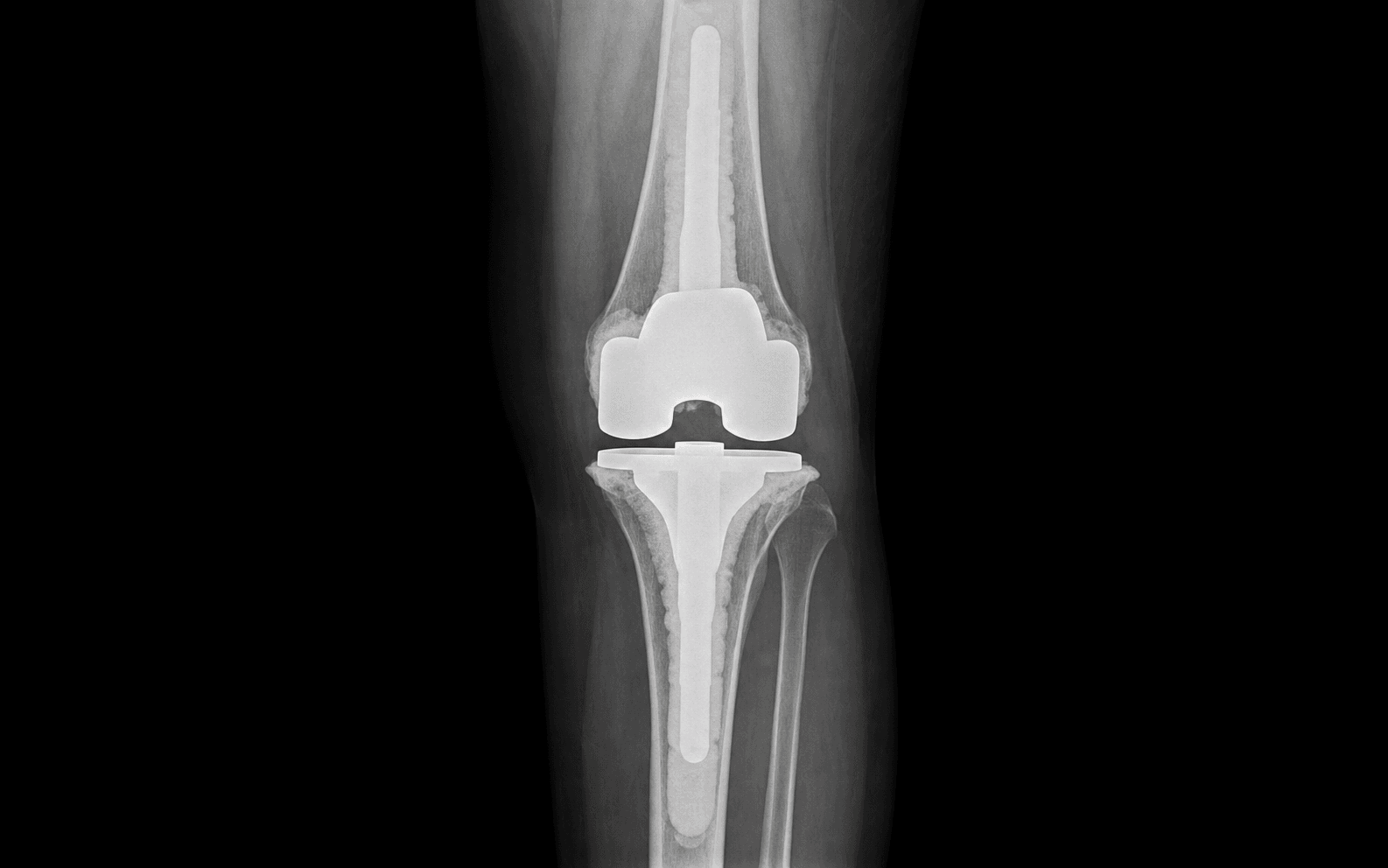
Stage 2 — reimplantation (minimum 6–8 weeks after Stage 1)
- Use the previous incision; the knee is often stiff, so plan extensile exposure again.
- Remove the antibiotic spacer carefully — an articulating spacer may be well fixed, and rough extraction wastes bone.
- Take 5–6 fresh tissue cultures before giving any antibiotics, just as in Stage 1. If frozen section is available, more than 5 PMN per high-power field suggests persistent infection — abort reimplantation, repeat the debridement and exchange the spacer.
- Grade bone loss with the AORI classification (see Background & Evidence). Disuse osteopenia, spacer erosion and the explantation itself mean there is usually more bone loss than at Stage 1.
- Plan stems to bypass the deficient metaphysis and achieve diaphyseal fixation, with augments, sleeves or cones for cavitary and segmental defects. Have a megaprosthesis available for AORI Type 3 defects.
- Assess ligament integrity (MCL/LCL) and the extensor mechanism — these drive the level of constraint.
- Establish tibial and femoral platforms on healthy bone.
- Address bone defects with augments, sleeves or cones, and add stems for diaphyseal fixation.
- Climb the constraint ladder based on ligament competency — condylar-constrained, then varus-valgus-constrained (VVC), then a hinge — rather than undersizing constraint in a knee with compromised collaterals.
- Cement the components; adding low-dose antibiotic to the fixation cement (e.g. 1 g vancomycin per 40 g) is practised by some but remains controversial. Handle osteopenic bone gently — intraoperative fracture is a real risk during impaction.
- Irrigate thoroughly, achieve meticulous haemostasis and close in layers (consider a drain).
- Antibiotic prophylaxis varies — 24–48 hours IV as a minimum, with some surgeons extending to 5 days or adding 3–6 months of oral suppression for resistant organisms or immunocompromised patients.
- Give extended pharmacological VTE prophylaxis (commonly to around 35 days after Stage 2, per AAOS and NICE/BOA guidance) given the prolonged immobility, balanced against bleeding and wound risk.
- Mobilise early — weight-bearing as tolerated unless a TTO or bone-loss reconstruction dictates otherwise.
- Location and risk
- 5–10 mm behind the posterior capsule, at risk during posterior cement removal
- How to protect it
- Keep the posterior retractor ON bone; vascular surgery backup if injured
- Location and risk
- Around the fibular neck, may be encased in scar in chronic infection
- How to protect it
- Careful lateral release; identify, protect and decompress if needed
- Location and risk
- Tibial tubercle insertion, high avulsion risk in scarred revisions
- How to protect it
- Use an extensile exposure (quadriceps snip or TTO) when the tendon is at risk
- Location and risk
- At the joint line, weakened by infection and debridement
- How to protect it
- Assess integrity and plan constraint (PS, VVC or hinge) accordingly
- Location and risk
- Multiple incisions, sinus tracts, poor vascularity
- How to protect it
- Tension-free closure; involve plastics early for a gastrocnemius flap
The popliteal artery lies only 5–10 mm behind the posterior capsule. During posterior cement removal and posterior capsular debridement, keep the posterior retractor on bone, not in the soft tissues, and work under direct vision. If it is injured, apply direct pressure, call for vascular surgery, and repair primarily where possible. Inadequate debridement is the commonest cause of treatment failure — do not compromise it, but protect the posterior structures while you debride.
Mix high-dose antibiotics (vancomycin 4 g plus tobramycin 4 g per 40 g cement) into the PMMA. Cement a metal femoral trial sized to the patient for a smooth articular surface and less cement wear, and form a flat or slightly dished cement tibial baseplate by hand or with a mould. Ensure adequate soft-tissue tension to prevent dislocation, and confirm the spacer is stable through a functional range. The metal femoral trial is reusable at Stage 2.
At reimplantation, take 5–6 fresh tissue cultures before any antibiotics and send frozen section. More than 5 PMN per high-power field suggests persistent infection — abort the reimplantation, repeat the debridement and exchange the spacer. Reimplanting into an infected joint will fail.
Aftercare & Complications
Rehabilitation Stage 1 (after explantation). Toe-touch or protected weight-bearing with a walker, depending on spacer stability — a knee immobiliser is used after a static spacer or if the spacer is unstable. Early gentle ROM is allowed with an articulating spacer but restricted with a static one. Give 6 weeks of IV organism-specific antibiotics, monitoring CRP and ESR weekly and checking antibiotic levels and renal function. Between the stages. Continue ROM exercises if an articulating spacer is in place. Optimise the host — albumin greater than 3.5 g/dL, total protein greater than 6 g/dL, smoking cessation, glucose control (HbA1c less than 8 percent), and treat any distant source (dental, urinary). Observe the 2-week antibiotic holiday, then reaspirate. Stage 2 (after reimplantation). Weight-bearing as tolerated unless a TTO or bone-loss reconstruction dictates otherwise. Begin CPM or early passive ROM, targeting 0–90 degrees by 2 weeks. Antibiotic practice varies (some add 3–6 months of oral suppression). Extended pharmacological VTE prophylaxis to around 35 days, per AAOS and NICE/BOA guidance. Review at 2 weeks (wound check), 6 weeks (radiograph), then 3, 6 and 12 months, and annually. Long-term surveillance. There is a lifelong risk of reinfection — any dental procedure warrants antibiotic prophylaxis, and the patient is monitored for recurrent infection with an annual CRP. Complications
- Recognition
- Continued pain, drainage, elevated CRP, positive cultures at Stage 2
- Prevention
- Thorough debridement, appropriate antibiotics, confirm eradication before Stage 2
- Management
- Repeat two-stage, arthrodesis, suppression or amputation
- Recognition
- New infection after a successful two-stage, same or different organism
- Prevention
- Optimise nutrition and immunity; avoid high-risk exposures
- Management
- Repeat two-stage or salvage procedure
- Recognition
- Dislocation (10–20%), fracture, migration
- Prevention
- Appropriate sizing, balanced tension, patient compliance
- Management
- Closed reduction for dislocation, or spacer exchange
- Recognition
- Persistent drainage, dehiscence, necrosis
- Prevention
- Tension-free closure, NPWT for high-risk wounds, early plastics input
- Management
- Wound care, debridement, gastrocnemius flap cover
- Recognition
- Inability to extend, palpable gap, patella alta
- Prevention
- Protect the tendon during exposure; TTO when it is at risk
- Management
- Primary repair, allograft reconstruction, or brace/fusion
- Recognition
- Significant defects at Stage 2, osteopenia
- Prevention
- Preserve bone at Stage 1; early weight-bearing
- Management
- Augments, sleeves/cones, structural allograft, megaprosthesis
- Recognition
- ROM less than 90 degrees at Stage 2
- Prevention
- Use an articulating spacer; early ROM
- Management
- Manipulation before Stage 2; extensile exposure at Stage 2
- Recognition
- Renal dysfunction, ototoxicity, marrow suppression
- Prevention
- Monitor levels and renal function; ID consultation
- Management
- Dose adjustment or alternative agent
- Recognition
- Calf pain, swelling, dyspnoea, hypoxia
- Prevention
- Mechanical and extended chemical prophylaxis
- Management
- Anticoagulation; IVC filter if recurrent
- Recognition
- Cardiac, respiratory, renal events
- Prevention
- Preoperative optimisation; staged surgery reduces stress
- Management
- ICU management; delay Stage 2 if needed
Viva & Exam Focus
MSISMSIS — diagnosing PJI
SPACERSPACER — the staged protocol
Clinical Decision Scenarios
Practise clinical reasoning and management decisions out loud
“A 72-year-old man is 3 years post primary TKA with 6 weeks of worsening knee pain and swelling. His CRP is 45 mg/L and aspiration shows 15,000 WBC with 92% PMN. Cultures grow MSSA. Outline your management.”
“Compare static and articulating antibiotic spacers for infected TKA. What are the advantages and disadvantages of each?”
“You have completed Stage 1 two-stage exchange for infected TKA. The patient has finished 6 weeks of IV antibiotics and the CRP is now 8 mg/L. What are your criteria for proceeding to Stage 2?”
MSIS criteria for PJI
- Major criteria (either = definite PJI): two positive cultures with the same organism, or a sinus tract
- Minor criteria totalling 6 or more points = PJI: CRP greater than 10 (2), ESR greater than 30 (1), WBC greater than 3000 (3), PMN greater than 80% (2), alpha-defensin positive (3)
- Aspirate before starting antibiotics if the organism is unknown
- Culture-negative: use broad-spectrum cover — vancomycin plus gram-negative cover
Acute vs chronic PJI
- Acute: less than 3–4 weeks from symptom onset, or within 4 weeks of the primary
- Chronic: greater than 3–4 weeks — requires two-stage exchange
- Biofilm matures at 2–4 weeks and resists antibiotics and host defences
- DAIR only for acute PJI with a susceptible organism and a stable implant
Stage 1 essentials
- Remove ALL components and ALL cement — residual cement means biofilm persistence
- Take 5–6 tissue cultures before antibiotics, hold for 14 days
- Radical debridement to healthy bleeding tissue
- Antibiotic cement: vancomycin 2–4 g plus tobramycin 2–4 g per 40 g PMMA
- An articulating spacer preserves ROM and eases Stage 2
Interim period
- 6 weeks of IV organism-specific antibiotics with ID consultation
- Monitor CRP weekly — it should normalise
- A 2-week antibiotic holiday before reaspiration is critical
- Optimise nutrition, stop smoking, control glucose
Eradication criteria for Stage 2
- CRP normalised (less than 10 mg/L)
- Wound healed, no clinical signs of infection
- Aspiration after the 2-week holiday: WBC less than 2000, culture negative
- If in doubt, delay — never reimplant into an infected joint
Stage 2 reconstruction
- Fresh cultures before antibiotics — frozen section if available (more than 5 PMN/HPF = abort)
- Expect more bone loss than at Stage 1 — plan stems and augments
- Use the AORI classification to grade bone loss
- Constraint ladder: condylar-constrained to PS to VVC to hinge, by ligament integrity
Outcomes & complications
- Two-stage eradication rate 85–90%
- Persistent infection 10–15% — repeat two-stage or salvage
- Spacer complications: dislocation 10–20%, fracture
- Wound complications are common — plastics input may be needed
- Registry data (NJR, AJRR, AOANJRR): revision for infection carries the highest subsequent re-revision rate of all revision indications
Background & Evidence
Epidemiology. Periprosthetic joint infection complicates roughly 1–2 percent of primary knee arthroplasties and is among the leading reasons for revision. Two-stage exchange eradicates infection in 85–90 percent of cases, with persistent infection in 10–15 percent and reinfection in 5–10 percent at 5 years. Registry data (the UK NJR, the American AJRR, the Australian AOANJRR, and the Swedish and New Zealand registries) consistently show that revision for infection carries the highest subsequent re-revision rate of all revision indications — which is why confirming eradication before reimplantation, and lifelong surveillance afterwards, matter so much. Diagnosing PJI — the MSIS/ICM criteria. Either major criterion makes the diagnosis definite. Otherwise, the minor criteria are scored and a total of 6 or more points equals PJI.
- Threshold
- Major criterion
- Score
- Definite PJI
- Threshold
- Major criterion
- Score
- Definite PJI
- Threshold
- Greater than 10 mg/L
- Score
- 2 points
- Threshold
- Greater than 30 mm/hr
- Score
- 1 point
- Threshold
- Greater than 3000 cells/µL
- Score
- 3 points
- Threshold
- Greater than 80%
- Score
- 2 points
- Threshold
- Positive
- Score
- 3 points
- Threshold
- —
- Score
- 2 points
- Threshold
- —
- Score
- 3 points
Grading bone loss at Stage 2 — the AORI (Anderson Orthopaedic Research Institute) classification.
- Metaphyseal bone
- Intact metaphysis
- Reconstruction
- Standard revision components
- Metaphyseal bone
- Damaged metaphysis (femur and/or tibia)
- Reconstruction
- Augments, sleeves or cones, plus stems
- Metaphyseal bone
- Deficient metaphysis
- Reconstruction
- Structural allograft or megaprosthesis
Key evidence. Insall's 1983 series established the modern two-stage protocol — complete removal of all components and cement, 6 weeks of parenteral antibiotics, then delayed reimplantation — with no recurrence of the original organism at a mean 34-month follow-up. Fehring's comparison of static versus tobramycin-laden articulating spacers found reinfection in 12 percent (static) versus 7 percent (articulating), with interstage bone loss in 15 of 25 static-spacer knees but in none of the articulating-spacer knees — supporting the articulating spacer as the default. Gomez's series of 504 PJI cases tempers the headline success rate: only 82.7 percent reached reimplantation, 11.9 percent needed an interim spacer exchange, and 36 patients died in the interstage period, so intention-to-treat success is lower than per-protocol figures imply. The 2018 evidence-based MSIS/ICM criteria (sensitivity 97.7 percent, specificity 99.5 percent on external validation) provide the diagnostic framework, and the IDSA guideline underpins the multidisciplinary antibiotic strategy.
References
Two-stage reimplantation for the salvage of infected total knee arthroplasty
- Foundational series of 11 two-stage reimplantations defining the modern protocol: removal of all components and cement, 6 weeks of parenteral antibiotics, then delayed reimplantation
- No recurrence of the original organism at a mean 34-month follow-up; the single later infection was a hematogenous event with a different organism
- Established that antibiotic therapy alone is inadequate and that complete removal of implant plus cement is essential
The 2018 Definition of Periprosthetic Hip and Knee Infection: An Evidence-Based and Validated Criteria
- Weighted scoring system: serum CRP greater than 1 mg/dL (2), D-dimer greater than 860 ng/mL (2), ESR greater than 30 mm/hr (1); synovial WBC greater than 3000/µL (3), alpha-defensin (3), leukocyte esterase (3), PMN greater than 80% (2), synovial CRP (1)
- A preoperative aggregate score of 6 or more is infected; 2 to 5 requires intraoperative findings (histology 3, purulence 3, single positive culture 2)
- Sensitivity 97.7%, superior to the 2011 MSIS (79.3%) and ICM definitions, with specificity 99.5% on external validation
Articulating versus static spacers in revision total knee arthroplasty for sepsis (The Ranawat Award)
- Comparative series: 25 static versus 30 tobramycin-laden articulating spacers; reinfection 12% (static) versus 7% (articulating)
- Unexpected interstage bone loss occurred in 15 of 25 static-spacer knees but in no articulating-spacer knees
- Final ROM averaged 98 degrees (static) versus 105 degrees (articulating); articulating spacers did not increase the reinfection risk
The Fate of Spacers in the Treatment of Periprosthetic Joint Infection
- 504 PJI cases (326 knees, 178 hips) treated with resection and spacer; only 82.7% reached reimplantation
- Interim spacer exchange was required in 11.9%; 36 patients died in the interstage period and some never underwent reimplantation
- Of reimplanted patients with one-year follow-up, 81.4% were treated successfully
Diagnosis and management of prosthetic joint infection: clinical practice guidelines by the Infectious Diseases Society of America
- Provides the evidence- and opinion-based framework for DAIR, resection with staged reimplantation, one-stage exchange and amputation
- Recommends organism-directed parenteral therapy with infectious-diseases co-management throughout the interstage period
- Defines the diagnostic and treatment pathways that align with the global named-society approach to PJI
Further primary references - Hofmann AA, Kane KR, Tkach TK, Plaster RL, Camargo MP. Treatment of infected total knee arthroplasty using an articulating spacer. Clin Orthop Relat Res. 1995;321:45–54. PMID: 7497685.
- Zimmerli W, Trampuz A, Ochsner PE. Prosthetic-joint infections. N Engl J Med. 2004;351(16):1645–1654. PMID: 15483283.
- Tan TL, Kheir MM, Tan DD, Parvizi J. Polymicrobial periprosthetic joint infections: outcome of treatment and identification of risk factors. J Bone Joint Surg Am. 2016;98(24):2082–2088. PMID: 28002371.
- Parvizi J, Zmistowski B, Berbari EF, et al. New definition for periprosthetic joint infection: from the Workgroup of the Musculoskeletal Infection Society. Clin Orthop Relat Res. 2011;469(11):2992–2994. PMID: 21938532.
- National joint replacement registries (NJR for England/Wales, AJRR, AOANJRR, Swedish/SHAR, NZJR). Annual Reports. Revision for infection consistently carries the highest subsequent re-revision rate among revision indications.