Clotting Factor Deficiency and Joint Disease
- Hemophilia A: Factor VIII deficiency - most common (85% of cases)
- Hemophilia B: Factor IX deficiency (Christmas disease, 15%)
- Hemophilic Arthropathy: Iron from blood causes synovitis leading to progressive cartilage destruction
- Prophylaxis: Regular factor replacement prevents joint disease - standard of care
- Target Joints: Knee (most common), ankle, elbow - hinge joints more affected
- Perioperative: Raise factor to 100% pre-op, maintain 50-80% post-op, check inhibitor status
- “Factor VIII = Hemophilia A
- “X-linked recessive inheritance
- “Iron toxicity causes arthropathy
- “Prophylaxis prevents joint disease
- “Check inhibitor status preoperatively
Orthopaedic surgery in hemophilia requires meticulous factor replacement:
- Pre-operative: Raise factor level to 100%
- Post-operative: Maintain at 50-80% for 7-14 days depending on procedure
- Coordinate with haematology - essential for all procedures
- Check inhibitor status (Bethesda assay) - affects treatment strategy
- Inhibitors present: May need bypassing agents (FEIBA, rFVIIa)
Overview and Epidemiology
Hemophilia is an X-linked recessive bleeding disorder affecting males.
- Hemophilia A: Factor VIII deficiency (85% of cases)
- Hemophilia B: Factor IX deficiency (15%, "Christmas disease")
- Hemophilia A: 1 in 5,000 male births
- Hemophilia B: 1 in 30,000 male births
- X-linked recessive
- Males affected (XY - single X chromosome)
- Females are carriers (usually asymptomatic)
- 30% are new mutations (no family history)
- Severe (under 1% factor activity): Spontaneous joint/muscle bleeds
- Moderate (1-5%): Bleeding after minor trauma
- Mild (5-40%): Bleeding after significant trauma/surgery
Pathophysiology
- Factor VIII (intrinsic pathway) accelerates Factor X activation
- Factor IX activates Factor X (with Factor VIII as cofactor)
- Deficiency leads to impaired thrombin generation and unstable clots
- Hemarthrosis: Bleeding into joint space
- Iron deposition: Hemoglobin breakdown releases iron (hemosiderin)
- Synovitis: Iron-induced chronic synovial inflammation
- Enzyme release: Synovial enzymes degrade cartilage
- Cartilage destruction: Progressive chondrolysis
- Secondary changes: Subchondral cysts, osteopenia, osteophytes
- End-stage arthropathy: Joint destruction, contractures, disability
- Hinge joints (knee, ankle, elbow) more affected than ball-and-socket
- Synovium is highly vascular - susceptible to bleeding
- Repeated bleeds create vicious cycle: bleed to synovitis to more bleeds
Hemarthrosis induces synovitis, which causes synovial proliferation and neovascularisation. The fragile new vessels bleed more easily, leading to recurrent hemarthroses and progressive joint destruction.
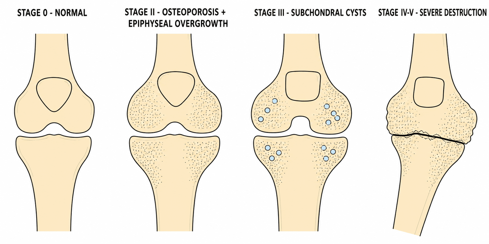
Imaging Findings in Hemophilic Arthropathy
Classification
Hemophilia Severity Classification
Classification based on factor activity level determines bleeding phenotype and treatment strategy.
- Factor Level
- Under 1%
- Bleeding Pattern
- Spontaneous joint/muscle bleeds from infancy
- Factor Level
- 1-5%
- Bleeding Pattern
- Bleeding after minor trauma, occasional spontaneous
- Factor Level
- 5-40%
- Bleeding Pattern
- Bleeding after significant trauma or surgery
Most orthopaedic pathology occurs in severe hemophilia due to recurrent spontaneous hemarthroses.
Clinical Presentation
Acute Hemarthrosis
- Acute onset joint pain (often atraumatic)
- Warmth and swelling
- Joint held in position of comfort (usually flexion)
- Limited range of motion
- May have prodromal tingling sensation ("aura")
- May recall minor trauma
- May have been undertreated (missed prophylaxis)
- Frequency of bleeds important for prognosis
- Tense effusion
- Increased warmth
- Tenderness on palpation
- Guarding and muscle spasm
- Check for other sites of bleeding
Acute hemarthrosis requires urgent factor replacement to arrest bleeding and minimise joint damage.
Muscle Bleeds: Iliopsoas Hematoma and Compartment Syndrome
Hemophilia causes "joint AND muscle bleeds" — the muscle bleeds are their own orthopaedic emergencies and must not be forgotten alongside the hemarthroses.
- Iliopsoas hematoma - the great mimic. A spontaneous bleed into the iliopsoas presents with groin, lower-abdominal or hip pain, the hip held flexed and externally rotated, and pain on extension — mimicking a septic hip, an acute abdomen or appendicitis. Expansion in the iliacus compartment can compress the femoral nerve (quadriceps weakness, lost patellar reflex, anterior-thigh numbness). Diagnose with ultrasound or CT; treat with urgent factor replacement, rest and graded mobilisation — NOT primarily surgery.
- Compartment syndrome from a muscle bleed. A bleed into a closed fascial compartment (forearm, calf) can raise pressure enough to cause an acute compartment syndrome; the flexor-forearm bleed is the hemophilic route to a Volkmann ischaemic contracture. Treat with immediate factor correction; fasciotomy is reserved for an established compartment syndrome and only under full factor cover.
- Why it matters. These are the muscle counterparts of the hemarthrosis — treat with factor first, image second — and recognise the femoral-nerve palsy of the iliopsoas bleed and the contracture risk of an untreated forearm/calf bleed.
Q: A boy with severe hemophilia has groin pain, holds the hip flexed and externally rotated, and has quadriceps weakness with a lost knee reflex - what is it? A: An iliopsoas hematoma compressing the femoral nerve (it mimics a septic hip or acute abdomen). Confirm with ultrasound/CT and treat with urgent factor replacement, rest and graded mobilisation - not primary surgery. Its muscle-compartment counterpart is a forearm/calf bleed causing compartment syndrome (and, if neglected, a Volkmann contracture).
Investigations
Laboratory Investigations
- Factor VIII assay: Reduced in Hemophilia A
- Factor IX assay: Reduced in Hemophilia B
- APTT: Prolonged (intrinsic pathway)
- PT/INR: Normal (extrinsic pathway intact)
- Bleeding time: Normal (platelet function intact)
- Inhibitor screen (Bethesda assay): Critical - antibodies to factor
- Factor level and recovery study
- Blood group and crossmatch
- Hepatitis B, C and HIV status (historical transfusion risk)
- Full blood count
- Liver function (may have hepatitis-related liver disease)
- Factor levels during treatment
- Trough levels if on prophylaxis
Inhibitor development occurs in 25-30% of severe Hemophilia A patients and significantly complicates treatment.
Hemophilia A vs B:
- Hemophilia A
- Factor VIII
- Hemophilia B
- Factor IX
- Hemophilia A
- 85% of cases
- Hemophilia B
- 15% of cases
- Hemophilia A
- Classic hemophilia
- Hemophilia B
- Christmas disease
- Hemophilia A
- Factor VIII concentrate
- Hemophilia B
- Factor IX concentrate
- Hemophilia A
- 25-30% in severe
- Hemophilia B
- 3-5% in severe
Differential Diagnosis
A swollen, painful joint in a young male is not always hemophilic hemarthrosis. The key orthopaedic discriminator is the bleeding history and coagulation profile.
- Distinguishing Features
- Recurrent atraumatic bleeds, target joint, prodromal aura, known family history
- Coagulation / Key Test
- Prolonged APTT, normal PT, reduced FVIII or FIX
- Distinguishing Features
- Fever, systemic upset, exquisite pain on micro-movement; can coexist with hemophilia
- Coagulation / Key Test
- Raised CRP/WCC, aspirate for Gram stain and culture (with factor cover)
- Distinguishing Features
- Mucocutaneous bleeding predominates, hemarthrosis rare except severe type 3
- Coagulation / Key Test
- Reduced vWF antigen/activity, variable FVIII
- Distinguishing Features
- Symmetrical or polyarticular, morning stiffness, no bleeding history
- Coagulation / Key Test
- Normal coagulation, raised inflammatory markers, ANA
- Distinguishing Features
- Single joint, recurrent blood-stained effusions, no clotting defect
- Coagulation / Key Test
- MRI low-signal hemosiderin (blooming), normal coagulation
- Distinguishing Features
- Clear injury mechanism, rapid effusion, normal clotting
- Coagulation / Key Test
- Normal APTT/PT, MRI shows structural injury
Management

Primary Prevention (Prophylaxis)
- Regular factor replacement prevents hemarthroses
- Start before or soon after first joint bleed
- Aim: Maintain trough level above 1-5%
- Hemophilia A: Factor VIII 25-40 IU/kg every other day or 3x/week
- Hemophilia B: Factor IX 25-40 IU/kg twice weekly
- Manco-Johnson et al, NEJM 2007 (severe Hemophilia A, boys under 30 months)
- Prophylaxis vs enhanced episodic treatment
- Prophylaxis group: 93% index joints normal on MRI at age 6
- Episodic group: 55% normal (relative risk of damage 6.1)
- Prophylaxis is now standard of care
- EHL FVIII/IX (Fc or albumin fusion, PEGylation) reduce dosing frequency and raise troughs
- Emicizumab (subcutaneous bispecific antibody) for Hemophilia A with or without inhibitors - HAVEN 3 showed 96-97% bleed reduction
- Improved adherence and quality of life
Early prophylaxis prevents the development of hemophilic arthropathy.
Perioperative Factor Dosing: the Calculation Behind the Targets
The topic repeatedly quotes target factor levels (100% pre-op, 50-80% post-op) — here is the pharmacology that turns a target into a dose.
- The FVIII rule. 1 IU/kg of factor VIII raises the plasma level by about 2%. So to correct a severe patient (baseline near 0%) to 100%, give roughly 50 IU/kg. FVIII half-life is about 8-12 hours, so it is redosed roughly every 8-12 hours (or by continuous infusion) to hold the level.
- The FIX rule. 1 IU/kg of factor IX raises the level by about 1% (FIX distributes into the extravascular space, so more is needed) — roughly 100 IU/kg to reach 100%. FIX half-life is longer (about 18-24 hours), allowing less frequent dosing.
- Worked target. Major orthopaedic surgery: correct to ~100% immediately pre-op, then hold 50-80% for the first 1-2 weeks (tapering as healing progresses); minor procedures need lower peaks and shorter duration.
- The caveat. These arithmetic rules apply only to standard factor concentrates — they do NOT translate to emicizumab (not measured in factor units, and it interferes with one-stage FVIII assays) or to a patient with inhibitors (where bypassing agents, not factor arithmetic, are used). Always confirm the plan with haematology and a recovery study.
Q: How much factor do you give a severe hemophilia A patient to reach 100% before major surgery, and how does hemophilia B differ? A: FVIII: 1 IU/kg raises the level ~2%, so ~50 IU/kg reaches 100% (half-life ~8-12 h, redose 8-12-hourly). FIX: 1 IU/kg raises only ~1% (larger volume of distribution), so ~100 IU/kg is needed (longer half-life ~18-24 h). These rules apply to standard concentrates only - NOT to emicizumab or inhibitor patients (bypassing agents), which need haematology guidance and a recovery study.
Complications
- Progressive joint destruction (arthropathy)
- Fixed flexion contractures
- Angular deformity (valgus/varus)
- Muscle wasting and weakness
- Limb length discrepancy
- Pseudotumours (rare - encapsulated haematomas in soft tissue/bone)
- Intraoperative bleeding
- Postoperative haematoma
- Infection (higher rate than general population)
- Wound healing problems
- Stiffness (especially knee)
- DVT/PE (paradoxical - still occurs)
- Inhibitor development: Antibodies to factor (25-30% Hemophilia A)
- Transfusion-transmitted infections (historical - HCV, HIV)
- Allergic reactions to factor concentrates
- Central venous catheter complications (if port present)
- Rare but serious
- Encapsulated haematoma in muscle or bone
- Progressive enlargement if untreated
- May erode bone
- Treatment: Factor replacement, surgery if large
Outcomes
- Joint disease markedly reduced
- Joint Outcome Study: 93% index joints normal on MRI with prophylaxis at age 6
- Life expectancy approaching normal
- Quality of life dramatically improved
- Severe arthropathy by adolescence
- Wheelchair dependence common
- Multiple joint replacements
- Significant disability
- Pain relief: 85-90% satisfied
- Function improvement: Significant
- Survivorship: 85-90% at 10 years
- Complications higher than general population
- Revision rate higher
- Severity of hemophilia (severe worse)
- Adherence to prophylaxis (critical)
- Inhibitor status (inhibitors worsen prognosis)
- Age at first bleed (earlier worse)
- Number of target joints
Guidelines, Registries & Global Practice
Global Epidemiology:
- Hemophilia A affects approximately 1 in 5,000 male births; Hemophilia B approximately 1 in 30,000 male births
- Hemophilia A accounts for roughly 80-85% of cases worldwide; Hemophilia B for 15-20%
- The World Federation of Hemophilia (WFH) Annual Global Survey identifies over 200,000 people with hemophilia, but case identification remains far below expected prevalence in low-income regions
- Around 30% of cases arise from de novo mutations with no family history
Side-by-Side Guidelines:
- Emphasis
- Comprehensive global standard
- Key Recommendation
- Prophylaxis for all severe hemophilia; integrated multidisciplinary care including musculoskeletal assessment
- Emphasis
- Treatment products and prophylaxis
- Key Recommendation
- Early prophylaxis from first joint bleed or young age; supports EHL and non-factor agents
- Emphasis
- National protocols and inhibitor management
- Key Recommendation
- Standardised perioperative factor dosing and structured inhibitor surveillance
- Emphasis
- Surgical decision-making
- Key Recommendation
- Mandatory haematology co-management; confirmed factor/inhibitor plan before any elective procedure
- WFH World Bleeding Disorders Registry collects longitudinal outcome and treatment data across countries
- National arthroplasty registries (NJR, AJRR, AOANJRR, Swedish/Norwegian) capture small but informative volumes of hemophilic joint replacements - consistently showing higher revision and infection rates than osteoarthritis
- Patient-reported and joint-health tools (Haemophilia Joint Health Score, HAL, FISH) increasingly standardised for global comparison
- High-resource settings: primary prophylaxis from infancy, EHL and non-factor agents (emicizumab), point-of-care ultrasound, elective arthroplasty for end-stage disease
- Limited-resource settings: factor supply is the dominant constraint; care is often on-demand or low-dose ("intermediate-dose") prophylaxis, with later presentation and more advanced arthropathy
- Lower-cost radionuclides (e.g. rhenium-188) and synovectomy are emphasised where repeated arthroplasty is not feasible
- Gene therapy (adeno-associated virus FVIII/FIX transfer) is emerging but currently limited to high-resource, highly selected populations
Controversies & Areas of Uncertainty
- Optimal prophylaxis target. Traditional regimens aim for troughs above 1%, but breakthrough bleeds still occur at this level. There is ongoing debate about individualised, higher-trough or pharmacokinetic-guided dosing versus fixed regimens, balanced against cost and venous access.
- Role of emicizumab in non-inhibitor patients. Non-factor therapy dramatically lowers bleeding rates, but long-term joint-protection data and its interaction with breakthrough bleeds requiring factor or bypassing agents are still maturing.
- Joint aspiration in acute hemarthrosis. Aspiration may relieve a tense effusion and reduce iron load, but carries infection and re-bleed risk; indications remain debated and it should never precede factor cover.
- Timing and choice of synovectomy. Radiosynovectomy, arthroscopic and open synovectomy all reduce bleeding, but comparative long-term superiority and the threshold for intervention are not firmly established; radionuclide availability drives practice regionally.
- Implant choice in arthroplasty. The balance between cemented vs cementless fixation and constrained vs less-constrained knee implants in young, often osteopenic hemophilic patients with deformity is unresolved.
- VTE prophylaxis after major surgery. Hemophilia is not fully protective against venous thromboembolism, especially with factor correction; whether and how to give chemical thromboprophylaxis perioperatively is genuinely uncertain and individualised with haematology.
- Gene therapy durability. AAV-mediated factor gene transfer can achieve sustained factor expression, but durability, hepatotoxicity, immune responses and long-term joint outcomes remain under active study.
Mnemonics for Exam Recall
ABXHemophilia Types
Hook:ABX - A is VIII, B is IX, X-linked inheritance.
KAETarget Joints: KAE
Hook:KAE - Knee, Ankle, Elbow in order of frequency.
CHIPPerioperative Protocol: CHIP
Hook:CHIP away at perioperative planning.
Self-Assessment Quiz
Viva Scenarios
Practise clinical reasoning and management decisions out loud
“12-year-old boy with severe Hemophilia A presents with acute left knee swelling and pain after playing soccer. He is on regular prophylaxis but missed yesterday's dose.”
“35-year-old man with severe Hemophilia A, no inhibitors, presents with severe right knee arthropathy. Fixed 30-degree flexion contracture, valgus deformity, pain limiting mobility. Conservative management has failed.”
“8-year-old boy with moderate Hemophilia A has recurrent right knee hemarthroses despite prophylaxis. X-ray shows early arthropathy with widened intercondylar notch. What do you recommend?”
“20-year-old with severe Hemophilia A needs elbow synovectomy for recurrent hemarthroses. Inhibitor screen shows Bethesda titre of 12 BU. How does this change your approach?”
TYPES
- Hemophilia A: Factor VIII (85%)
- Hemophilia B: Factor IX (15%)
- X-linked recessive
- Severe: under 1% factor
TARGET JOINTS (KAE)
- Knee most common
- Ankle second
- Elbow third
- Hinge joints vulnerable
PREVENTION
- Prophylaxis standard of care
- JOS: 93% joints normal on MRI
- Start soon after first joint bleed
- Trough above 1-5%
PERIOPERATIVE (CHIP)
- Coordinate haematology
- Hundred percent factor pre-op
- Inhibitor check (Bethesda)
- Post-op 50-80% for 2 weeks
Evidence Base
- RCT of 65 boys under 30 months with severe Hemophilia A: prophylactic recombinant FVIII vs enhanced episodic therapy
- At age 6, normal index-joint structure on MRI in 93% (prophylaxis) vs 55% (episodic), P=0.006
- Relative risk of MRI joint damage with episodic therapy 6.1 (95% CI 1.5-24.4)
- Joint and total hemorrhage rates significantly lower with prophylaxis
- Cohort of 76 patients with severe hemophilia, median follow-up to age 19
- Median age at first joint bleed 2.2 years; prophylaxis started median age 6
- Pettersson score was 8% higher (95% CI 1-16%) for every year prophylaxis was postponed after the first joint bleed
- Effect independent of age at scoring and prophylactic dose
- Phase 3 RCT of subcutaneous emicizumab in 152 patients with hemophilia A WITHOUT inhibitors
- Annualised bleeding rate 1.5 (weekly) and 1.3 (fortnightly) vs 38.2 with no prophylaxis - 96-97% reduction
- Intra-individual comparison: 68% lower bleeding than prior FVIII prophylaxis
- No thrombotic events or new FVIII inhibitors; main adverse event mild injection-site reaction