Age 4-10 Years
- Malignant Progression: Very high risk of becoming severe before skeletal maturity.
- MRI Mandatory: 20% have intraspinal pathology (Arnold-Chiari, Syrinx, Tethered Cord).
- Lung Development: Alveolar multiplication continues until age 8 (avoid early fusion!).
- Casting Limit: Rarely effective after age 4 due to stiffness.
- Treatment Gap: Often requires 'Growth Friendly' surgery (growing rods).
- “Full neurological exam is critical (Abdominal reflexes)
- “Look for cafe-au-lait spots (NF1)
- “Assess flexibility (Bending films)
- “Check lung function (if cooperative)
Overview and Epidemiology
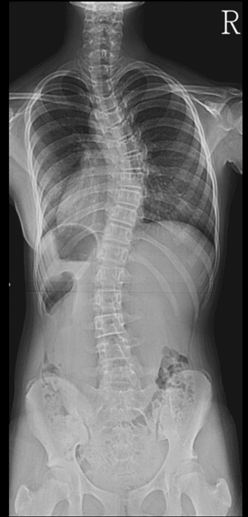
Juvenile idiopathic scoliosis (JIS) is scoliosis diagnosed between the ages of 4 and 10. It sits in a grey zone between infantile scoliosis, which has the potential to resolve, and adolescent scoliosis, whose patterns are predictable. It accounts for approximately 10-15% of all idiopathic scoliosis.
Who. As in AIS, girls are more commonly affected. The curve pattern begins to resemble the adolescent one, with a right thoracic curve.
Why it is called malignant. The child has a huge growth potential remaining, with the whole pubertal growth spurt still ahead. A 30-degree curve at age 5 faces that spurt untreated, and at the Robinson-McMaster rates of 4.5 to 11 degrees a year after age 10 it can exceed 100 degrees by maturity.
- Juvenile (4-10)
- High (20%)
- Adolescent (10+)
- Low (2-4%)
- Juvenile (4-10)
- Very High ('Malignant')
- Adolescent (10+)
- Variable (Depends on Risser)
- Juvenile (4-10)
- Female predominance (modest)
- Adolescent (10+)
- Female predominance (marked)
- Juvenile (4-10)
- Moderate (Age 4-8 critical)
- Adolescent (10+)
- Low (Lungs developed)
Pathophysiology and Mechanisms
Lung development. From birth to age 8 the alveoli multiply in number; after 8 they increase in size but not in number. Early fusion, or a severe deformity, before age 8 results in a permanent reduction in alveolar number, which is true pulmonary hypoplasia. Fusion after age 10 typically has minimal impact on pulmonary function.
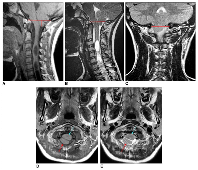
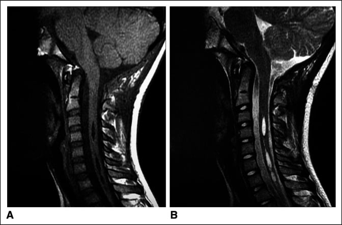
Neural axis abnormalities. Approximately 20% of patients with "idiopathic" juvenile scoliosis have a neural axis abnormality. They include the Chiari 1 malformation, herniation of the cerebellar tonsils by more than 5 mm, and syringomyelia, a fluid-filled cyst in the spinal cord.
How a syrinx bends the spine. The syrinx expands the cord preferentially on one side and damages the anterior horn cells that innervate the paraspinal muscles. The resulting muscle imbalance drives the scoliosis.
Classification Systems
Lenke. Designed for AIS, the Lenke classification is often applied to older juvenile patients (age 8-10) to describe the curve pattern. Its utility is limited, because triple major patterns are less common here than long neurological C-curves or simple thoracic curves.
C-EOS. More useful for juvenile patients, it records:
- Aetiology: I, idiopathic
- Cobb: the magnitude of the angle
- Kyphosis: greater than 50 is unfavourable
- Progression: P2, the malignant group, progresses more than 20 degrees a year
Clinical Assessment
History. Night pain or back pain is a red flag for tumour or syrinx. Headaches or a change in bowel or bladder function point to a Chiari malformation or a tethered cord. Record any strong family history of scoliosis.
Examination. Look for cutaneous markers: hairy patches, dimples and haemangiomas. Cavovarus feet suggest Charcot-Marie-Tooth disease or dysraphism. The Adams forward bend test, with a scoliometer, quantifies the rib hump.
Abdominal reflexes. Stroking the abdomen should make the umbilicus deviate towards the stimulus. An asymmetric response is a syrinx until proven otherwise.
Red flags. The four in a juvenile curve, with what each suggests:
- Left thoracic curve: check the MRI
- Significant night pain: tumour or infection
- Abnormal reflexes: syrinx
- Rapid progression: more than 10 degrees in 6 months
Investigations
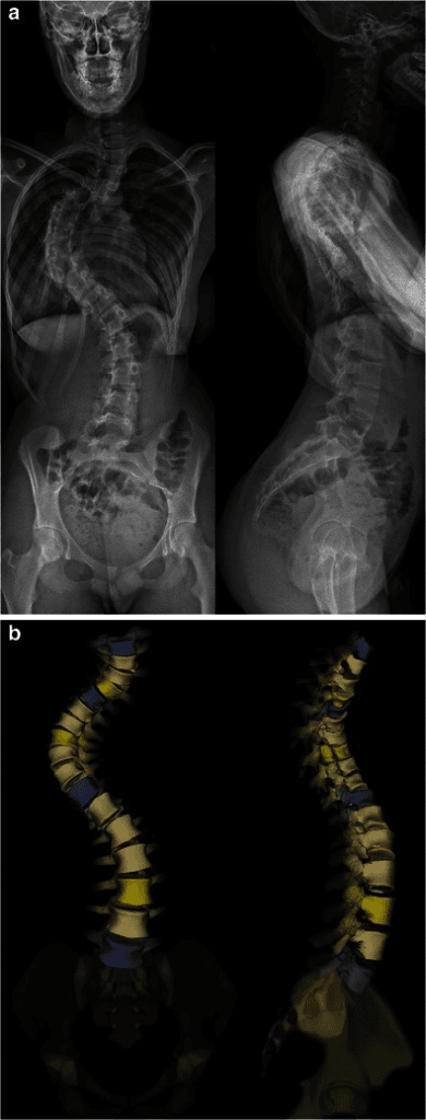
Radiographs. PA and lateral whole-spine radiographs are the baseline. Supine and bending films assess flexibility, which determines whether bracing is viable (a flexible curve) or a release is needed (a stiff one).
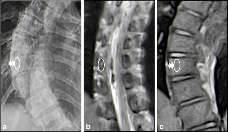
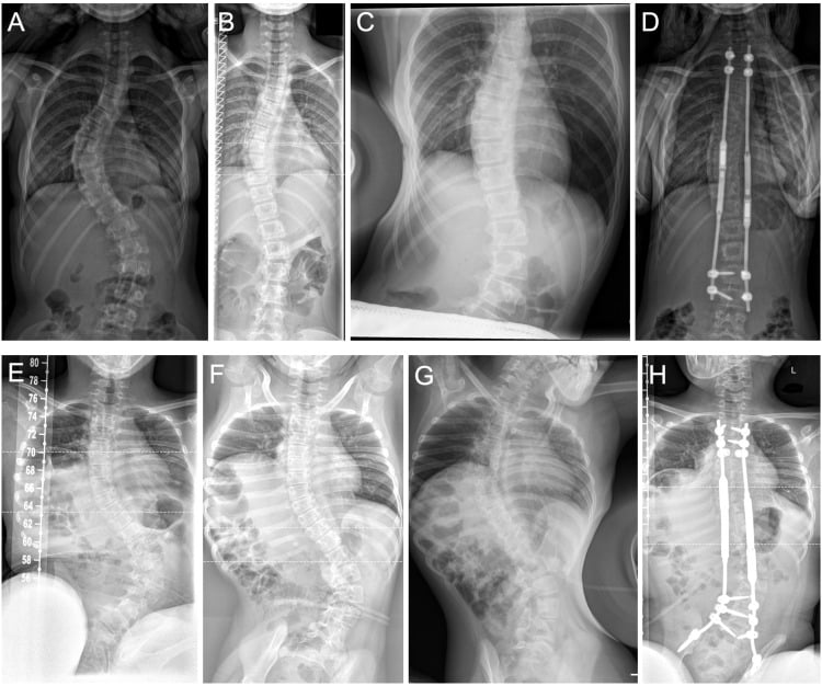
Juvenile scoliosis = MRI spine. In adolescent idiopathic scoliosis, MRI is reserved for red flags. In juvenile scoliosis, the condition itself is a red flag.
MRI. Whole-spine MRI is mandatory for all juvenile scoliosis greater than 20 degrees, and it must image from the brainstem to the sacrum.
Differential Diagnosis
A curve presenting in the 4-10 year window is "idiopathic" only after structural, neurological and syndromic causes are excluded.
- Key Discriminators
- Flexible, no congenital anomaly, normal neurology, right thoracic pattern emerging
- Decisive Test
- Diagnosis of exclusion after normal MRI
- Key Discriminators
- Left thoracic curve, asymmetric/absent abdominal reflexes, cavovarus feet, pain
- Decisive Test
- Whole-spine MRI
- Key Discriminators
- Short, sharp, rigid curve; failure of formation/segmentation; possible VACTERL
- Decisive Test
- Plain films plus CT; renal/cardiac screen
- Key Discriminators
- Cafe-au-lait spots/dystrophic curve (NF1), arachnodactyly (Marfan), disproportion
- Decisive Test
- Genetic/clinical phenotyping, MRI
- Key Discriminators
- Painful, rapidly progressive, night pain, constitutional symptoms, rigid
- Decisive Test
- MRI plus inflammatory markers
Management Algorithm
Goals. Treatment in this age group aims to:
- Prevent severe deformity, greater than 90 degrees
- Preserve lung growth, preventing thoracic insufficiency syndrome
- Postpone definitive fusion until age 10-12
Bracing. A TLSO is indicated for flexible curves of 25-45 degrees at Risser 0. It is less effective than in AIS: it usually cannot prevent surgery eventually, and its goal is to delay it until age 10-12, when the lungs have matured. The prescription is full-time wear of 20-23 hours a day; whether a night-time bending brace can substitute is unsettled (see Controversies).
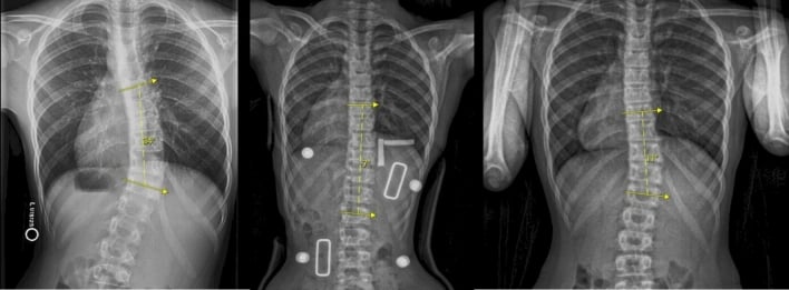
Casting. Casting is rarely used after age 5, but it is useful to buy time when a brace is failing.
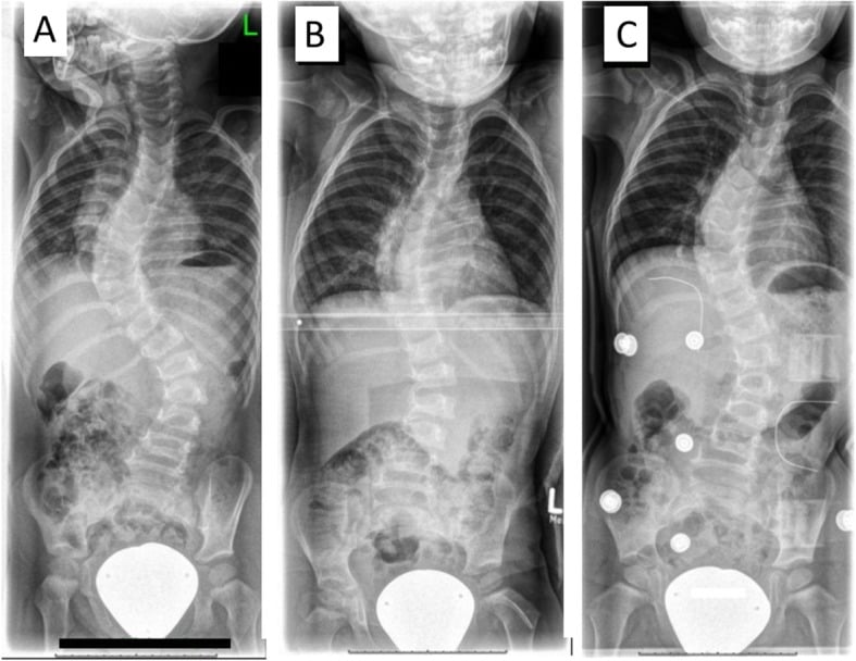
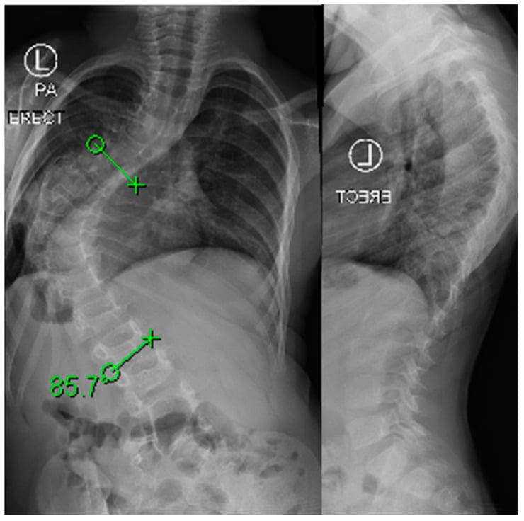
Growth-friendly surgery. It is indicated for a curve greater than 50 degrees, or progression despite bracing. The goal is to control the curve and allow growth; the options are described under Surgical Techniques.
Definitive fusion. The goal is one surgery and one recovery. The indications are:
- Age greater than 10-12
- Risser 1 or above
- Menarche
Timing Spinal Surgery After Neuraxis Decompression
When the MRI finds a Chiari malformation, syrinx or tethered cord, the practical question is how long to wait before instrumenting the spine.
Decompress first. The neurosurgical procedure (foramen-magnum decompression for Chiari, cord detethering, or syrinx drainage or shunt) is generally performed before any scoliosis correction. Operating on a cord that is under tension or contains a syrinx risks neurological injury.
Then give the curve time. After decompression the scoliosis may stabilise or partially improve, particularly in younger children with smaller, flexible curves. A period of observation, commonly around 3-6 months with interval radiographs, shows whether the curve settles before instrumentation is committed to.
Then treat the residual curve on its own merits. If the curve continues to progress or remains large despite decompression, proceed with the age-appropriate scoliosis treatment: brace, growth-friendly surgery or fusion.
Clarify the neural axis before the metal goes in. Metallic growing-rod constructs create MRI artefact that hampers later syrinx surveillance. The neural axis should be clarified and, where possible, addressed before definitive metalwork is placed; once the rods are in, the question "has the syrinx enlarged?" cannot easily be answered.
Surgical Techniques
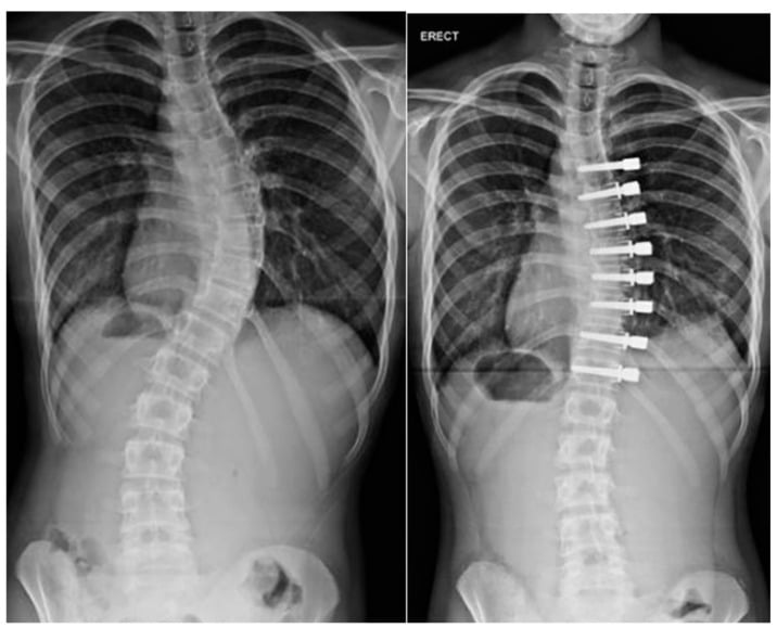
Four strategies are available for the growing spine: distraction with growing rods (MCGR or traditional growing rods, TGR), growth guidance (Shilla), chest expansion (VEPTR, in the next section) and anterior growth modulation (VBT).
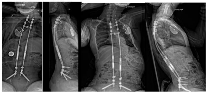
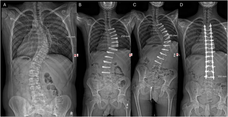
Growing rods. Proximal anchors (hooks or screws at T2/3) and distal anchors (screws at L3/4) are joined by submuscular rods. In a magnetically controlled growing rod (MCGR), an external magnet lengthens the rod 3-5 mm in clinic, usually every 3 months. The disadvantages are metal artefact on MRI, which makes syrinx monitoring difficult, and cost.
The law of diminishing returns. With traditional distraction-based surgery, every operation to lengthen the rods scars and stiffens the spine ("auto-fusion"), and the length gained per surgery decreases over time. By the 5th or 6th lengthening the spine may be completely stiff, yielding no length at all while every risk of surgery remains. The solution is to minimise interventions, which MCGR does by avoiding the repeated open surgeries, or to delay initial implantation as long as possible.
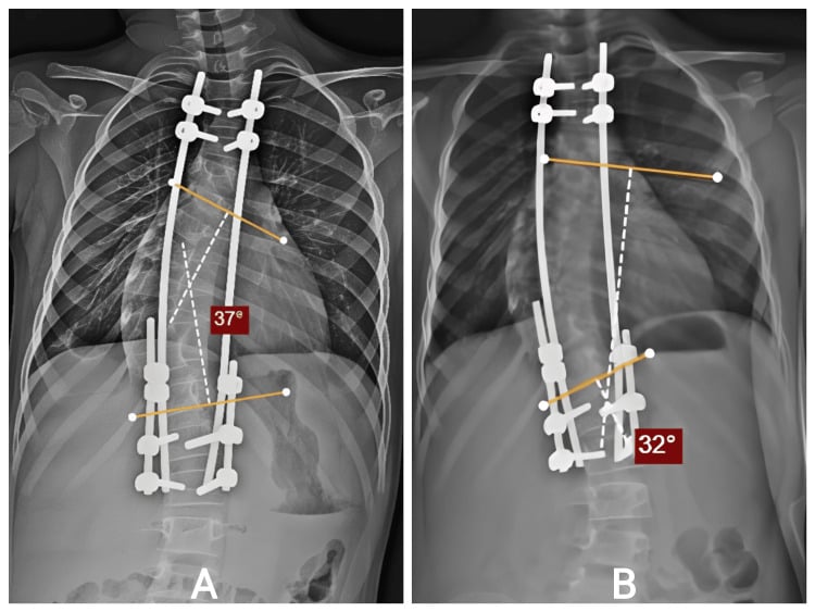
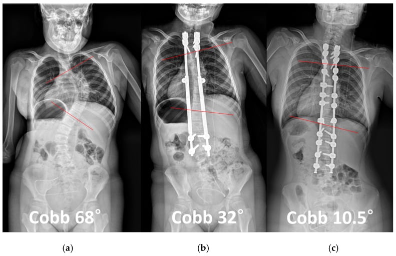
Shilla growth guidance. Only the apex is fused (for example T8-T10). The proximal and distal screws are not locked to the rod, which slides through them as the child grows, like a trombone. In theory it is "one and done" surgery; the disadvantages are implant prominence, screw pull-out and breakage.
Vertebral body tethering (VBT). Screws placed thoracoscopically into the vertebral bodies on the convex side are connected by a flexible cord. The principle is the Hueter-Volkmann law: compression slows growth on the convexity, allowing the concavity to catch up. Its role is controversial, but it is gaining popularity for flexible curves at age 8-12.
VEPTR and Thoracic Insufficiency Syndrome
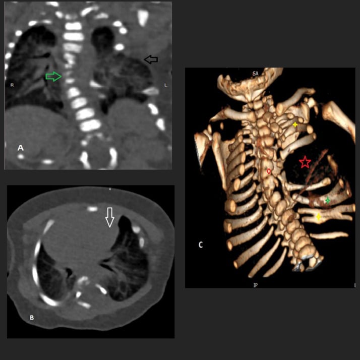
Thoracic insufficiency syndrome (TIS). Campbell defined it as the inability of the thorax to support normal respiration or lung growth. A short, stiff, deformed thorax, from a severe early curve, fused or absent ribs, or early spinal fusion, cannot expand, so the underlying lung cannot grow; this is why the whole topic stresses preserving thoracic height and avoiding early definitive fusion. It is assessed clinically (chest excursion, and the thumb-excursion test of hemithorax expansion) and radiographically (space available for lung).
VEPTR. The Vertical Expandable Prosthetic Titanium Rib is Campbell's rib-based distraction device. It is anchored rib-to-rib (and rib-to-spine or rib-to-pelvis), is periodically lengthened as the child grows, and is designed to expand the thorax directly rather than only correct the spinal curve.
Where it fits. Its classic indication is TIS with a chest-wall abnormality: fused ribs with congenital scoliosis, or other conditions where the thorax itself is deficient. For a purely spinal idiopathic curve, spine-based growing rods (MCGR) or the other options above are usually preferred. Like all distraction-based systems it is subject to the law of diminishing returns and to device and wound complications. The broader early-onset scoliosis picture is developed in that topic.
Complications
- Rate
- High
- Prevention/Management
- MCGR rods jam. Anchors pull out.
- Rate
- Moderate
- Prevention/Management
- Increased with multiple surgeries (less with MCGR).
- Rate
- Common
- Prevention/Management
- Proximal Junctional Kyphosis. Avoid stopping at kyphotic apex.
- Rate
- Unknown
- Prevention/Management
- Titanium debris from sliding rods (Shilla).
- Rate
- Rare
- Prevention/Management
- Monitor cord during lengthening.
Postoperative Care
Most growing-rod patients require a brace after surgery to protect the proximal anchors, and contact sports are restricted. The lengthening schedule must be adhered to strictly.
Outcomes and Prognosis
Untreated. The result is severe disability, restrictive lung disease and cor pulmonale.
Treated. Most children require definitive fusion at maturity. The goal is a "straight-ish" spine with adequate lung volume; final height is usually short, with a short trunk, but functional. Adults with treated JIS have higher rates of back pain than AIS patients.
Guidelines, Registries & Global Practice
Global epidemiology
- Juvenile-onset (age 4-10) accounts for roughly 10-15% of all idiopathic scoliosis; infantile, juvenile and adolescent together form the "early-onset" (under age 10) spectrum that carries the highest pulmonary and progression risk.
- Female predominance increases with age at onset; the curve pattern transitions toward the adolescent right-thoracic pattern by the upper juvenile age range.
Society guidance — side by side
- Position relevant to juvenile / early-onset scoliosis
- Whole-spine MRI for early-onset and atypical curves; growth-friendly instrumentation (MCGR, traditional dual rods) preferred over early definitive fusion; C-EOS used for risk stratification.
- Position relevant to juvenile / early-onset scoliosis
- Specialist paediatric spinal MDT review; MRI before any intervention in young-onset curves; serial low-dose imaging to limit cumulative radiation.
- Position relevant to juvenile / early-onset scoliosis
- Endorses growth-sparing surgery to protect thoracic and pulmonary development; emphasises avoidance of repeated open lengthenings.
- Position relevant to juvenile / early-onset scoliosis
- Provides conservative-management (bracing / scoliosis-specific exercise) consensus; bracing in young-onset curves aims to delay surgery rather than guarantee avoidance.
Registry & device notes
- MCGR adoption was tempered worldwide by reports of metallosis and limited true distraction over time; several registries and national bodies (including UK device alerts) prompted closer surveillance and explant analysis.
- Vertebral body tethering carries device-specific regulatory status that differs by region (e.g. US FDA Humanitarian Device Exemption), and is offered selectively within specialist centres.
High- vs limited-resource practice
- Well-resourced settings: routine whole-spine MRI, EOS or other low-dose biplanar imaging for serial monitoring, MCGR and VBT availability, neuromonitoring for instrumented correction.
- Limited-resource settings: later presentation with larger, stiffer curves; greater reliance on bracing, casting and definitive fusion; halo-gravity traction used to reduce neurological risk when advanced imaging or neuromonitoring is unavailable.
- Radiation stewardship is universal: cumulative imaging over years of growth raises breast-tissue dose concerns, so low-dose biplanar systems and limited-frequency radiography are recommended wherever available.
Deep Dive: The Crankshaft Phenomenon
Definition. Progressive rotational and angular deformity that occurs after posterior fusion in a skeletally immature patient.
Mechanism. The fused posterior elements act as a tether while the anterior vertebral bodies (neurocentral synchondrosis) continue to grow. The spine bulges anteriorly and twists around the posterior tether.
The risk group. This is agreed: Risser 0 with an open triradiate cartilage, which is the single best marker of remaining anterior spinal growth. Triradiate closure, not the Risser sign alone, is what tells you the danger has passed.
The classic rule, and why it is no longer absolute. The classic rule held that in this group posterior fusion alone is inadequate, and a circumferential (anterior plus posterior) fusion is needed to arrest every growth plate. It comes from the era of hook-and-wire constructs, which controlled rotation poorly. With modern segmental pedicle-screw constructs, which grip all three columns and control rotation far better, both the true incidence and the clinical significance of crankshaft after posterior-only fusion are much less certain than the historical literature implies. Most surgeons no longer add an anterior fusion routinely for this indication alone, given its real pulmonary and approach-related cost in a small child.
How to answer it in the exam. State the mechanism, name the open triradiate cartilage as the at-risk marker, and quote the circumferential teaching as the classic teaching. Then say that the modern alternative is a rigid segmental posterior construct, or, better still in this age group, avoiding definitive fusion altogether in favour of a growth-friendly strategy, which sidesteps the problem rather than solving it.
Controversies & Areas of Uncertainty
VBT durability. Vertebral body tethering offers fusionless correction but carries high reoperation rates (tether rupture, overcorrection). Long-term curve behaviour into adulthood and ideal patient selection remain unresolved.
MCGR longevity and metallosis. Magnetically controlled rods reduce open lengthenings, but real-world distraction falls short of predicted gains over time and explant studies show titanium/metal wear debris (metallosis). The optimal lengthening frequency and total service life are debated, and device alerts have prompted closer surveillance.
Bracing efficacy in JIS. Bracing can alter natural history and buy time, but it rarely prevents surgery in larger juvenile curves. The threshold and protocol (full-time versus night-time bending brace) are not standardised for this age group. The management above prescribes 20-23 hours a day, yet in Jarvis's juvenile series a night-time bending brace successfully managed 19 of 37 curves.
When to obtain MRI. Universal MRI in juvenile curves is widely endorsed given the neural axis yield, yet cost-effectiveness, sedation risk in young children, and exactly which lower-magnitude curves warrant imaging are still discussed.
Timing of definitive fusion. Balancing crankshaft and progression risk against pulmonary and spinal-height loss means the "right age" to convert from growth-friendly to definitive fusion is individualised, not fixed.
Deep Dive: Vertebral Body Tethering (VBT)
The New Kid on the Block VBT is a "growth modulation" technique (like 8-plates for knees).
- Indication: Skeletally immature (Risser 0-2), Flexible curve, Age 8-12.
- Mechanism: Screws placed laterally in vertebral bodies on the convex side. A flexible polyethylene cord connects them. Tension is applied.
- Result: Compression of the convex growth plate slows growth. Concave side keeps growing. The spine straightens as the child grows.
- Controversy: High revision rate (tether rupture, over-correction). Long term results unknown. FDA approved (HDE) but still considered "innovative" in many guidelines.
MCQ Practice Points
Q: At what age does the risk of true pulmonary hypoplasia (loss of alveolar number) significantly decrease? A: Age 8. Fusion after age 8 generally affects lung volume (size) but not alveolar count.
Q: What is the most common cause of growing rod failure? A: Anchor failure (Hook/Screw pullout) or Rod Fracture.
Q: What clinical sign is most specific for a syrinx in a child with scoliosis? A: Absent abdominal reflexes. (Asymmetrical superficial abdominal reflex).
Q: A 5-year-old has a 60 degree congenital scoliosis (Unilateral unsegmented bar with contralateral hemivertebra). Treatment? A: Fusion. Congenital curves DO NOT respond to bracing. A short fusion of the congenital anomaly is required to stop the "evil" growth mismatch. Growing rods are for long curves (idiopathic/NM).
Q: What percentage of patients with juvenile idiopathic scoliosis have a neural axis abnormality (Chiari/Syrinx) on MRI? A: Approximately 20%. This is why MRI is mandatory for all juvenile scoliosis patients.
Self-Assessment Quiz
Viva Scenarios
Practise clinical reasoning and management decisions out loud
“6-year-old female. Right Thoracic curve 35 degrees. MRI is normal. Risser 0.”
“8-year-old male. Left Thoracic curve. MRI shows a large thoracic syrinx.”
“A 9-year-old underwent posterior fusion for severe scoliosis 2 years ago. Now the deformity is recurring and she is twisting.”
BASICS
- Age 4-10
- High Progression
- MRI Mandatory
- The Grey Zone
RED FLAGS
- Left curve
- Pain
- Neuro Signs
- Rapid Increase
THE LUNGS
- Alveoli Age 8
- TIS Risk
- Avoid Early Fusion
- Volume is Life
SURGERY
- Growing Rods
- Shilla
- Tethering (VBT)
- Delay Fusion
Evidence Base
Robinson & McMaster — Curve Patterns and Prognosis
- 109 consecutive juvenile idiopathic scoliosis patients (67 girls, 42 boys; mean age 6 years 10 months)
- 104 of 109 curves were progressive; only 5 (5%) resolved spontaneously
- Progression was 1 to 3 degrees per year before age 10, accelerating to 4.5 to 11 degrees per year after age 10
- Single/double mid-thoracic curves (Groups 1 and 2) carried the worst prognosis; thoracolumbar and lumbar curves (Groups 3 and 4) were more benign
Zhang et al — Intraspinal Anomalies in Infantile & Juvenile Scoliosis (504 patients)
- Largest MRI series of 'presumed idiopathic' infantile/juvenile scoliosis (n=504)
- Neural axis abnormality found in 94 patients (18.7%); Arnold-Chiari with/without syringomyelia accounted for 64.8% of these
- Male sex, left thoracic curve and right lumbar curve were independently associated with intraspinal pathology
- Confirms a routine whole-spine MRI is warranted in scoliosis presenting before age 10
Karol — Early Definitive Fusion: Pulmonary Consequences
- Systematic review of early-onset scoliosis treated by definitive fusion
- Restrictive pulmonary disease (FVC less than 50% predicted) occurred in 43% to 64% of patients fused young
- Extensive and proximal thoracic fusions carried the highest pulmonary risk
- Post-fusion thoracic growth averaged only 50% of that in non-fused scoliotic children; reduced thoracic height correlated with reduced FVC
Bess et al — Complications of Growing-Rod Treatment (140 patients)
- Multicenter Growing Spine Study Group; 140 patients, 897 growing-rod procedures
- 81 patients (58%) sustained at least one complication
- Complication risk rose 24% with every additional surgical procedure and fell 13% for each year initial implantation was delayed
- Dual rods and submuscular placement reduced implant and wound complications versus single/subcutaneous rods
Akbarnia et al — Magnetically Controlled Growing Rod (14 patients)
- Prospective multicenter series of MCGR with outpatient, anaesthesia-free distractions
- Mean Cobb angle corrected from 60 to 34 degrees after index surgery, maintained at 31 degrees at follow-up
- Dual rods achieved greater spinal height gain than single rods (T1-S1: 3.09 vs 1.27 mm/month)
- No neurological deficit or implant failure during early follow-up
Cheung et al — First-in-Human MCGR Series
- Prospective case series introducing the magnetically controlled growing rod (5 patients)
- In the 2 patients with 24-month follow-up, mean Cobb angle fell from 67 to 29 degrees
- Each non-invasive monthly distraction lengthened the instrumented segment by approximately 1.9 mm
- No MCGR-related complications; improved quality-of-life and cost-effectiveness versus traditional growing rods
Jarvis et al — Part-Time Bracing in Juvenile Idiopathic Scoliosis
- Retrospective review of JIS treated with a nighttime bending brace (curves over 20 degrees, Risser 0)
- 19 of 37 curves (51%) were successfully managed in-brace; 7 patients ultimately required fusion
- Success correlated with greater in-brace correction rather than initial curve magnitude
- Part-time bracing performed better than the natural history with psychosocial/compliance advantages
Samdani et al — Anterior Vertebral Body Tethering, 2-Year Results
- First clinical cohort of anterior VBT in skeletally immature idiopathic scoliosis (11 patients)
- Thoracic Cobb angle corrected from 44 to 13.5 degrees (70% correction) over 2 years via growth modulation
- Axial trunk rotation improved from 12.4 to 6.9 degrees; no major complications
- 2 of 11 patients returned to theatre for tether loosening to prevent overcorrection