Osteochondromas Throughout the Skeleton
- EXT1/EXT2: Tumor suppressor genes.
- Malignant Transformation: 1-5% to chondrosarcoma.
- Warning Signs: New growth, pain, size greater than 5cm in adults.
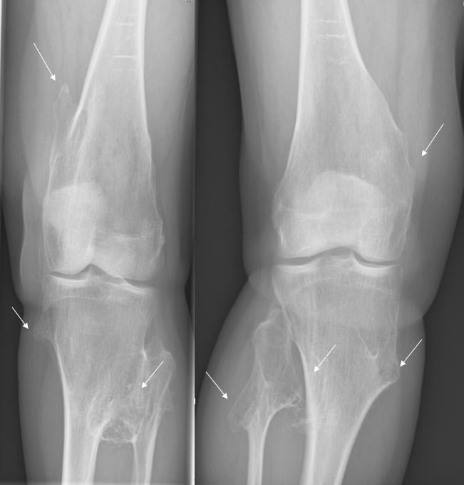
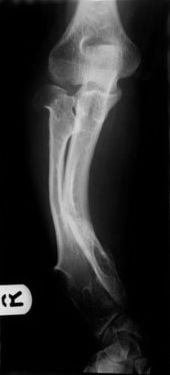
- Forearm Deformity: Common (ulnar shortening).
- Excision: For symptomatic lesions.
- “EXT1/EXT2 mutations
- “1-5% malignant transformation
- “Pain/growth in adults = concerning
- “Forearm deformity common
Warning signs of malignant transformation to chondrosarcoma:
- New pain in a previously painless lesion.
- Growth after skeletal maturity.
- Size greater than 5cm cartilage cap on imaging.
- Irregular margins, scattered calcifications.
- Obtain MRI. If concerning, biopsy or excise with wide margins.
- Details
- 1-5% to chondrosarcoma
- Management
- Wide excision if suspected
- Details
- Ulnar shortening, radial bowing
- Management
- Ulnar lengthening, osteotomy
- Details
- Pain, nerve compression
- Management
- Excision
MHEMHE Features
Hook:EMM - EXT, Multiple, Malignant risk.
PGSMalignant Warning Signs
Hook:PGS - Pain, Growth, Size.
Overview/Epidemiology
Multiple Hereditary Exostoses (MHE) is characterized by multiple osteochondromas.
- Genetics: Autosomal dominant. EXT1 or EXT2 mutations (tumor suppressor genes).
- Incidence: 1 in 50,000.
- Pathophysiology: Loss of EXT function leads to abnormal cartilage growth at physes → osteochondromas.
- EXT1 vs EXT2: EXT1 mutations (chromosome 8) typically cause more severe phenotype than EXT2 (chromosome 11).
- Natural History: Lesions grow until skeletal maturity, then stop. Malignancy risk persists lifelong.
Pathophysiology and Mechanisms
Osteochondroma Structure
- Cartilage-capped bony outgrowth arising from the metaphysis.
- Continuous with host bone cortex and medulla.
- Grows away from the adjacent joint (important for diagnosis).
- Cartilage cap: Normally less than 1cm in adults, greater than 2cm concerning for malignancy.
Why Deformity Occurs
- Osteochondromas at the distal ulna → ulnar shortening → radial bowing.
- Tethering effect on growth plate.
- Similar mechanism at ankle (fibular lesions) → ankle valgus.
Common Sites
- Distal femur (most common).
- Proximal tibia.
- Proximal humerus.
- Distal ulna/radius (causes forearm deformity).
Molecular Mechanism: EXT → Heparan Sulphate → Hedgehog Signalling
The topic states that EXT1/EXT2 are "tumour-suppressor genes" encoding "heparan sulphate glycosyltransferases" and that loss of EXT function causes "abnormal cartilage growth at physes" — but the mechanism that links the gene to the lesion is worth developing, because it is the classic basic-science question for this condition.
From gene to signalling. EXT1 and EXT2 form a complex of glycosyltransferases that synthesise and elongate heparan-sulphate (HS) chains on heparan-sulphate proteoglycans. HS proteoglycans act as a scaffold that shapes the gradient and signalling of growth-plate morphogens — most importantly Indian Hedgehog (Ihh) (and also FGFs, BMPs and Wnts). In the normal physis, the Ihh/PTHrP negative-feedback loop sets the pace of chondrocyte proliferation and keeps proliferating chondrocytes in orderly longitudinal columns.
From signalling to osteochondroma. Defective HS disrupts this Ihh signalling, so chondrocytes at the periphery of the growth plate lose their normal polarity and proliferate laterally, escaping the perichondrial groove of Ranvier to form a cartilage-capped outgrowth. That osteochondroma then grows by endochondral ossification from its own cartilage cap and — because it is driven by the physis — ceases at physeal closure, exactly the natural history described above.
The tumour-suppressor (second-hit) angle. As EXT1/EXT2 are tumour suppressors, osteochondromas behave as clonal lesions arising after a somatic "second hit" (loss of the remaining wild-type EXT allele) in a cartilage-cap cell; progression to secondary peripheral chondrosarcoma involves further genetic change in that clone — the molecular basis of the lifelong transformation risk. (The general biochemistry of heparan-sulphate proteoglycans is developed in the proteoglycans-collagen topic, and the Ihh/PTHrP loop in the physis-growth-plate-anatomy topic.)
EXT1/EXT2 encode glycosyltransferases that build heparan-sulphate chains; HS proteoglycans shape the Indian Hedgehog (Ihh)/PTHrP gradient governing orderly growth-plate proliferation. Defective HS lets peripheral chondrocytes lose polarity and bud laterally → a cartilage-capped osteochondroma growing by endochondral ossification until physeal closure. As tumour-suppressor genes, a somatic second hit underlies the clonal lesion and the step toward secondary chondrosarcoma.
Classification Systems
Masada Classification (Forearm Deformity)
- Type I: Ulnar shortening with bowing of the radius, secondary to distal ulnar osteochondroma (most common).
- Type IIA: Dislocation of the radial head with osteochondroma of the proximal radius.
- Type IIB: Dislocation of the radial head secondary to more distal involvement (no proximal radial lesion).
- Type III: Relative radial shortening due to osteochondromas at the distal radius.
Clinical Assessment
- Age of first lesion.
- Symptomatic lesions (pain, cosmesis, nerve compression).
- Family history.
- Any new pain or growth in adults (malignancy concern).
- Palpable Masses: Typically at metaphyses.
- Deformity: Forearm (short ulna), ankle (valgus).
- ROM: Limited by impingement.
- Neurovascular: Peroneal nerve at knee, etc.
Investigations
- X-ray: Multiple osteochondromas, broad-based or pedunculated.
- MRI: If malignancy suspected (cartilage cap thickness greater than 2cm or irregular).
- EXT1/EXT2 testing if diagnosis uncertain.
Differential Diagnosis
Multiple Cartilaginous Lesions:
- Key Features
- Multiple osteochondromas
- Differentiator
- Continuous cortex, metaphyses, EXT mutation
- Key Features
- Multiple enchondromas
- Differentiator
- Medullary lesions, not cortical
- Key Features
- Enchondromas + hemangiomas
- Differentiator
- Soft tissue vascular lesions
- Key Features
- Osteochondromas + enchondromas
- Differentiator
- Both lesion types present
- Key Features
- Epiphyseal osteochondroma
- Differentiator
- Single limb, epiphysis involved
Key Distinguishing Points:
- MHE: Metaphyseal, cortex continuous with host bone.
- Enchondromas (Ollier): Medullary, radiolucent with stippled calcifications.
- DEH (Trevor Disease): Single limb, epiphyseal involvement.
- Malignancy risk: MHE 1-5%, Ollier 25-30%, Maffucci 25-30%.
Management Algorithm
Observation
- Asymptomatic lesions: Watch and wait.
- Regular clinical follow-up.
- Educate on warning signs for malignancy.
Surgical Techniques
Osteochondroma Excision
Indications: Symptomatic lesions (pain, nerve compression, impingement).
Technique:
- Marginal excision at the base.
- Include entire cartilage cap to prevent recurrence.
- Careful of adjacent neurovascular structures.
Pearl: Incomplete excision of cartilage cap leads to recurrence.
Lower-Limb Deformity & Guided Growth
The surgical sections develop the forearm in detail, but MHE causes major lower-limb deformities that the management above does not address — and these are high-yield. They arise from osteochondromas tethering or asymmetrically loading the peri-articular physes.
- Genu valgum — from peri-knee osteochondromas (and relative fibular overgrowth/tethering); one of the commonest MHE angular deformities.
- Ankle valgus — the classic MHE ankle deformity, from relative distal fibular shortening (distal fibular/tibial lesions) producing lateral talar tilt.
- Leg-length discrepancy and disproportionate (mesomelic) short stature — from physeal involvement; coxa valga and acetabular dysplasia also occur at the hip.
- Guided growth (hemiepiphysiodesis with tension-band / "8-plates") while the physes remain open is the least-invasive first line for angular deformity — medial distal femur and/or proximal tibia for genu valgum, and medial distal tibia for ankle valgus.
- Corrective osteotomy when growth remaining is insufficient or the deformity is severe/rigid; fibular lengthening for marked ankle valgus.
- Excise the tethering osteochondroma at the affected physis, and manage leg-length discrepancy as for other causes (epiphysiodesis of the long limb or lengthening).
The general technique of guided growth, genu valgum correction and leg-length-discrepancy management is developed in the guided-growth-angular-deformity-correction, genu-valgum-varum and limb-length-discrepancy-epiphysiodesis topics.
Beyond the forearm, MHE causes genu valgum (peri-knee lesions), ankle valgus (relative distal fibular shortening → lateral talar tilt), leg-length discrepancy and short stature. While physes are open, guided growth (hemiepiphysiodesis / 8-plates — medial distal femur/proximal tibia for genu valgum, medial distal tibia for ankle valgus) is first-line; reserve corrective osteotomy / fibular lengthening for the mature or severe deformity, and excise the tethering osteochondroma.
MHEForearm Deformity in MHE
Hook:URD - Ulnar short, Radial bow, Dislocation risk.
Complications
- Context
- 1-5%, lifelong risk
- Management
- Surveillance, wide excision
- Context
- Incomplete excision
- Management
- Complete cartilage cap removal
- Context
- Peroneal at knee
- Management
- Careful dissection
- Context
- Childhood growth
- Management
- Early intervention
- Context
- Popliteal region
- Management
- Pre-op imaging
Postoperative Care
- Simple Excision: Early mobilization, wound care.
- Forearm Reconstruction: Splinting, protected ROM.
- Lengthening: Fixator care, daily adjustments.
- All Patients: Continue surveillance for other lesions.
Outcomes/Prognosis
- Most lesions remain benign.
- Malignant transformation: 1-5%.
- Significant deformity may limit function.
Guidelines, Registries & Global Practice
Global epidemiology
- Prevalence approximately 1 in 50,000 worldwide, with a male predominance (~1.5:1) and near-complete penetrance (~96%); roughly 10% of cases arise de novo without a family history.
- Mean number of osteochondromas per patient is 15-18; lesions appear and enlarge in the first decade and cease at physeal closure.
Consensus and society guidance (side by side)
- Position
- Confirm diagnosis clinically/radiologically; offer EXT1/EXT2 testing and genetic counselling; lifelong awareness of transformation; refer suspicious lesions to a bone-tumour centre
- Position
- Surveillance of accessible lesions; MRI for new pain or growth; biopsy/wide resection only for suspected malignancy
- Position
- Suspected secondary chondrosarcoma referred to a sarcoma MDT; en-bloc resection in a specialist centre
- Position
- Pediatric deformity correction in tertiary units; pooled registry/biobank data through European rare-disease networks
Registry and network evidence
- No dedicated arthroplasty-style registry exists; long-term data derive from population databases (e.g. Schmale's Washington kindreds), tertiary bone-tumour registries and rare-disease networks (Orphanet, EuroBoNeT/European sarcoma networks).
High- vs limited-resource practice
- Well-resourced settings: MRI surveillance, genetic testing, guided growth (8-plates), staged forearm reconstruction and limb salvage for transformation.
- Limited-resource settings: diagnosis is clinical/radiographic; emphasis on educating patients about warning signs (new pain, growth after maturity, enlarging cartilage cap) and timely referral when malignancy is suspected, since chondrosarcoma is chemo- and radio-resistant and depends on adequate surgical margins.
Controversies and Areas of Uncertainty
- Routine surveillance imaging: No consensus on whether asymptomatic adults need scheduled whole-body MRI/screening. Some advocate baseline imaging of axial/proximal lesions (pelvis, shoulder) that are hard to monitor clinically; others rely on symptom-driven imaging and patient education. Porter's data suggest EXT1 carriers may justify more proactive screening.
- Timing of forearm reconstruction: Whether early excision and ulnar lengthening prevent radial head dislocation, versus observation until deformity declares itself, remains debated; high-quality comparative evidence is limited.
- True malignant transformation rate: Quoted figures range from 0.5% to 5%; older series likely overestimated risk through referral bias, while genotype (EXT1) and lesion location modify individual risk.
- Guided growth vs osteotomy: Optimal first-line correction of knee/ankle valgus (hemiepiphysiodesis versus corrective osteotomy) depends on remaining growth and deformity magnitude, without firm thresholds.
- Emerging medical therapy: Heparan-sulphate pathway and palovarotene-type signalling research is experimental; no disease-modifying drug is established in practice.
MCQ Practice Points
Q: What genes are mutated in MHE? A: EXT1 and EXT2.
Q: What is the malignant transformation rate? A: 1-5% to chondrosarcoma.
Q: What are warning signs for malignancy? A: Pain, growth after skeletal maturity, cartilage cap greater than 2cm.
Q: What causes forearm deformity in MHE? A: Distal ulnar osteochondroma causes ulnar shortening, leading to radial bowing and potential radial head dislocation.
Q: What is the Masada classification used for? A: Forearm deformity in MHE. Types I-III based on ulnar shortening, radial deformity, and radial head status.
Q: Which nerve is commonly compressed at the knee in MHE? A: Common peroneal nerve (fibular head osteochondroma).
Self-Assessment Quiz
Additional Quiz Questions
Viva Scenarios
Practise clinical reasoning and management decisions out loud
“30-year-old with known MHE. One of his knee lesions has become painful and appears to have grown over the past year.”
“10-year-old with MHE has progressive forearm deformity with limited pronation/supination.”
“12-year-old with MHE presents with foot drop. X-ray shows large osteochondroma at proximal fibula. How do you manage?”
GENETICS
- EXT1/EXT2
- Autosomal Dominant
- Tumor suppressors
- 1 in 50,000
CLINICAL
- Multiple osteochondromas
- Metaphyses
- Forearm deformity
- Knee most common
MALIGNANCY
- 1-5% transformation
- Pain in adults
- Growth after maturity
- Cap greater than 2cm
TREATMENT
- Observe if asymptomatic
- Excise if symptomatic
- Wide excision if malignant
- Correct deformity
FOREARM
- Ulnar shortening
- Radial bowing
- Masada classification
- Early intervention prevents RH dislocation
NERVES
- Peroneal at fibular head
- Sciatic at hip
- Foot drop = excision
- Pre-op imaging
Evidence Base
- Prevalence ~1 in 50,000; male predominance (~1.5:1); mean 15-18 osteochondroma sites
- EXT1/EXT2 germline mutations found in ~90% of patients (heparan sulphate glycosyltransferases)
- Secondary peripheral chondrosarcoma estimated at 0.5-5%; resect en-bloc with tumour-free margins
- Characteristic deformities: short stature, limb-length discrepancy, knee/ankle valgus, radial bowing with ulnar wrist deviation, radiocapitellar subluxation
- EXT mutations disrupt chondrocyte proliferation and maturation
- Surgery can prevent progression and correct selected deformities; slight sarcomatous risk
- 36 forearms in 30 patients classified into 3 types (the Masada classification)
- Type I ulnar shortening + radial bowing; Type II radial head dislocation (IIa/IIb); Type III distal radial involvement
- 92% satisfactory results after type-directed surgery (excision, ulnar lengthening, radial osteotomy)
- Population database: 46 kindreds, 113 affected; prevalence at least 1 in 50,000; penetrance 96%
- Median age at diagnosis 3 years; 39% had obvious forearm deformity, 8% knee, 2% ankle
- Average of two operations per surgically treated patient
- Prospective genotype-phenotype study of 172 individuals (78 families); mutation identified in 83%
- EXT1 carriers significantly worse than EXT2 in stature, deformity and function
- 7 sarcomas in EXT1 carriers vs 1 in EXT2; EXT1 sarcoma risk comparable to screened breast-cancer risk
- T2-weighted MRI best depicts and differentiates the cartilage cap from adjacent soft tissue
- Cap thickness is essential to assessing malignant transformation; CT shows matrix calcification and bone destruction
- MRI is the most reliable technique for locoregional staging of malignant cartilage tumours