The Stiffness is the Key
- Definition: A flatfoot with restricted subtalar motion and no arch reconstitution
- Peroneal Spastic Flatfoot: A clinical description (not a diagnosis) usually caused by Coalition
- Differential: Tarsal Coalition, Vertical Talus, Septic Arthritis, JIA, Trauma
- Workup: Weight-bearing X-rays are first line; CT is gold standard for bony coalition; MRI for fibrous
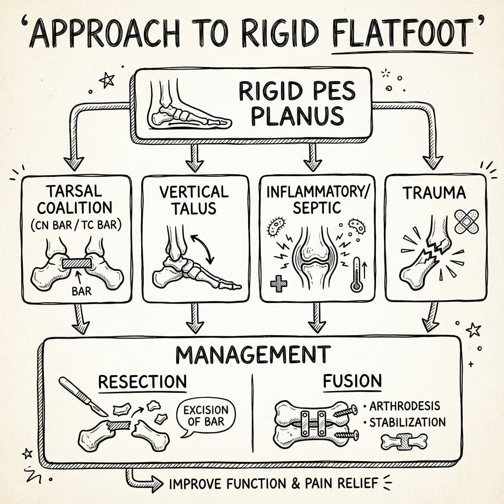
- Management: Depends on cause (Resection vs Fusion)
- “If the heel stays in valgus when they go on tiptoes, think RIGID
- “Always check subtalar motion - if it's stiff, get a CT
- “Unilateral flatfoot is suspicious
- “Pain in the sinus tarsi or medial malleolus suggests coalition
"Peroneal Spastic Flatfoot" is a historical term describing the clinical appearance of a rigid flatfoot where the peroneal tendons are tight/spastic.
- It is NOT a diagnosis in itself.
- It is a sign of an underlying pathology (usually Tarsal Coalition).
- Do not stop at "Peroneal Spastic Flatfoot" - always find the underlying cause!
- Flexible Flatfoot
- Heel Inverts (Varus)
- Rigid Flatfoot (e.g. Coalition)
- Heel Remains Valgus
- Flexible Flatfoot
- Arch Reconstitutes
- Rigid Flatfoot (e.g. Coalition)
- Arch Remains Flat
- Flexible Flatfoot
- Normal
- Rigid Flatfoot (e.g. Coalition)
- Restricted / Absent
- Flexible Flatfoot
- Usually Asymptomatic
- Rigid Flatfoot (e.g. Coalition)
- Often Painful
- Flexible Flatfoot
- Normal Anatomy (just flat)
- Rigid Flatfoot (e.g. Coalition)
- Bony/Cartilaginous Abnormalities
- Typical Age / Clue
- Child-adolescent, recurrent sprains
- Discriminating Feature
- C-sign / anteater nose, restricted subtalar motion
- Confirmatory Test
- CT (bony) or MRI (fibrous)
- Typical Age / Clue
- Newborn, rocker-bottom sole
- Discriminating Feature
- Irreducible dorsal navicular dislocation
- Confirmatory Test
- Forced plantarflexion lateral X-ray
- Typical Age / Clue
- Any age, acute, febrile
- Discriminating Feature
- Refusal to weight-bear, raised CRP/ESR
- Confirmatory Test
- Aspiration / MRI, washout
- Typical Age / Clue
- Child, insidious, may be bilateral
- Discriminating Feature
- Morning stiffness, synovitis, warmth
- Confirmatory Test
- MRI synovitis, inflammatory markers
- Typical Age / Clue
- Adolescent, night pain
- Discriminating Feature
- Pain relieved by NSAIDs/aspirin
- Confirmatory Test
- CT nidus
- Typical Age / Clue
- Adult, unilateral, progressive
- Discriminating Feature
- Flexible early then rigid, too-many-toes sign
- Confirmatory Test
- MRI / dynamic exam
- Typical Age / Clue
- Any age, history of injury
- Discriminating Feature
- Prior fracture, deformity
- Confirmatory Test
- X-ray / CT
TCCoalition Locations
Hook:The two main bars.
ACBRadiographic Signs
Hook:The Zoo of Radiology.
Overview/Epidemiology
Rigid Flatfoot is a descriptive term for a foot that lacks a medial longitudinal arch and is stiff. Unlike the ubiquitous flexible flatfoot, a rigid flatfoot is almost always pathological.
Historically, this described a foot held in rigid valgus by spasm of the peroneal muscles. We now know that the peroneal spasm is usually a protective reflex to splint a painful, stiff subtalar joint (typically due to a coalition). It may also be seen in inflammatory conditions (Subtalar arthritis).
- Infant: Congenital Vertical Talus (CVT).
- Child: Tarsal Coalition (Calcaneonavicular - ossifies earlier).
- Adolescent: Tarsal Coalition (Talocalcaneal - ossifies later).
- Any Age: Septic Arthritis, Osteomyelitis, Trauma, Tumor (Osteoid Osteoma).
Anatomy/Biomechanics
Allows inversion/eversion. Essential for accommodating uneven ground. The "Torque Converter" of the foot.
- Block to Motion: A bony or cartilaginous bar (coalition) or dislocation (CVT) prevents subtalar inversion/eversion.
- Fixed Valgus: The heel is locked in valgus.
- Midfoot Unlock: Because the hindfoot cannot invert, the midfoot cannot lock (via the locking wedge mechanism of the transverse tarsal joint) to become a rigid lever for push-off.
- Peroneal Overdrive: The peroneals shorten over time or spasm to prevent painful inversion against the bar.
- Calcaneonavicular (CN): Connection between the anterior process of the calcaneus and the navicular.
- Talocalcaneal (TC): Connection typically at the middle facet of the subtalar joint.
Pathophysiology: Tarsal Coalition
A failure of mesenchymal segmentation during fetal development. The bar is initially cartilaginous (synchondrosis) or fibrous (syndesmosis) and allows some motion, hence why young children are asymptomatic. As the child grows and the bar ossifies (synostosis), stiffness increases, micro-fractures occur across the rigid bar during activity, and pain develops.
- Calcaneonavicular (CN): Ossifies between 8-12 years. Symptoms appear at this age.
- Talocalcaneal (TC): Ossifies between 12-16 years. Symptoms appear later in adolescence.
Why does it cause Spasm? The rigid subtalar joint loses its ability to invert/evert. When walking on uneven ground, the ground reaction force attempts to invert the heel. The rigid subtalar joint cannot invert. This places stress on the peroneal muscles which fire reflexively to hold the foot in eversion (valgus) to protect the stiff joint from forced inversion stress.
Associated Anomalies:
- Fibular Hemimelia (often associated with Tarsal Coalition).
- Apert Syndrome.
- Nievergelt-Pearlman Syndrome.
CAVETCauses of Rigid Flatfoot
Hook:Don't CAVE To the rigid foot.
Deep Dive: Inflammatory and Septic Causes
- Presentation: Acute onset rigid flatfoot, refusal to bear weight, fever, elevated CRP/ESR.
- Mechanism: Pus in the joint causes severe spasm (splinting) of the surrounding muscles (Peroneals).
- Urgency: Surgical emergency requiring washout.
- Presentation: Insidious onset stiffness, often bilateral (but starts unilateral). Subtalar joint is a common target.
- Signs: Warmth, swelling, morning stiffness.
- Natural History: If uncontrolled, leads to spontaneous fusion (ankylosis) of the subtalar joint. This causes a permanent rigid flatfoot in adulthood.
- Classic Site: Talar neck or Subtalar joint.
- Mechanism: The tumor secretes prostaglandins which cause intense local inflammation and reflexive muscle spasm (Peroneal Spastic Flatfoot).
- Key Symptom: Night pain relieved by Aspirin/NSAIDs.
- Imaging: CT reveals the "nidus".
Detail: Coalition Resection
- CT Scan: Essential. Assess the size of the coalition. Rule of thumb: if the bar involves greater than 50% of the joint surface, resection is likely to fail (instability/pain). Fusion is preferred.
- Hindfoot Valgus: If severe (greater than 15-20 degrees), resection alone won't correct alignment. Need calcaneal osteotomy.
- Incision: Lateral oblique incision in the lines of tension skin.
- Protection: Sural nerve (posterior) and Superficial Peroneal Nerve (dorsal).
- Exposure: Elevate the Extensor Digitorum Brevis (EDB) from its origin.
- Identification: The bar is palpable between the calcaneus and navicular.
- Resection: Using an osteotome or burr. The resection must be generous (rectangular block).
- Check: Visualize the talar head (medial) and cuboid (lateral) to ensure full width resection.
- Interposition: The EDB muscle belly is sewn into the defect with absorbable suture to act as a spacer.
- Incision: Medial curvilinear incision over the sustentaculum tali.
- Protection: Tibialis Posterior tendon, FDL, FHL, and Neurovascular bundle (retract posteriorly).
- Exposure: Open the sheath of FDL/Tib Post. Identify the middle facet.
- Resection: High speed burr to remove the bony bridge.
- Safety: Do not penetrate too deeply into the posterior facet (lateral) or sinus tarsi.
- Interposition: Fat graft (from Kager's triangle or local fat) or bone wax.
Classification Systems
Anatomical Classification of Coalition
- Syndesmosis: Fibrous union (Stiff but maybe some motion).
- Synchondrosis: Cartilaginous union (Stiffer).
- Synostosis: Bony union (Rigid).
Detail: Advanced Classification of Coalitions
- Upasani Classification:
- Type 1: Fibrous/Cartilaginous (Irregular joint line).
- Type 2: Bony (Solid bar).
Based on CT scan morphology and percentage of posterior facet involvement.
- Type I: Linear (straight bond).
- Type II: Linear with posterior hook.
- Type III: Shingled (overlapping).
- Type IV: Complete bony block.
- Type V: Posterior facet involvement greater than 50%. (Poor prognosis for resection).
- Coronal Plane: Best for TC coalition (Middle facet).
- Sagittal Plane: Best for C-Sign.
- Oblique/Axial: Best for CN coalition.
- Impingement Signs: Check for dorsal "beaking" on the talus (Traction spur, not OA) vs true joint space narrowing (OA). Resection is contraindicated if greater than 50% joint narrowing.
Clinical Assessment
- Pain: Often "vague" ankle pain or sinus tarsi pain. Worse with activity or uneven ground.
- Sprains: Recurrent ankle sprains (because the subtalar joint can't accommodate, the ankle rolls).
- Stiffness: "My foot doesn't move like the other one."
- Look:
- Flattened arch (Pes Planus).
- Heel Valgus (Hindfoot Valgus).
- "Too Many Toes" Sign (Forefoot abduction).
- Feel:
- Tender Sinus Tarsi (CN Coalition/Arthritis).
- Tender Medial Malleolus/Sustentaculum (TC Coalition).
- Tight Peroneal Tendons (bowstringing behind lateral malleolus).
- Move:
- Subtalar ROM: Lock the talus in the mortise (dorsiflex ankle) and swing the heel. Restricted or Absent in rigid flatfoot.
- Tiptoe Test: Heel fails to invert.
- Jack's Test: Arch fails to rise.
Investigations
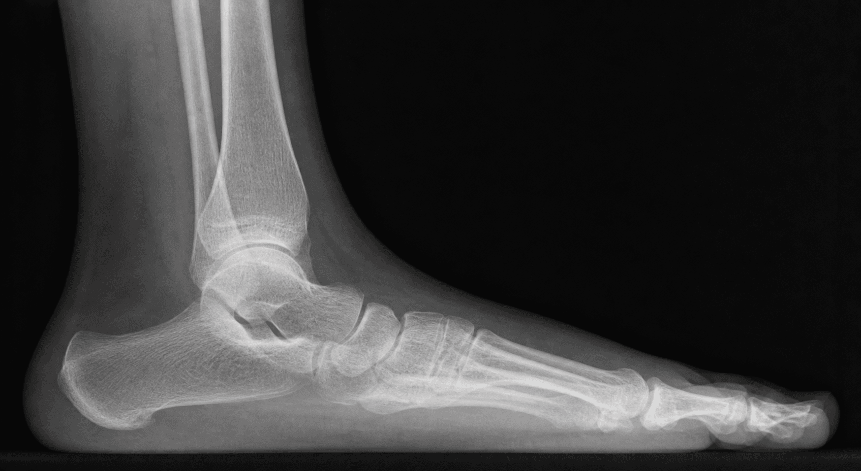
- AP Foot: "Talonavicular uncoverage".
- Lateral Foot:
- C-Sign: Continuous C-shaped line from talar dome to sustentaculum (TC Coalition).
- Anteater Nose Sign: Elongated anterior process of calcaneus (CN Coalition).
- Talar Beak: Dorsal osteophyte on talar head (traction spur from navicular capsule).
- Vertical Talus: Talus axis points to sole, Navicular dorsal.
- Harris Heel View: Special view to see the posterior and middle facets.
- CT Scan: Gold Standard for defining bony anatomy and mapping coalitions. Essential for surgical planning (size of bar, hindfoot valgus angle).
- MRI: Useful for Fibrous or Cartilaginous coalitions (which may be invisible on CT) and for assessing soft tissue/inflammatory causes (synovitis).
Management Algorithm
Detail: Triple Arthrodesis
Indication: Severe rigid flatfoot with degenerative changes (arthritis) or failure of coalition resection. The "Gold Standard" salvage.
Principle: Fusion of the Talonavicular (TN), Calcaneocuboid (CC) and Subtalar (TC) joints to create a rigid, stable, plantigrade foot.
- Incision: Lateral Ollier's incision (extended).
- Exposure: EDB reflected. Retract peroneals plantarwards.
- Joint Prep (Resection):
- CC Joint: Resect cartilage to bleeding bone.
- Subtalar Joint: Resect posterior and anterior facets. Remove the coalition.
- TN Joint: Exposed from lateral side (challenging) or separate medial incision. Remove cartilage.
- Correction:
- Reduce the Talonavicular joint first (key to alignment).
- Correct Valgus at the Subtalar joint.
- Correct Abduction at the CC joint.
- Fixation:
- Subtalar: large screw (6.5mm/7.0mm) from heel to talus.
- TN: screws or staples.
- CC: screws or staples.
- Closure: Layered closure over a drain.
- Position: Supine, sandbag under ipsilateral hip.
- Tourniquet: Thigh.
- Post-op: NWB for 6-12 weeks until union.
Surgical Technique
Calcaneonavicular Bar Excision
Approach: Lateral Ollier's incision (over sinus tarsi). Technique:
- Identify Extensor Digitorum Brevis (EDB).
- Reflect EDB distally.
- Identify the bar between Anterior Calcaneus and Navicular.
- Resect the bar thoroughly (rectangular block).
- Check motion (should improve immediately).
- Interposition: Sew the EDB belly into the defect to prevent regrowth.
Complications
- Risk Factors
- Inadequate resection, no interposition.
- Prevention/Management
- Prevention: Generous resection and EDB/Fat interposition.
- Risk Factors
- Missed second coalition, underlying arthritis.
- Prevention/Management
- Prevention: Pre-op CT to scan whole foot. Management: Fusion.
- Risk Factors
- Lateral approach.
- Prevention/Management
- Prevention: Identify and protect.
- Risk Factors
- Medial approach (TC coalition).
- Prevention/Management
- Prevention: Careful handling of skin.
- Risk Factors
- Prolonged immobilization.
- Prevention/Management
- Prevention: Early ROM if stable.
Postoperative Care
- Weeks 0-2: Splint/Cast, Non-Weight Bearing (NWB). Elevate significantly to prevent wound breakdown.
- Weeks 2-6:
- Motion: Start active ROM exercises (writing alphabet with foot).
- Physio: Focus on peroneal strengthening and subtalar eversion/inversion.
- Weight: Touch down weight bearing in a boot.
- Weeks 6+:
- Weight bearing as tolerated in shoes.
- Continue physio for 3-6 months.
- Return to sport at 3-4 months.
- Weeks 0-2: Backslab, strictly NWB. Elevation.
- Weeks 2-6: Conversion to lightweight fibreglass cast or CAM boot (locked). Still NWB to protect the fusion mass.
- Weeks 6-12:
- Progressive weight bearing in CAM boot.
- X-ray at 6 weeks to check alignment.
- X-ray at 12 weeks to confirm union.
- Months 3-6:
- Wean out of boot into stiff-soled shoe.
- Gait retraining (expect stiff gait).
- No impact sports.
- CRPS (Complex Regional Pain Syndrome): High risk in foot surgery. Early movement and desensitization are key prevention strategies. Vitamin C 500mg daily is often prescribed.
- Stiffness: Failure to mobilize after resection leads to fibrosis.
Outcomes/Prognosis
- Tarsal Coalition:
- Resection yields good results in young patients (~75-80% relief).
- Poorer results in older patients or large bars.
- Vertical Talus:
- Dobbs technique gives excellent functional results and avoids stiff, small feet associated with extensive releases.
- Untreated Rigid Flatfoot:
- Leads to progressive degenerative arthritis of the triple joint complex.
- May require Triple Arthrodesis in adulthood.
Surgical Tips and Tricks
- Headlight: Essential for visualization, especially medial approach.
- Bone Wax: Use liberally on the raw bone surfaces after resection to prevent hematoma and re-ossification.
- Fat Graft: Don't skimp. Harvest a large plug from the Kager's triangle (retro-calcaneal fat pad). It has a robust blood supply.
- Intra-op Fluoroscopy: Use it to confirm the amount of bone removed. The "Harris Line" (middle facet) must be clear.
- Dynamic Check: After resection, the subtalar motion should return immediately. If it's still stiff, you haven't taken enough bone, or there's another coalition.
- Order of Fixation:
- Talonavicular (TN): This sets the version of the foot. Reduce this first.
- Subtalar (TC): Corrects the valgus/varus.
- Calcaneocuboid (CC): Follows the others.
- Screw Position:
- Subtalar screw should aim for the talar dome but NOT penetrate it.
- TN screws should be placed from navicular into talar head (or vice versa), avoiding the joint surface.
- Bone Graft: Use local autograft from the resected wedges to pack the fusion sites.
Guidelines, Registries & Global Practice
Global Epidemiology:
- Tarsal coalition: Classically quoted at under 1% symptomatic prevalence, but cadaver/CT studies show non-osseous coalition in roughly 11-13% of feet - most are asymptomatic. Bilateral in 50-60%. Calcaneonavicular and talocalcaneal account for the vast majority; up to 20% of patients have more than one coalition.
- Congenital vertical talus: Rare (around 1 in 10,000 live births). Roughly half are syndromic (arthrogryposis, myelomeningocele, chromosomal anomalies).
Side-by-Side Practice (no single national framework):
- Convergent global practice
- Weight-bearing radiographs for every rigid flatfoot
- Where opinion still differs
- CT vs MRI as the next study (CT for bony mapping; MRI better for fibrous/cartilaginous bars and marrow oedema)
- Convergent global practice
- Minimally invasive reverse-Ponseti (Dobbs) method is now the international standard of care
- Where opinion still differs
- Role and extent of soft-tissue release in non-idiopathic/syndromic feet
- Convergent global practice
- Resection + interposition for symptomatic, non-arthritic bars
- Where opinion still differs
- The 50% posterior-facet "rule" - newer data (Khoshbin) suggest larger TC bars can still do well
- Convergent global practice
- Address hindfoot valgus, not just the bar
- Where opinion still differs
- Calcaneal lengthening osteotomy vs subtalar/triple fusion for severe valgus
High- vs Limited-Resource Settings:
- Well-resourced: Routine CT/MRI, motion-preserving reconstruction (resection plus lengthening osteotomy), early Dobbs-method CVT casting in infancy.
- Limited-resource: Diagnosis often delayed to symptomatic adolescence or neglected CVT; reliance on plain films; later presentation shifts the balance toward arthrodesis/salvage. Naviculectomy with limited release has been described as an affordable "third way" for neglected/complex CVT.
- Registries: There is no dedicated coalition or CVT registry; evidence is drawn from single-centre series and small cohorts, which is itself a key limitation (see Controversies).
Deep Dive: Congenital Vertical Talus (CVT)
Also known as "Rocker Bottom Foot". It is a dorsolateral dislocation of the talonavicular joint. The talus is locked in a vertical (plantarflexed) position, and the navicular is dislocated onto the dorsal neck of the talus. This is the hallmark of a rigid flatfoot in a newborn.
- Hindfoot: Severe valgus and equinus (calcaneus is plantarflexed).
- Midfoot: Dorsally dislocated.
- Forefoot: Abducted and dorsiflexed.
- Soft Tissue Contractures: Tight Achilles, Peroneals, Tibialis Anterior, and Extensor Digitorum Longus.
- Idiopathic: 50% of cases.
- Syndromic: 50% of cases. Highly associated with Arthrogryposis, Spina Bifida (Myelomeningocele), and Genetic Syndromes (Trisomy 13, 15, 18).
- CVT: Rigid. Navicular does not reduce on plantarflexion X-ray.
- Oblique Talus: Flexible/Reducible. Navicular slides back on plantarflexion.
Deep Dive: The Talar Beak
Definition: A dorsal osteophyte located on the head of the talus.
Clinical Pearl: The presence of a Talar Beak is often the first clue on a lateral X-ray that the subtalar joint is stiff, even if the coalition itself is not visible. It signifies abnormal mechanics.
Pathomechanics:
- It is NOT a sign of osteoarthritis of the Talonavicular joint.
- It is a Traction Spur.
- Because the subtalar joint is rigid, the navicular overrides the talar head during dorsiflexion. This causes excessive tension on the dorsal talonavicular capsule/ligament.
- This tension pulls on the periosteum, leading to bone formation (Enthesophyte).
Radiographic Distinction:
- Talar Beak: Located proximal to the joint line. The joint space itself is preserved. Resection is still an option.
- Degenerative Spur: Located at the joint margin (lipping) and associated with joint space narrowing. Resection is contraindicated (Fusion needed).
Multiple Coalitions: Image the Whole Foot, and Beware Persistent Stiffness
The topic repeatedly warns to "rule out a second coalition" and notes that persistent stiffness after resection suggests a missed bar — worth consolidating, because it changes both the imaging and the operation.
- More than one coalition is common. Up to about 20% of patients have more than one coalition (in the same or the opposite foot), and coalitions are bilateral in 50-60%; a calcaneonavicular and a talocalcaneal bar can coexist in the same foot.
- Image the whole foot, not just the symptomatic bar. Because a second coalition is easily missed, the preoperative CT should map the entire hindfoot and midfoot (and image the other foot if it is symptomatic) - finding one bar does not exclude another.
- The intra-operative rule. After an adequate resection, subtalar motion should return immediately on the table. If the foot is still stiff after a technically complete resection, assume either an unaddressed second coalition or established degenerative arthritis - do not simply close and hope.
- The commonest reason a resection fails (persistent pain and stiffness) is therefore a missed second coalition or unrecognised arthritis - both are avoided by whole-foot preoperative imaging and an intra-operative motion check.
Q: You have completely resected a calcaneonavicular bar but subtalar motion has not returned on the table - what do you do? A: Do not close and hope. Persistent stiffness after a technically complete resection means either a missed second coalition (up to ~20% have more than one) or established arthritis. This is why the whole foot must be CT-mapped preoperatively and motion checked intra-operatively - re-examine for a second bar and reassess the joint surfaces.
Controversies & Areas of Uncertainty
- The 50% posterior-facet rule: The traditional teaching that talocalcaneal coalitions involving over 50% of the posterior facet require fusion rather than resection is being challenged. Khoshbin et al. found favourable long-term outcomes after resecting TC bars exceeding 50% of the facet with hindfoot valgus over 16 degrees, and Mosca's calcaneal lengthening offers a motion-preserving alternative even for "unresectable" coalitions.
- Treat the deformity or the bar?: Resecting the coalition without correcting fixed hindfoot valgus can leave a painful, poorly aligned foot. There is no consensus on the valgus threshold mandating an added calcaneal lengthening osteotomy versus resection alone.
- Interposition material: EDB, fat graft, bone wax and tendon have all been used to prevent reossification after resection; no high-level comparative evidence establishes one as superior.
- Pinning in CVT: Dobbs' own series showed recurrence only in feet that were NOT pinned, but the optimal duration of talonavicular K-wire fixation and casting is not standardised.
- Asymptomatic coalition: Whether an incidentally discovered coalition should ever be resected prophylactically (e.g. in a young athlete) remains unresolved - current practice treats symptoms, not the radiograph.
- Evidence quality: Nearly all data are Level III-IV retrospective series from single centres with no registry; there are no randomised trials comparing resection strategies or CVT techniques.
Parent's Guide: Frequently Asked Questions
Q: Will my child grow out of it? A: Unlike flexible flatfeet (which often improve), a rigid flatfoot (Tarsal Coalition) is a structural problem. The "bar" between the bones will not disappear. Symptoms might fluctuate, but the stiffness remains.
Q: Is surgery always needed? A: No. If the foot is not painful, we leave it alone. We treat the symptoms, not the X-ray. Many adults have coalitions they don't know about.
Q: Can they play sports after surgery? A: Yes. After resection, most children return to sports. If a fusion (Triple Arthrodesis) is performed, high-impact sports (running, soccer) may be difficult, but cycling and swimming are excellent.
Q: Why is the cast on for so long? A: To allow the swelling to settle (after resection) or to allow the bones to knit together (after fusion). Rushing rehabilitation can lead to persistent pain.
MCQ Practice Points
Q: The 'Anteater Nose' sign is pathognomonic for which condition? A: Calcaneonavicular Coalition. It represents the elongated anterior process of the calcaneus.
Q: Which facet is most commonly involved in Talocalcaneal coalitions? A: Middle Facet. It is often hard to see on standard lateral views (requires Harris view or CT).
Q: What is the primary contraindication to coalition resection? A: Degenerative Changes (Arthritis) in the subtalar or talonavicular joint. If arthritis is present, resection will fail; fusion is required.
Q: What is the characteristic finding of Peroneal Spastic Flatfoot? A: Rigid Valgus that does not correct on tiptoeing, with tight/bowstrung peroneal tendons.
Q: What is the gold standard imaging for diagnosing Talocalcaneal coalition? A: CT Scan. It best demonstrates bony anatomy and the extent of the coalition (less than 50% = resection, greater than 50% = fusion).
Q: What tissue is interposed after calcaneonavicular coalition resection? A: Extensor Digitorum Brevis (EDB) muscle belly. This prevents bony regrowth and maintains the resection gap.
Viva Scenarios
Practise clinical reasoning and management decisions out loud
“13-year-old boy, recurrent ankle sprains, painful flat feet. Exam shows restricted subtalar motion.”
“Newborn with a rigid flatfoot, convex sole (Rocker Bottom). Top of foot touches shin.”
“35-year-old male, sudden onset painful unilateral flatfoot. No trauma. History of 'minor' sprains.”
DEFINITION
- Restricted Subtalar Motion
- No Arch Reconstitution
- Fixed Valgus
- Peroneal Spasm (Reactive)
DIFFERENTIAL
- Tarsal Coalition
- Vertical Talus
- Arthritis (Septic/JIA)
- Trauma
- Tumor
WORKUP
- Tip-Toe Test
- Jack's Test
- X-ray (C-sign, Anteater)
- CT (Gold Standard for Bone)
MANAGEMENT
- Symptomatic: Resection
- Arthritic/Large: Fusion
- Infant (CVT): Casting + Pinning
- Always rule out 2nd coalition
RED FLAGS
- Unilateral
- Night Pain (Tumor)
- Systemic Symptoms (Sepsis)
- Fever / Elevated CRP
Evidence Base
- 11 patients (19 feet) with idiopathic congenital vertical talus, minimum 2-year follow-up
- Serial reverse-Ponseti casts (mean 5) then talonavicular pinning plus percutaneous Achilles tenotomy; no extensive releases needed
- Initial correction achieved in all 19 feet; recurrence in 3 feet - all of which had NOT had talonavicular pin fixation
- 24 children treated with the minimally invasive (Dobbs) method; radiographic recurrence in 7 (29%)
- Mean PROMIS scores within 1 SD of the reference population for pain interference, mobility and peer relations
- Children who started treatment after 12 months of age had significantly lower mobility scores
- 24 patients (32 resections: 19 calcaneonavicular, 13 talocalcaneal), mean follow-up to age 25-27 years
- CN and TC resections produced similar long-term function and satisfaction scores
- Favourable results were attained even for TC coalitions over 50% of the posterior facet and hindfoot valgus over 16 degrees
- 8 patients (13 painful talocalcaneal coalitions) treated with calcaneal lengthening osteotomy, with or without resection
- Osteotomy corrected valgus and relieved pain even when the coalition was unresectable, preserving talonavicular/calcaneocuboid motion
- All feet underwent concurrent gastrocnemius or Achilles lengthening
- 78 patients / 97 coalition resections (49 talocalcaneal, 47 calcaneonavicular, plus rarer types)
- Mean return to desired activity 18.3 weeks; mean post-operative Roles & Maudsley score 1.3 (excellent/good)
- Most patients returned to their desired activity level after excision
- 100 cadaver feet dissected after spiral CT: non-osseous coalition prevalence 12.7% (CN most common single type, 9.1%)
- True prevalence is far higher than the classically quoted under 1%, because most coalitions are asymptomatic
- Spiral CT detected only ~55% of coalitions and missed 4 non-osseous bars
- 27 adults (31 feet) with non-paralytic disease, mean 14-year follow-up; 93% satisfied with the result
- Severe adjacent-joint arthrosis developed in the ankle (7 ankles), naviculocuneiform and tarsometatarsal joints; 3 later needed ankle fusion
- 74% reported moderate-to-severe difficulty on uneven ground; SF-36 physical score well below the population mean