Orthopaedic Emergency | Staphylococcus aureus | Urgent Washout | Prevent AVN
- True Orthopaedic Emergency: Pus under pressure tamponades the blood supply (AVN) and enzymes destroy cartilage (Chondrolysis).
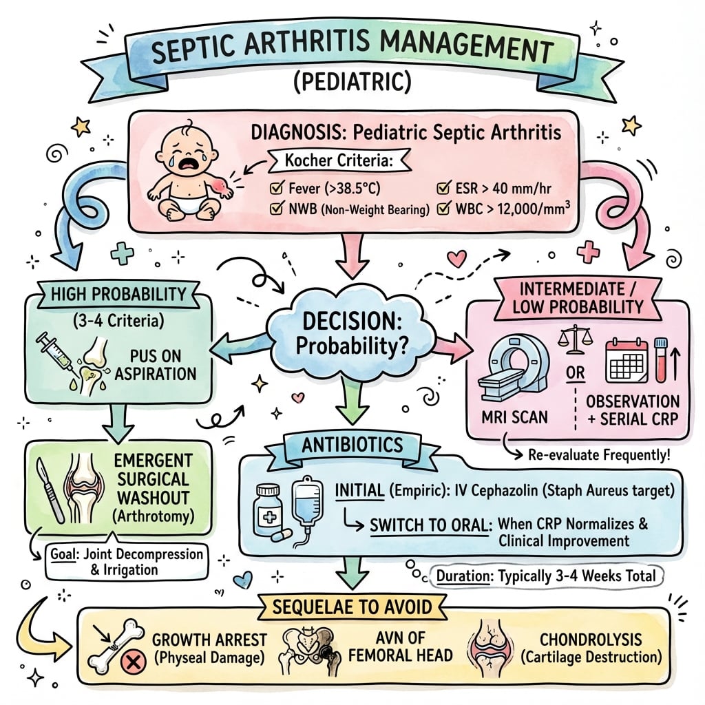
- Kocher Criteria: Fever over 38.5, NWB, ESR over 40, WCC over 12,000. (Caird added CRP over 20).
- Investigation: Ultrasound confirms effusion but not infection. Aspiration is Gold Standard.
- Management: Urgent surgical drainage (Arthrotomy or Arthroscopy) and IV antibiotics.
- Kingella kingae: Increasing prevalence in younger children (under 4y), often culture negative (requires PCR).
- “Never delay washout for MRI if clinical suspicion is high (Kocher 4/4).
- “Aspirate BEFORE antibiotics to guide treatment.
- “In neonates, septic hip can coexist with osteomyelitis (Tom Smith Arthritis).
- “Cartilage destruction begins within 8 hours in animal models - time is cartilage.
A missed septic hip is a career-ending error. It leads to permanent joint destruction, limb length discrepancy, and disability. Have a low threshold for aspiration and washout.
Neonates and Immunocompromised patients may NOT mount a fever or raised WCC. Clinical signs (pseudoparalysis, pain with nappy change) are key.
Do not start antibiotics before obtaining a sample (blood culture or fluid). Sterilizing the joint before culture makes targeted therapy impossible.
This is an after-hours case. Do not wait for the morning list. Intra-articular pressure exceeds systolic pressure leading to AVN.
- Septic Arthritis
- Bacterial Infection (Emergency)
- Transient Synovitis
- Inflammatory / Post-viral (Benign)
- Septic Arthritis
- High (usually over 38.5)
- Transient Synovitis
- Low grade or absent
- Septic Arthritis
- Refusal (NWB)
- Transient Synovitis
- Limping but may weight bear
- Septic Arthritis
- Elevated (over 12,000)
- Transient Synovitis
- Normal or mild elevation
- Septic Arthritis
- Significantly Elevated
- Transient Synovitis
- Normal or Mild
- Septic Arthritis
- Pus, WCC over 50k, PMN over 75%
- Transient Synovitis
- Straw coloured, WCC under 15k
- Septic Arthritis
- Surgical Drainage + Abs
- Transient Synovitis
- Rest + NSAIDs
FENWKocher Criteria
Hook:FENW - Four criteria predicting septic arthritis.
SKINGOrganisms by Age
Hook:SKING - The bugs that get under the SKING.
Overview and Epidemiology
Key Concepts
Septic arthritis is a bacterial infection of the joint space. In the hip, it is unique because the femoral metaphysis is intracapsular, allowing osteomyelitis to break directly into the joint.
- Hematogenous Spread: Bacteremia seeds the synovium (most common).
- Direct Extension: From osteomyelitis of the proximal femur (common in neonates).
- Direct Inoculation: Traumatic or iatrogenic (rare).
- Chondrolysis: Proteolytic enzymes from WBCs and bacteria digest articular cartilage.
- Avascular Necrosis (AVN): Increased intracapsular pressure tamponades retinacular vessels.
Pathophysiology and Mechanisms
Blood Supply at Risk
The femoral head blood supply is unique and precarious.
- Medial Circumflex Femoral Artery: Gives off retinacular vessels.
- Retinacular Vessels: Travel along the femoral neck (intracapsular) to supply the head.
- Intracapsular Pressure: Normal hip pressure is low. In septic arthritis, effusion pressure can exceed arterial perfusion pressure, leading to tamponade and AVN.
Implication: Urgent decompression (aspiration/arthrotomy) is essentially a "fasciotomy of the hip".
Classification Systems
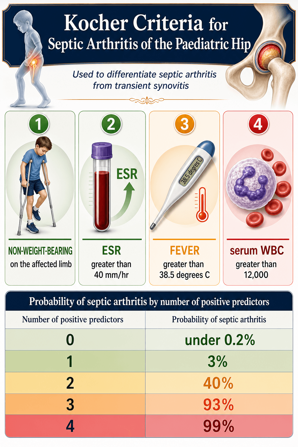
Kocher Criteria (1999)
A validated prediction rule for differentiating septic arthritis from transient synovitis.
- Probability of Septic Arthritis
- 3%
- Action
- Observe
- Probability of Septic Arthritis
- 40%
- Action
- Aspirate or MRI
- Probability of Septic Arthritis
- 93%
- Action
- Urgent Aspiration/Washout
- Probability of Septic Arthritis
- 99%
- Action
- Emergency Washout
The Criteria:
- Fever (Temp over 38.5°C)
- Non-weight bearing
- ESR over 40 mm/hr
- WCC over 12,000 cells/mm³
The probability increases exponentially with each added factor.
Clinical Assessment
- Pain: Acute onset, severe groin/thigh/knee pain.
- Function: Refusal to walk or move leg (Pseudoparalysis).
- Systemic: Fever, malaise, irritability, poor feeding (neonates).
- Trauma: Absence of trauma history.
- Position: Hip held in Flexion, Abduction, External Rotation (FABER) - maximum volume position.
- ROM: "Log roll" is extremely painful. Any movement causes distress.
- Tenderness: Anterior joint line.
- Neonates: Pain with nappy change is a key sign.
Neonates are deceptive. They may present with no fever and normal WCC. The only signs may be irritability, poor feeding, and pseudoparalysis (holding one leg still). High index of suspicion is required.
Investigations
Imaging Protocol
- Usually normal early.
- Look for: Widened joint space (Waldenstrom sign over 2mm asymmetry), osteomyelitis changes (rare early), subluxation.
- Gold standard screening. Detects fluid.
- Cannot reliably distinguish sterile vs infected fluid (though turbidity helps).
- Facilitates guided aspiration.
- Diagnostic dilemma solver (e.g., Psoas abscess vs Septic Hip vs Osteomyelitis).
- Excellent for evaluating concomitant osteomyelitis.
MRI should be reserved for cases where the diagnosis is unclear or Psoas abscess is suspected.
Detailed Differential Diagnosis
Septic Arthritis vs Transient Synovitis
Transient synovitis is the most common cause of hip pain in this age group, but is a diagnosis of exclusion.
- Septic Arthritis
- Toxic, high fever
- Transient Synovitis
- Well child, mild fever
- Septic Arthritis
- Usually over 12,000
- Transient Synovitis
- Usually normal
- Septic Arthritis
- Over 40 mm/hr
- Transient Synovitis
- Under 20 mm/hr
- Septic Arthritis
- Progressive worsening
- Transient Synovitis
- Improves with NSAIDs
Rule of Thumb: If the child can walk into the clinic (even with a limp), it is unlikely to be septic arthritis.
The Adolescent Hip: Disseminated Gonococcal Arthritis
The organism tables on this page list Neisseria gonorrhoeae as the adolescent pathogen, but it behaves so differently from a Staphylococcus or Kingella hip that it deserves separate treatment — it is the commonest cause of infectious arthritis in a sexually active adolescent or young adult, and getting it wrong means missing both a sexually transmitted infection and, in a child, possible sexual abuse.
Two faces of disseminated gonococcal infection (DGI)
- Arthritis-dermatitis syndrome (bacteraemic phase): migratory polyarthralgia, tenosynovitis (classically of the wrist, hand and ankle) and scattered painless pustular or vesiculopustular skin lesions, usually without a frankly purulent joint. Blood cultures may be positive while joint fluid is sterile.
- Purulent (septic) arthritis: a true pus-filled mono- or oligoarthritis, the knee most common, but it can localise to the hip — the form that overlaps with a typical septic hip.
Diagnosis and why it is missed
N. gonorrhoeae is fastidious: Gram stain and routine culture of blood and joint fluid are frequently negative. Yield improves with chocolate or Thayer-Martin medium and, decisively, nucleic-acid amplification (PCR / NAAT) of genital, pharyngeal, rectal and joint specimens. Always screen for co-infection (chlamydia, syphilis, HIV).
Management — where it diverges from a Staph hip
- Disseminated gonococcal
- Sexually active adolescent / young adult
- Staph / Kingella pyogenic
- Infant or young child
- Disseminated gonococcal
- Migratory polyarthralgia, tenosynovitis, dermatitis
- Staph / Kingella pyogenic
- Acute monoarticular, toxic child
- Disseminated gonococcal
- Often sterile; NAAT/PCR positive
- Staph / Kingella pyogenic
- Pus; organism usually grows (except Kingella)
- Disseminated gonococcal
- Ceftriaxone (plus STI co-treatment)
- Staph / Kingella pyogenic
- Anti-staphylococcal agent
- Disseminated gonococcal
- Often resolves on antibiotics alone; drain only if frank pus
- Staph / Kingella pyogenic
- Urgent surgical decompression of the hip
A frankly purulent gonococcal hip still needs drainage on the same emergency principle as any pus-under-pressure hip, but the bacteraemic arthritis-dermatitis syndrome typically settles with ceftriaxone and does not mandate washout — a key contrast with the Staphylococcus hip.
Gonococcal infection in a prepubertal child is a sentinel sign of sexual abuse and mandates child-protection evaluation. In a neonate, consider peripartum transmission (ophthalmia neonatorum, and rarely gonococcal arthritis).
Management Algorithm
Core Principles
- Decompression: Urgent removal of pus to reduce pressure and enzymatic damage.
- Antibiotics: High dose IV therapy to sterilize blood and tissues.
- Rest: Immobilization for symptom control and stability.
Empirical Antibiotics (organism- and age-directed, always after cultures):
- Default (over 3 months): Anti-staphylococcal agent — flucloxacillin or cefazolin; clindamycin if penicillin-allergic (also covers most Kingella).
- Neonate: Anti-staphylococcal penicillin PLUS a third-generation cephalosporin (cefotaxime) to cover Group B Streptococcus and Gram-negatives.
- Unimmunised / Hib risk: Add a third-generation cephalosporin (ceftriaxone or cefotaxime).
- MRSA risk (high-prevalence region or severe sepsis): Vancomycin or clindamycin per local antibiogram.
The goal is rapid sterilisation of the joint fluid; rationalise to a narrow-spectrum agent once cultures and PCR return.
Late Management of Sequelae
Management by Choi Type
Treatment follows the Choi grade defined above.
- Type I (transient ischaemia ± mild coxa magna): No reconstruction; observe and allow remodelling.
- Type II (epiphysis/physis/metaphysis deformity): Operate to prevent subluxation — improve acetabular coverage and abductor efficiency (greater-trochanter transfer or epiphysiodesis) and equalise leg length.
- Type III (neck malalignment or pseudarthrosis): Realignment proximal femoral osteotomy or bone-grafting of the neck pseudarthrosis; a valgus osteotomy is used for a coxa vara with a neck-shaft angle less than 110 degrees.
- Type IV (head and neck destruction): Salvage — pelvic support osteotomy or arthrodesis, with total hip arthroplasty in adulthood.
Treatment must be individualised based on age and deformity severity.
Surgical Technique
Anterior Approach (Smith-Petersen)
Preferred for Septic Hip. Allows direct access to the joint and easy drainage.
- Incision: Bikini line or longitudinal from ASIS.
- Interval: Sartorius/Tensor Fascia Lata (Superficial), Rectus Femoris/Gluteus Medius (Deep).
- Capsulotomy: Longitudinal or T-shaped incision in capsule.
- Washout: Copious saline irrigation. Inspect head.
- Closure: Leave drain? (Controversial, many close over drain). Loosely close capsule.
Pros: Excellent exposure, classic approach. Cons: Risk to Lateral Cutaneous Nerve of Thigh (LCNT).
Complications
- Mechanism
- Vessel Tamponade
- Outcome
- Collapse, Deformity
- Mechanism
- Enzymatic Destruction
- Outcome
- Pain, Stiffness
- Mechanism
- Physeal Damage
- Outcome
- Leg Length Discrepancy
- Mechanism
- Osteomyelitis Sequestrum
- Outcome
- Recurrent Sepsis
- Mechanism
- Capsular damage
- Outcome
- Dislocation/Subluxation
SEPTICComplications of Septic Hip
Hook:SEPTIC hips have SEPTIC complications.
Classifying the Residual Deformity (Choi & Hunka)
When septic arthritis damages the immature hip, the residual deformity is graded radiographically — and that grade drives the reconstructive decisions in the next section. Two systems are quoted in the literature. Note that there is no validated "Suk" classification for post-septic hip sequelae; the recognised systems are Choi and Hunka.
Choi classification (1990) — by radiographic damage
According to PubMed (Choi et al., in the Evidence Base below), 34 infantile septic hips were sorted into four types, with the satisfactory-result rate falling steadily from Type I to Type IV:
- Radiographic deformity
- Transient epiphyseal ischaemia, with or without mild coxa magna
- Implication
- Best prognosis; no reconstruction (5 of 5 satisfactory)
- Radiographic deformity
- Deformity of the epiphysis, physis and metaphysis
- Implication
- Operate to prevent subluxation (coverage, abductor efficiency, length)
- Radiographic deformity
- Femoral neck malalignment (extreme ante/retroversion) or neck pseudarthrosis
- Implication
- Realignment osteotomy or grafting of the pseudarthrosis
- Radiographic deformity
- Destruction of head and neck (only a medial neck remnant)
- Implication
- Worst prognosis (only 4 of 13 satisfactory); salvage reconstruction
Hunka classification (1982) — by the epiphysis and stability
The complementary Hunka system grades the severe sequelae by the presence or absence of the capital femoral epiphysis and by hip stability (this is the genuine source of the "Type I to Type V" range sometimes seen quoted). In Hunka's own series of severely destroyed heads, the worst results were the Type III hips with a femoral-neck pseudarthrosis, which needed the most operations to salvage.
Q: How is the post-septic hip deformity classified? A: By the Choi types (I-IV, by radiographic damage — prognosis worsens from I to IV) and the Hunka types (by the capital epiphysis and hip stability). There is no "Suk" classification for this. The grade decides whether you observe, perform a coverage/realignment osteotomy, or proceed to salvage.
Follow-Up Protocol
Post-Op Recovery
Keep NBM until repeated washouts unlikely. Continue IV antibiotics until CRP normalizes/improves significantly (usually 3-5 days).
Switch to oral when: Afebrile for 24-48h, CRP decreasing, tolerating oral. Total duration 3-4 weeks (6 weeks if Osteomyelitis).
Touch weight bearing initially. Full weight bearing as tolerated once pain free and inflammatory markers normal.
X-rays at 3, 6, 12 months to monitor for AVN or growth disturbance.
Outcomes and Prognosis
Time to Treatment is Critical
Prognosis correlates directly with delay in drainage.
- Treated under 4 days: Low risk of sequelae.
- Treated over 4 days: High risk of cartilage damage and AVN.
Classifying the sequelae: residual deformity is graded by the Choi (Type I-IV, by radiographic damage) and Hunka (by the capital epiphysis and hip stability) systems — see "Classifying the Residual Deformity" above. The worst grades range up to complete head-and-neck destruction with hip dislocation. (There is no validated "Suk" classification for this.)
Guidelines, Registries & Global Practice
Global Epidemiology
- Annual incidence of paediatric septic arthritis is roughly 4-10 per 100,000 children in high-income settings, with the hip and knee the most commonly affected joints.
- Roughly half of cases occur in children under 3 years; Staphylococcus aureus dominates overall, while Kingella kingae is the leading cause between 6 and 48 months in regions that use PCR.
- Incidence and severity are higher in limited-resource settings, where late presentation and untreated osteomyelitis drive a greater burden of destructive sequelae (Tom Smith hip).
- Common high-income recommendation
- Aspirate/culture before antibiotics (AAOS, BOA-BOAST, ESPID consensus)
- Practical note
- Universal; only delayed if child is septic/unstable
- Common high-income recommendation
- PCR or blood-culture-bottle inoculation of joint fluid in under-4s
- Practical note
- Markedly raises yield where available
- Common high-income recommendation
- Short IV then early oral switch once afebrile and CRP falling; ~2-4 weeks total
- Practical note
- ESPID/UK trials support early oral switch in uncomplicated cases
- Common high-income recommendation
- Urgent decompression (arthrotomy or arthroscopy)
- Practical note
- Hip effusion under pressure is treated as an emergency worldwide
High-resource: Routine ultrasound, joint-fluid PCR, MRI for diagnostic dilemmas, early IV-to-oral switch. Limited-resource: Reliance on clinical findings, aspiration and plain radiographs; later presentation means a higher rate of AVN, growth arrest and salvage surgery.
There is no dedicated implant registry for paediatric septic arthritis. National arthroplasty registries (NJR, AJRR, AOANJRR, SHAR, NZJR) become relevant only decades later, capturing the small cohort needing total hip arthroplasty for a destroyed hip in adulthood.
CA-MRSA is an increasingly important pathogen in many regions (parts of North America, Oceania, and high-prevalence Indigenous and remote communities worldwide). Where local MRSA prevalence is high or sepsis is severe, empirical cover with clindamycin or vancomycin should be guided by the local antibiogram rather than a single national protocol.
Controversies and Areas of Uncertainty
Arthroscopic washout offers a smaller incision and faster recovery, but is technically demanding in the infant hip and lacks comparative trial data. Open anterior arthrotomy remains the default in the very young; choice is currently surgeon- and centre-dependent.
Traditional 4-6 week IV courses have been challenged by trials supporting a short IV phase then early oral switch (guided by clinical response and falling CRP) in uncomplicated cases. The optimal total duration, especially with concomitant osteomyelitis, is still debated.
With a high Kocher/Caird probability some advocate proceeding directly to arthrotomy, while others aspirate first to confirm pus and obtain culture. Both are defensible; the key is not to delay decompression in a clear-cut case.
MRI is excellent for concomitant osteomyelitis, psoas abscess and pyomyositis, but adds no reliable infection-specific sign and may require sedation. Recent data show MRI enhancement cannot replace clinical scores, so it should not delay urgent washout.
MCQ Practice Points
Q: What is the probability of septic arthritis with 3 Kocher criteria? A: 93%. (0= under 0.2%, 1=3%, 2=40%, 3=93%, 4=99%).
Q: What is the overall most common organism in pediatric septic arthritis? A: Staphylococcus aureus. However, in the 6 months to 4 years age group, Kingella kingae is increasingly identified as a major pathogen.
Q: What is the appropriate empirical antibiotic regimen for a neonate? A: Anti-Staphylococcal penicillin (Flucloxacillin) + 3rd Gen Cephalosporin (Cefotaxime/Ceftriaxone). This covers Staph, Group B Strep, and Gram Negatives.
Q: Why is the hip joint uniquely susceptible to osteomyelitis spreading into the joint? A: The proximal femoral metaphysis is intracapsular. In other joints (knee), the metaphysis is extracapsular, so osteomyelitis tracks outwards, not into the joint.
Exam Cheat Sheet
Diagnosis
- Kocher: Fever, NWB, ESR over 40, WCC over 12k
- 4/4 = 99% Septic
- Aspirate if doubt (WCC over 50k)
- Ultrasound showing effusion is SCREENING only
Organisms
- Overall: Staph aureus
- Under 4y: Kingella kingae (PCR)
- Neonate: GBS, Gram negatives
- Adolescent: N. gonorrhoeae
Management
- EMERGENCY Washout
- Anterior Arthrotomy
- IV Abs (Fluclox) AFTER culture
- Monitor CRP
Complications
- AVN (Tamponade)
- Chondrolysis
- Growth Arrest
- Dislocation
Viva Scenarios
Practise clinical reasoning and management decisions out loud
“A 4-year-old child presents with a fever of 39°C and refusal to walk. ESR is 60, WCC is 18. X-ray is normal. What is your management?”
“A 3-week-old neonate is irritable and not moving the right leg. There is no fever. WCC is normal. Examination is difficult. How do you investigate?”
“You wash out a hip in a 2-year-old. There was frank pus. 48 hours later, the cultures (Gram stain and standard culture) are negative. The child is improving. Why?”
Evidence Base
Kocher Clinical Prediction Algorithm
- Four independent predictors: fever, non-weight-bearing, ESR at least 40 mm/hr, WCC over 12,000
- Predicted probability of septic arthritis: 0.2% (0), 3% (1), 40% (2), 93.1% (3), 99.6% (4 predictors)
- Retrospective cohort of children with an acutely irritable hip at one tertiary centre
Caird Prospective Modification (adds CRP)
- Prospective study of 53 children undergoing hip aspiration (48 analysed)
- CRP over 20 mg/L was the strongest independent predictor on multivariate analysis
- Probability with 3, 4 and 5 predictive factors was 83%, 93% and 98% respectively
Kingella kingae: an Emerging Pathogen
- Over 95% of K. kingae infections occur between 6 and 48 months of age
- Presentation is often subtle with normal acute-phase reactants
- Recovery requires inoculation into blood-culture bottles or nucleic-acid amplification (PCR)
Choi Sequelae Classification (Infantile Septic Hip)
- 34 hips classified into four deformity types based on radiographic damage
- Satisfactory functional result fell from 5/5 (Type I) to only 4/13 (Type IV head/neck destruction)
- Provides the framework for reconstructive decision-making in late sequelae
MRI Femoral Head Enhancement Cannot Replace Clinical Judgement
- 34 children (14 septic arthritis, 20 transient synovitis) with hip effusion on contrast MRI
- Decreased femoral head enhancement did not reliably distinguish the two (71% vs 50%, p=0.296)
- Higher Kocher and modified Kocher scores remained significantly associated with septic arthritis
Tractionless Arthroscopic Washout
- Describes a tractionless 1-2 portal hip arthroscopy technique for the small child
- Allows joint irrigation, debridement and drain placement without a distraction set
- Presented as a safe, minimally invasive alternative to open arthrotomy