Extension Type | Pulseless Pink Hand | Gartland Classification
- Most common elbow fracture in children (peak age 5-7)
- Extension type (97%): FOOSH with hyperextension
- Brachial artery at risk (pulseless with pink/white hand)
- AIN most common nerve injured (extension type)
- Gartland III = surgical emergency
- “Pink pulseless hand: Reduce urgently, reassess perfusion
- “If pink and perfused post-reduction: May observe
- “If white/non-perfused post-reduction: Explore brachial artery
- “Cubitus varus (gunstock deformity) = malunion complication

Brachial artery at risk. Pink pulseless hand: artery kinked but collateral perfusion. White pulseless hand: True ischemia = emergency. Reduce urgently then reassess.
Extension type: AIN (anterior interosseous nerve) most common - test OK sign. Median nerve next. Flexion type: Ulnar nerve at risk. Most recover spontaneously.
Posterolateral or posteromedial displacement. Posteromedial is the more common direction (approximately three-quarters; radial nerve at risk). Posterolateral is less common but is the pattern that threatens the median/AIN and brachial artery. Surgical emergency.
Crossed K-wires (lateral and medial) or lateral only. Medial wire risks ulnar nerve. Flex elbow minimally to protect ulnar nerve when placing medial wire.
Overview and Classification
Supracondylar humerus fracture is the most common elbow fracture in children. Peak age 5-7 years.
Mechanism
Extension Type (97%): FOOSH (fall on outstretched hand) with elbow hyperextension. Distal fragment displaces posteriorly.
Flexion Type (3%): Direct blow to posterior elbow. Distal fragment displaces anteriorly.
Gartland Classification (Extension Type)
Type I: Undisplaced or minimally displaced. Posterior cortex intact. Fat pad sign may be only clue.
Type II: Displaced with posterior cortex intact (hinged). May be angulated.
Type IIa: Extension angulation only. Type IIb: Rotation or translation (more unstable).
Type III: Completely displaced, no cortical contact.
Type IIIa: Posteromedial displacement (the more common direction, approximately three-quarters; radial nerve at risk). Type IIIb: Posterolateral displacement (less common; median/AIN and brachial artery at risk).
Elbow Anatomy in Children
Distal Humerus Anatomy
- CRITOE mnemonic: Capitellum (1), Radial head (3), Internal (medial) epicondyle (5), Trochlea (7), Olecranon (9), External (lateral) epicondyle (11)
- Ages approximate - useful for identifying fracture patterns
- Thin bone above condyles - weak point for fracture
- Anterior humeral line normally passes through middle third of capitellum
- Baumann angle (shaft to capitellar physis): 70-75° normal
- Location
- Anterior, crosses fracture site
- Risk with Displacement
- Posterolateral (extension) displacement
- Location
- Anterior, with artery
- Risk with Displacement
- Posterolateral (extension) displacement
- Location
- Lateral, near lateral column
- Risk with Displacement
- Posteromedial displacement
- Location
- Medial, posterior to epicondyle
- Risk with Displacement
- Flexion type, medial wire insertion
Pathophysiology
The supracondylar region is the thinnest, weakest part of the distal humerus — a flat segment of bone between the medial and lateral columns, bounded by the coronoid and olecranon fossae. In the 5-7 year age group this region is undergoing remodelling and is mechanically vulnerable, which is why it fails before the ligaments rupture (the reverse of the adult elbow, where dislocation predominates).
Mechanism and fragment behaviour:
- Extension type (~97%): a fall on the outstretched hand transmits a hyperextension force through a locked olecranon, levering the distal fragment posteriorly. The proximal (shaft) fragment is driven anteriorly into the brachialis and the overlying neurovascular bundle.
- The anteriorly migrating proximal spike is the source of the classic complications: it tents or buttonholes the brachialis (pucker sign), and stretches/kinks the brachial artery and the closely applied median nerve / AIN.
- Displacement direction predicts the injured nerve: posterolateral displacement tethers the anteromedial structures (median/AIN), while posteromedial displacement threatens the laterally placed radial nerve.
- Flexion type (~3%): a direct blow to the flexed elbow displaces the distal fragment anteriorly, drawing the ulnar nerve taut behind the medial epicondyle.
Why cubitus varus occurs: malunion is driven chiefly by uncorrected internal rotation and medial column collapse at reduction, not by simple varus angulation — hence rotation control intra-operatively is critical.
LeMONDisplacement Predicts the Nerve at Risk
Hook:Posterolateral hits Median/AIN; posteromedial hits Radial; flexion hits Ulnar.
Classification
Gartland Classification (Extension Type)

- Undisplaced or minimally displaced
- Posterior cortex intact
- May see fat pad sign only
- Treatment: Above-elbow cast, 90° flexion
- Displaced with posterior cortex hinge
- IIa: Angulated only
- IIb: Rotated/translated (less stable)
- Completely displaced, no cortical contact
- IIIa: Posteromedial (more common, approximately three-quarters; radial nerve risk)
- IIIb: Posterolateral (less common; median/AIN and brachial artery risk)
- Displacement
- Undisplaced
- Treatment
- Cast, 90° flexion, 3-4 weeks
- Displacement
- Angulated only
- Treatment
- Closed reduction + K-wires
- Displacement
- Rotated/translated
- Treatment
- Closed reduction + K-wires
- Displacement
- Complete, no cortex
- Treatment
- Urgent closed reduction + K-wires
Clinical Assessment
Neurovascular Examination
CRITICAL: Assess before any manipulation.
Vascular:
- Brachial pulse, radial pulse
- Capillary refill
- Hand color (pink or white)
Nerve Function:
- AIN: OK sign (FPL, FDP to index)
- Median: Thenar power, sensation
- Radial: Wrist/finger extension, sensation
- Ulnar: Interossei, little finger sensation
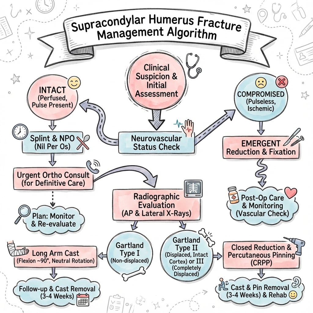
Pulseless Hand Algorithm
Pink pulseless hand: Reduce fracture urgently. If perfusion improves → observe. Remain pink without pulse → may observe with close monitoring.
White pulseless hand: True ischemia. Reduce urgently. If still white → explore brachial artery (may be trapped, kinked, or injured).
Investigations
Radiographic Assessment
- AP elbow
- True lateral elbow (essential for classification)
- Fat pad sign: Posterior fat pad always abnormal; anterior fat pad displaced ("sail sign") suggests effusion/occult fracture
- Anterior humeral line: Should pass through middle third of capitellum
- Baumann angle: Shaft to capitellar physis angle (70-75° normal)


- Normal
- Through middle 1/3 capitellum
- Abnormal
- Anterior to capitellum (extension)
- Normal
- 70-75°
- Abnormal
- Compare to opposite side
- Normal
- Not visible
- Abnormal
- Visible = occult fracture
- Normal
- Small, close to bone
- Abnormal
- Displaced = sail sign
Differential Diagnosis
The displaced supracondylar fracture is rarely subtle, but the undisplaced fracture and the swollen, painful paediatric elbow have several mimics. Distinguishing these changes management entirely.
- Distinguishing features
- Anterior humeral line anterior to capitellum; transcondylar tenderness; both columns involved
- Key pitfall
- Type I seen only as raised fat pad — easily missed
- Distinguishing features
- Tenderness lateral; Salter-Harris IV; risk of non-union and progressive valgus
- Key pitfall
- Internal oblique view needed; under-treated displacement
- Distinguishing features
- Medial tenderness; check for incarceration in joint after dislocation
- Key pitfall
- Fragment trapped in joint mistaken for trochlear ossification
- Distinguishing features
- Infant/toddler; whole epiphysis displaces medially; consider non-accidental injury
- Key pitfall
- Mistaken for elbow dislocation (rare in young children)
- Distinguishing features
- Radiocapitellar line disrupted; older child/adolescent
- Key pitfall
- Coexisting fracture missed post-reduction
- Distinguishing features
- Toddler, axial-traction mechanism, arm held pronated, NO swelling
- Key pitfall
- Imaging normal — clinical diagnosis, reduces with supination/flexion
Management
Undisplaced fracture.
Treatment: Long arm cast in 90° flexion. Avoid hyperflexion (compromises circulation).
Duration: 3-4 weeks.
Follow-up: X-ray at 1 week to confirm no displacement.
Surgical Technique
Closed Reduction and K-Wire Fixation
- Longitudinal traction with elbow extended
- Correct medial/lateral displacement
- Correct rotation (pronation for posteromedial, supination for posterolateral)
- Flex elbow while milking distal fragment anteriorly
- Apply varus/valgus correction as needed
- Check reduction on fluoroscopy
- 2 Lateral divergent: Safer (avoids ulnar nerve), biomechanically adequate for most
- Crossed wires (lateral + medial): More stable, but medial wire risks ulnar nerve
- If medial wire: Flex elbow minimally (20-30°), palpate nerve, small stab incision
- Advantages
- Avoids ulnar nerve
- Disadvantages
- Less rotational stability
- Advantages
- Maximum stability
- Disadvantages
- Ulnar nerve risk (2-5%)
- Advantages
- Good stability, no nerve risk
- Disadvantages
- More wires, more time
Complications
Early
- Vascular injury (brachial artery)
- Nerve injury (AIN most common in extension type)
- Compartment syndrome
- Volkmann's ischemic contracture (missed ischemia)
Late
- Cubitus varus (gunstock deformity): Most common complication. Malunion with varus tilt. Cosmetic deformity. May need late supracondylar osteotomy.
- Stiffness: Usually temporary. Avoid aggressive physiotherapy.
- Myositis ossificans: Rare in children.
Cubitus Varus and the Corrective Supracondylar Osteotomy
The topic names cubitus varus as "the most common complication" and mentions a "late supracondylar osteotomy" and "dome or closing wedge" options, but never develops the deformity or its correction - a classic viva progression.
- It is a triplanar malunion, not simple varus. Although called cubitus "varus", the deformity is a combination of varus angulation, extension and (crucially) internal rotation of the distal fragment - the same rotational malreduction that also causes it. The "gunstock" appearance is what the child and family notice; the carrying angle is lost or reversed.
- Mostly cosmetic - but not entirely benign. It usually does not limit function, but recognised sequelae include posterolateral rotatory instability, tardy (late) posterolateral rotatory or ulnar/PIN nerve palsy, an increased risk of a later lateral condyle fracture, and snapping triceps/medial elbow pain - which is why a symptomatic or severe deformity is corrected rather than simply observed.
- When to correct. Indications are a cosmetically unacceptable or progressive deformity, functional limitation, or symptomatic instability/nerve compromise, usually deferred until the deformity is established and remodelling has plateaued (typically at least a year, and with adequate remaining bone stock).
- How to correct. A distal humeral (supracondylar) osteotomy re-aligns all three planes. Options include a lateral closing-wedge osteotomy (simplest and commonest, but can leave a prominent lateral condyle / "bump"), a dome osteotomy (better cosmetic contour, technically harder), step-cut and complex multiplanar/oblique osteotomies, and increasingly CT-planned patient-specific guides for the rotational component. Fixation is typically K-wires, a plate or a lateral external fixator; the radial and ulnar nerves are at risk and the rotational and lateral-prominence components must be addressed, not just the varus angle.
Q: Why does cubitus varus occur and how is it corrected? A: It is a triplanar malunion (varus + extension + internal rotation) driven chiefly by rotational malreduction, not simple varus angulation - so preventing it means controlling rotation intra-operatively. It is largely cosmetic but can cause posterolateral rotatory instability, tardy nerve palsy and later lateral condyle fracture. Correction is a distal humeral (supracondylar) osteotomy - lateral closing-wedge (simple, risks a lateral prominence), dome (better contour, harder) or a multiplanar/CT-planned osteotomy - addressing all three planes, with the radial and ulnar nerves at risk.
The Flexion-Type Supracondylar Fracture (the other 3%)
The topic repeatedly flags the flexion type (~3%) - ulnar nerve at risk, often needs open reduction - but never develops it, and it behaves as the mirror image of the extension fracture.
- Reversed mechanism and displacement. A direct blow to the flexed elbow (or a fall onto the point of the elbow) drives the distal fragment anteriorly, the opposite of the FOOSH extension injury. On the lateral radiograph the anterior humeral line therefore passes posterior to the capitellum (the reverse of the extension malalignment).
- The ulnar nerve, not the AIN. Because the distal fragment displaces anteriorly and often into valgus, the ulnar nerve is the nerve most at risk in flexion-type fractures (whereas the AIN/median dominates in extension type). Assess ulnar motor (interossei, froment) and sensation specifically.
- Reduce in the opposite direction. The reduction manoeuvre is essentially reversed: after traction and correction of translation/rotation, the fragment is controlled and the elbow is often held/pinned in relative extension rather than the deep flexion used for extension-type fractures - so the closed reduction can be awkward and the fracture is prone to losing reduction in flexion.
- Higher open-reduction rate. Flexion-type fractures are more often irreducible closed (soft-tissue interposition, an unstable pattern, or ulnar nerve tethering) and thus have a higher rate of open reduction than extension types; fixation is still K-wires, with the same medial-pin ulnar-nerve caution.
Q: How does the flexion-type supracondylar fracture differ from the extension type? A: It is the mirror image - a blow to the flexed elbow drives the distal fragment anteriorly (anterior humeral line passes posterior to the capitellum), the ulnar nerve (not the AIN) is most at risk, reduction is achieved and held in relative extension rather than deep flexion, and it has a higher open-reduction rate because it is more often irreducible closed.
Postoperative Care
Immediate Postoperative
- Above-elbow backslab or cast
- Elbow at 60-80° flexion (not hyperflexed - risks circulation)
- Forearm neutral or slight pronation
- Elevate limb
- Hourly neurovascular checks for first 24 hours
- Monitor for compartment syndrome (pain with passive finger extension)
- Check cast not too tight
- Target
- Present
- Action if Abnormal
- Urgent review, check cast
- Target
- Less than 2 seconds
- Action if Abnormal
- Loosen cast, elevate
- Target
- Controlled
- Action if Abnormal
- If severe - compartment syndrome?
- Target
- Active
- Action if Abnormal
- Document, reassure if nerve injury
Outcomes
Expected Outcomes
- Most children achieve excellent results
- Full ROM typically restored within 6-12 weeks
- Nerve injuries usually recover (90%+ neurapraxias)
- Low malunion rate with anatomic reduction
- Cubitus varus (5-15% with malreduction)
- Stiffness (usually temporary)
- Nerve palsy (5-10%, most recover)
- Volkmann's contracture (rare but devastating)
- Rate
- 90%+
- Key Factor
- Anatomic reduction, no aggressive PT
- Rate
- 5-15%
- Key Factor
- Malreduction (rotation, varus tilt)
- Rate
- 90%+
- Key Factor
- Most are neurapraxias
- Rate
- Rare
- Key Factor
- Missed compartment syndrome
Guidelines, Registries & Global Practice
Global Epidemiology
- Most common elbow fracture in children; accounts for roughly 55-70% of paediatric elbow fractures worldwide
- Peak incidence age 5-7 years; non-dominant (left) arm slightly more often affected
- Extension type ~97-98%; flexion type 2-3% (flexion type skews slightly older and female, often higher-energy mechanism)
- Common mechanisms globally: falls from playground equipment, monkey bars, trampolines and bunk beds
- Displaced (II/III) fixation
- Closed reduction + percutaneous pinning; lateral OR medial-lateral acceptable
- Vascular emphasis
- Urgent reduction for pulseless, poorly perfused (at-risk) limb
- Displaced (II/III) fixation
- Timely reduction and K-wire fixation; consultant-led decision
- Vascular emphasis
- Document perfusion; immediate surgery for the threatened limb
- Displaced (II/III) fixation
- CRPP standard; lateral entry first-line, mini-open for medial pin
- Vascular emphasis
- Reduce first; explore only the persistently ischaemic (white) hand
- Displaced (II/III) fixation
- Lateral-entry preferred to minimise iatrogenic ulnar injury
- Vascular emphasis
- Expectant approach to the well-perfused pink pulseless hand
Controversies & Areas of Uncertainty
Crossed pins resist loss of reduction slightly better (Xing meta-analysis) but triple the iatrogenic ulnar nerve risk (Dekker). Most surgeons default to lateral entry; a mini-open medial pin neutralises the nerve risk when extra stability is needed.
No randomised evidence. Consensus favours urgent reduction then close observation if the hand stays pink and perfused, even without a palpable pulse. Routine vascular exploration of every pulseless hand is NOT supported; the white/cold hand is explored.
The well-perfused, neurologically intact displaced fracture does not need overnight surgery — but delay over 8 hours raises the open reduction rate (Walmsley). Limb-threatening (white hand) or open fractures remain true emergencies.
Adjunctive imaging rarely changes acute decisions in the threatened limb and should not delay theatre. Duplex and angiography add little to the pink pulseless hand (Griffin).
MCQ Practice Points
Q: What is the most common mechanism and displacement pattern for pediatric supracondylar fractures?
A: Extension-type (95-97%): Fall on outstretched hand with elbow extended and hyperextended. Distal fragment displaces posteriorly. Flexion-type (3-5%): Fall on flexed elbow or direct blow; distal fragment displaces anteriorly - higher rate of ulnar nerve injury. Extension type further classified by displacement direction: Posteromedial (most common in extension type) - radial nerve at risk; Posterolateral - median/AIN at risk. Understanding displacement pattern predicts neurovascular injury risk.
Q: How do you assess reduction quality using radiographic parameters in supracondylar fractures?
A: Baumann's angle (AP view): Angle between humeral shaft axis and physeal line of capitellum; normal 70-75 degrees; should match opposite side. Anterior humeral line (lateral view): Line along anterior humeral cortex should pass through middle third of capitellum; if anterior to capitellum, extension malreduction. Coronoid line: Line along anterior coronoid should not pass posterior to anterior humeral cortex. Rotation: On lateral, assess teardrop of lateral column for symmetry. Intraoperative fluoroscopy essential to confirm reduction.
Q: What are the indications for open reduction in pediatric supracondylar fractures?
A: Open fractures - require debridement and stabilization. Vascular compromise not corrected by closed reduction - explore brachial artery. Irreducible fractures - soft tissue interposition (brachialis muscle, median nerve, brachial artery can become entrapped; "pucker sign" on skin indicates buttonholed structures). Neurological deficit worsening after reduction - nerve may be trapped. Open reduction via anterior approach allows visualization of neurovascular structures. Delayed presentation (greater than 5-7 days) with significant swelling may require open approach.
Q: What is the "pucker sign" and its clinical significance in supracondylar fractures?
A: The pucker sign is skin dimpling or puckering at the antecubital fossa indicating that the proximal fragment has buttonholed through the brachialis fascia. Structures at risk of entrapment: Brachialis muscle, brachial artery, median nerve. Significance: Suggests closed reduction may be impossible - the entrapped soft tissues block reduction. If pucker sign persists after reduction attempt, suspect soft tissue interposition and consider open reduction via anterior approach. Associated with higher rates of neurovascular injury.
Q: What is Volkmann's ischemic contracture and how does it develop after supracondylar fractures?
A: Volkmann's ischemic contracture is the devastating end-result of missed forearm compartment syndrome. Pathophysiology: Vascular injury or swelling leads to elevated compartment pressure, causing muscle ischemia and necrosis. As muscles fibrose, they shorten, causing flexion contracture of wrist and fingers (worse with elbow extension, MCP extension). Classic position: Flexed wrist, extended MCP, flexed IP joints. Prevention: Recognize compartment syndrome early (6 P's: Pain with passive stretch, Pallor, Pulselessness, Paresthesias, Paralysis, Pressure). Emergent fasciotomy if suspected.
At a Glance
Supracondylar humerus fractures are the most common elbow fracture in children (peak age 5-7 years). Extension type (97%) results from FOOSH with hyperextension; flexion type (3%) from direct blow. The Gartland classification guides management: Type I (undisplaced - cast), Type II (hinged on posterior cortex - K-wires), Type III (complete displacement - surgical emergency). Critical assessment: the 3Ps - Pulse, Perfusion, Paralysis. Brachial artery is at risk; pink pulseless hand indicates artery kinked but collaterals perfusing - reduce urgently and reassess. AIN is the most commonly injured nerve (test OK sign). Fixation with crossed or lateral-only K-wires; medial wire placement risks ulnar nerve.
3PsSupracondylar Assessment
Hook:3Ps = Pulse, Perfusion, Paralysis - check all immediately!
OK SignAIN Function Test
Hook:AIN = OK sign (FPL + FDP to index)!
Exam Viva Scenarios
Practise clinical reasoning and management decisions out loud
“A 6-year-old presents with a displaced supracondylar fracture and a pink but pulseless hand. How do you manage?”
“You are managing a 7-year-old with a Gartland Type III posterolateral supracondylar fracture. In theatre under general anesthesia, you notice a skin dimple (pucker sign) in the antecubital fossa. After two attempts at closed reduction, you cannot achieve adequate alignment - the distal fragment keeps subluxating posteriorly. Fluoroscopy shows persistent posterior displacement with the anterior humeral line passing anterior to the capitellum. The hand is pink and well perfused. What is your next step and how would you proceed?”
“You performed closed reduction and crossed K-wire fixation (one lateral, one medial wire) for a Gartland Type III supracondylar fracture in a 5-year-old girl last night. The fracture was perfectly reduced with good wire position on post-operative X-rays. This morning on ward rounds, the mother reports the child cannot spread her fingers apart and has numbness in the little finger. Pre-operatively, all nerve function was documented as intact. On examination, you confirm ulnar nerve palsy with weak interossei, inability to abduct/adduct fingers, and diminished sensation in the ulnar distribution. What is your assessment and management?”
Gartland Classification
- I: Undisplaced - cast
- II: Hinged on posterior cortex
- III: Complete displacement - emergency
Neurovascular
- Brachial artery at risk
- AIN most common nerve (extension)
- Check OK sign (FPL, FDP index)
- 3Ps: Pulse, Perfusion, Paralysis
Pink Pulseless Algorithm
- Reduce urgently
- Pink post-reduction: Observe
- White post-reduction: Explore artery
Fixation
- Lateral K-wires (2 divergent) - safer
- Crossed wires - stronger, ulnar nerve risk
- Avoid hyperflexion in cast
Evidence Base
Key Evidence
- Original three-type classification
- Foundation for current management
- Modified by Wilkins (Type IV added)
- Multiple studies show no significant difference in stability for most fractures
- Lateral-only avoids ulnar nerve injury (0% vs 2-5%)
- Crossed preferred for very unstable (Type IV) fractures
- Finding
- Similar outcomes, lateral safer
- Evidence Level
- Level II (meta-analyses)
- Finding
- Within 8 hours if possible
- Evidence Level
- Level IV
- Finding
- Reduce first, most perfuse
- Evidence Level
- Level IV
Landmark Evidence
Crossed versus lateral-only pinning: meta-analysis of 19 RCTs
- Meta-analysis of 19 randomised controlled trials, 1297 Gartland type II and III fractures
- Medial-lateral crossed pinning had LOWER loss of reduction (RR 0.70, 95% CI 0.52-0.94) than lateral-only
- Crossed pinning had HIGHER iatrogenic ulnar nerve injury (RR 2.21, 95% CI 1.11-4.41)
- Mini-open medial pin technique abolished the excess ulnar nerve risk (RR 1.73, NS)
- No difference in Baumann angle, carrying angle, Flynn excellent grading or pin-tract infection
Crossed vs lateral entry: systematic review and meta-analysis
- 13 studies (7 RCTs, 6 prospective cohorts), 1158 displaced extension-type fractures
- No difference in Flynn outcome (RR 1.07) or loss of reduction (crossed 11.6% vs lateral 12.4%)
- Iatrogenic ulnar nerve injury: 4.1% (crossed) vs 0.3% (lateral entry) — roughly threefold higher
- Conclusion: lateral entry is safest if the surgeon wishes to avoid all ulnar nerve risk
Pink pulseless hand in Gartland III: therapeutic consensus
- 404 Gartland type III fractures; 68 (17%) had acute vascular injury, 63 pink pulseless and 5 white/cold
- Pink pulseless hands treated by urgent closed reduction and pinning, then close observation
- Radial pulse restored immediately in 42 and within hours-to-11 days in 18 of the pink pulseless group
- All 5 ischaemic (white) hands and 3 failed reductions underwent exploration — brachial artery incarcerated at fracture
- At mean 8.4 years all patients had a palpable radial pulse and full spontaneous nerve recovery
The pink pulseless hand: literature review of vascular management
- Review of case series on management of the pink pulseless hand after fracture reduction
- A pink pulseless hand after reduction can be managed expectantly with close observation
- Angiography and colour duplex add little to acute decision-making
- Exploration is indicated only if additional signs of vascular compromise develop
Delay to surgery increases the need for open reduction
- 171 closed Gartland III fractures without vascular compromise, retrospective comparison
- Surgery under 8 hours from presentation (126) versus over 8 hours (45)
- Delay over 8 hours raised the open reduction rate to 33.3% vs 11.2% (p less than 0.05)
- No difference in overall complication rate between early and delayed groups
Nerve injuries: spontaneous recovery and role of exploration
- 272 displaced Gartland II/III fractures; nerve injury in 48 (18%) overall
- Iatrogenic (post-treatment) nerve injury in 39 (14%), predominantly ulnar (34 of 39)
- All nerve injuries resolved clinically at a mean of 3.5 months (range 3 weeks to 8 months)
- Routine early wire removal or nerve exploration not indicated; mini-open pinning reduces nerve risk
AAOS Clinical Practice Guideline: pediatric supracondylar humerus fracture
- Closed reduction with percutaneous pinning recommended for displaced (type II/III) fractures
- Either lateral-entry or medial-and-lateral pin configurations are acceptable for displaced fractures
- If a medial pin is used, techniques to avoid iatrogenic ulnar nerve injury are advised
- Urgent closed reduction for a fracture with absent pulse and poor perfusion (limb at risk)