Defective Globin Chain Synthesis with Skeletal Manifestations
- Beta Thalassemia Major: Most severe form, transfusion-dependent from early childhood.
- Marrow Expansion: Causes widened medullary cavities, cortical thinning, pathological fractures.
- Osteoporosis: Multifactorial - marrow expansion, iron toxicity, hypogonadism, deferoxamine.
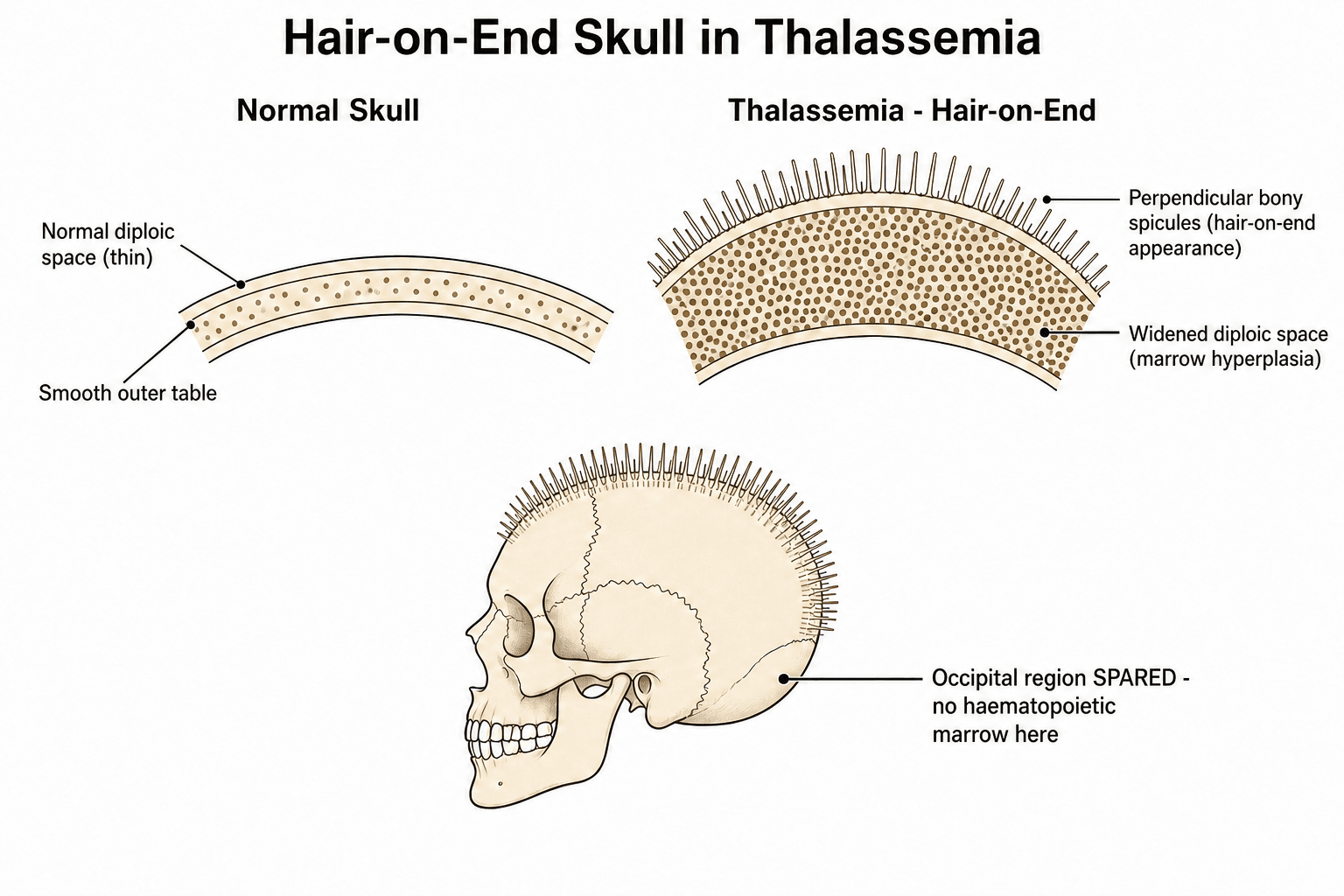
- Hair-on-end Skull: Classic radiographic finding from diploic expansion.
- Extramedullary Hematopoiesis: Can cause spinal cord compression.
- “Hair-on-end skull - occipital spared (no marrow)
- “Chipmunk facies from maxillary expansion
- “Osteoporosis common even in young patients
- “DEXA screening and bisphosphonates for bone health
- “EMH can compress spinal cord - surgical emergency
Osteoporosis is a major cause of morbidity, even in young patients.
- Causes: Marrow expansion, iron toxicity, deferoxamine, hypogonadism, vitamin D deficiency
- Prevalence: 40-80% of adult patients have osteoporosis
- Complications: Vertebral compression fractures (10-20%), long bone fractures
- Management: DEXA screening, bisphosphonates, vitamin D/calcium, hormone replacement
Overview/Epidemiology
Thalassemia is a group of inherited hemoglobin disorders characterized by reduced or absent synthesis of one or more globin chains.
- Autosomal recessive inheritance
- Alpha thalassemia: Deletion of 1-4 alpha globin genes (chromosome 16)
- Beta thalassemia: Point mutations in beta globin gene (chromosome 11)
- Most common inherited hemoglobin disorder worldwide
- Highest prevalence: Mediterranean, Middle East, Southeast Asia, Indian subcontinent, Africa
- Carrier frequency: Up to 30% in endemic areas
- Annual affected births: 60,000-70,000 globally
- Genotype
- β0/β0 or β0/β+
- Clinical Features
- Severe anemia, transfusion-dependent
- Genotype
- Variable
- Clinical Features
- Moderate anemia, variable transfusion needs
- Genotype
- β/β0 or β/β+
- Clinical Features
- Mild microcytic anemia, carrier state
- Genotype
- 1-4 gene deletions
- Clinical Features
- Variable: silent carrier to hydrops fetalis
Pathophysiology
Ineffective Erythropoiesis
The fundamental defect in thalassemia is imbalanced globin chain production:
- Beta thalassemia: Reduced or absent beta chains → excess alpha chains
- Unpaired alpha chains precipitate → damage RBC membrane
- Premature destruction of RBC precursors in bone marrow
- Chronic hemolytic anemia triggers compensatory marrow expansion
- Erythroid precursors increase up to 6-fold
- Medullary cavity expansion and cortical thinning result
This process drives the skeletal manifestations unique to thalassemia.
Skeletal Pathophysiology:
The orthopaedic manifestations result from:
- Marrow Expansion: Widened medullary cavities, thin cortices, increased fragility
- Bone Resorption: Cancellous bone loss from osteoclast activity
- Iron Toxicity: Direct damage to osteoblasts and osteocytes
- Chelation Effects: Deferoxamine may impair bone metabolism
- Endocrine Dysfunction: Hypogonadism, hypothyroidism, diabetes from iron deposition
Classification
Beta Thalassemia Classification
By Genotype and Severity:
- Genotype
- β0/β0, β0/β+, β+/β+ (severe)
- Clinical Phenotype
- Transfusion-dependent from infancy
- Genotype
- Variable combinations
- Clinical Phenotype
- Moderate anemia, variable transfusions
- Genotype
- β/β0 or β/β+
- Clinical Phenotype
- Mild microcytic anemia, asymptomatic
- Presents 6-12 months of age (after HbF decline)
- Severe anemia (Hb 3-6 g/dL untreated)
- Hepatosplenomegaly
- Growth retardation
- Skeletal changes from marrow expansion
- Variable severity
- May not require regular transfusions
- Still develop skeletal changes
- Iron overload from GI absorption (not transfusion)
Modern transfusion programs have reduced skeletal deformities.
Clinical Presentation
Craniofacial Changes
Skull:
- Hair-on-end appearance: Perpendicular spicules from diploe expansion
- Widened diploic space
- Thinned outer table
- Occipital sparing: No haematopoietic marrow in occipital bone
Face:
- Chipmunk facies: Maxillary hypertrophy
- Frontal bossing
- Prominent malar eminences
- Lateral orbital displacement
- Dental malocclusion
- Sinus obliteration (except ethmoid)
These changes are less common with modern transfusion protocols that suppress marrow expansion.
Systemic Manifestations Affecting Orthopaedics:
- Growth retardation: Short stature, delayed puberty
- Hypogonadism: Contributes to osteoporosis
- Splenomegaly: May need splenectomy (increases infection risk)
- Iron overload: Affects bone metabolism
Investigations
Laboratory Studies:
- Expected Finding
- Microcytic anemia (MCV low)
- Clinical Significance
- Severity indicates type
- Expected Finding
- Increased HbA2, HbF
- Clinical Significance
- Diagnostic for beta thalassemia
- Expected Finding
- High ferritin, high iron
- Clinical Significance
- Iron overload monitoring
- Expected Finding
- May be elevated
- Clinical Significance
- Hepatic iron deposition
- Expected Finding
- Hypogonadism, hypothyroidism
- Clinical Significance
- Secondary complications
- Expected Finding
- Often deficient
- Clinical Significance
- Contributes to osteoporosis
Plain Radiographs
- Hair-on-end appearance
- Widened diploic space
- Occipital sparing
- Osteopenia
- Striated vertebrae
- Compression fractures
- Biconcave deformities
- Widened medullary cavities
- Cortical thinning
- Pathological fractures
X-ray remains useful for initial assessment and fracture detection.
Management
Perioperative Considerations:
- Coordinate with hematology for transfusion timing
- Check cardiac function (iron cardiomyopathy)
- Risk of venous thromboembolism (post-splenectomy especially)
- Immunocompromised if splenectomized
- Delayed wound and fracture healing possible
Medical Management
- Beta major: Regular transfusions every 2-4 weeks
- Target: Pre-transfusion Hb of 9-10 g/dL
- Suppresses endogenous erythropoiesis
- Reduces marrow expansion and skeletal deformities
- Deferoxamine (Desferal): SC or IV infusion, traditional agent
- Deferasirox (Exjade): Oral, once daily
- Deferiprone (Ferriprox): Oral, for cardiac iron
- Target: Ferritin less than 1000 μg/L
- Calcium and vitamin D supplementation
- Bisphosphonates for osteoporosis (zoledronic acid, pamidronate)
- Hormone replacement for hypogonadism
- Regular DEXA monitoring
- Bone marrow transplant (HLA-matched sibling donor)
- Gene therapy (emerging)
Modern comprehensive care has dramatically improved outcomes.
Surgical Considerations in Thalassemia
Preoperative Planning
- Preoperative transfusion to Hb 10-11 g/dL
- Assess cardiac function (iron cardiomyopathy)
- Check ferritin levels and chelation status
- Optimize coagulation (platelets, liver function)
- DEXA scan for bone density
- CT/MRI for bone architecture if significant surgery planned
- Anticipate poor bone quality for fixation
- Target
- 10-11 g/dL
- Concern
- Transfuse if low
- Target
- EF greater than 50%
- Concern
- Iron cardiomyopathy risk
- Target
- Optimized chelation
- Concern
- Bleeding risk if high
- Target
- DEXA T-score
- Concern
- Implant choice affected
Complications
Skeletal Complications
- Most common orthopaedic complication
- Prevalence 40-80% in adults
- Affects quality of life significantly
- Vertebral compression fractures: 10-20%
- Long bone fractures: Increased risk
- Hip fractures: Major concern
- Delayed healing common
- Craniofacial changes (undertreated patients)
- Spinal deformity
- Short stature
- Height less than 3rd percentile common
- Delayed bone age
- Delayed puberty
Growth disturbance in thalassemia major reflects both marrow expansion diverting metabolic resources and endocrine dysfunction from iron overload.
Comparison: Thalassemia vs Sickle Cell Disease:
- Thalassemia
- Globin chain quantity (reduced)
- Sickle Cell Disease
- Globin chain quality (HbS)
- Thalassemia
- Marrow expansion, osteoporosis
- Sickle Cell Disease
- Vaso-occlusion, infarction
- Thalassemia
- Uncommon
- Sickle Cell Disease
- Very common (hip, shoulder)
- Thalassemia
- No
- Sickle Cell Disease
- Yes (vaso-occlusive)
- Thalassemia
- Not increased
- Sickle Cell Disease
- Increased (Salmonella)
- Thalassemia
- Classic finding
- Sickle Cell Disease
- Can occur but less common
Desferrioxamine (Deferoxamine) Bone Toxicity
Deferoxamine appears repeatedly in this topic as a cause of bone disease (the "D" of the MIDHE mnemonic and a recurring risk factor), yet the mechanism deserves explicit development because chelator-related skeletal injury is a distinct, potentially reversible entity separate from transfusional iron overload.
- Deferoxamine (DFO) chelates iron, but at high doses relative to the body iron burden it also depletes zinc and copper and directly inhibits DNA synthesis, fibroblast/osteoblast proliferation and collagen formation, injuring the growth-plate cartilage and metaphyseal bone.
- Toxicity is dose-dependent and greatest when the chelator dose is high relative to iron stores. Porter's therapeutic (toxicity) index — the mean daily DFO dose in mg/kg divided by the serum ferritin in µg/L — should be kept below 0.025; a persistent index greater than 0.025 is associated with skeletal and other toxicity.
- Young children are most susceptible, and risk rises paradoxically as iron control improves and ferritin falls (relative over-chelation).
- Metaphyseal changes — widening, cupping, irregularity, sclerosis and demineralisation, most obvious around the knees and wrists.
- Platyspondyly / vertebral body flattening producing disproportionate short stature with a shortened trunk (reduced sitting height), a characteristic clue that short stature is chelation-related rather than purely marrow-driven or endocrine.
- Genu valgum, pseudo-rickets and growth-plate cartilage dysplasia; growth velocity may fall.
recognise DFO dysplasia clinically and radiographically, reduce the DFO dose to hold the toxicity index below 0.025 (especially when ferritin falls), monitor growth and spine radiographs, and consider switching to or rotating oral chelators (deferasirox, deferiprone). Because the lesion is at least partly reversible, early recognition protects final height and spinal shape.
If asked why a well-chelated thalassaemic child is short with an abnormal spine, do not stop at "marrow expansion." Name desferrioxamine bone dysplasia — a metaphyseal and vertebral cartilage lesion of over-chelation — and state that it is managed by keeping the DFO dose-to-ferritin (toxicity) index below 0.025 rather than by escalating chelation. Recognising the disproportionate short trunk marks it out.
Genetic Determinants of Low Bone Mass
The Voskaridou & Terpos EvidenceCard explicitly names the COLIA1 gene polymorphism as playing "an important role" in thalassaemic bone loss, and the Baldini cohort found demineralisation in 92.7% despite optimal transfusion and chelation — yet the body of this topic never explains why bone mass varies so widely between similarly-treated patients. Genetic susceptibility is the missing piece: thalassaemic osteoporosis is a gene–environment interaction, not simply the sum of acquired insults.
- COL1A1 encodes the alpha-1 chain of type I collagen, the main organic matrix protein of bone. A polymorphism at the Sp1 transcription-factor binding site (the "s"/T allele) alters the collagen alpha-1 to alpha-2 ratio and is associated with lower bone mineral density and higher osteoporosis/fracture risk, both in the general population and in thalassaemia cohorts (heterozygous and homozygous "s" carriers fare worst).
- Vitamin D receptor (VDR) polymorphisms (e.g. FokI, BsmI) and variants in TGF-beta1 have also been linked to bone mass in thalassaemic series, reinforcing that inherited matrix and endocrine-signalling variation modulate the phenotype.
- It explains the residual, sometimes severe, osteoporosis in patients whose transfusion, chelation and endocrine care are otherwise excellent — a favourite examiner probe.
- It reframes the disease as inherited susceptibility layered on top of acquired drivers (marrow expansion, hypogonadism, iron and chelation toxicity), so bone protection cannot be assumed adequate just because iron and hormones are controlled.
- Genotyping is not yet routine for guiding therapy, but the concept justifies lifelong DEXA surveillance and a low threshold for anti-resorptive treatment in every patient.
Asked why a perfectly transfused, chelated and hormonally-replaced thalassaemic still has osteoporosis, cite the genetic axis: the COL1A1 Sp1 polymorphism (and VDR/TGF-beta1 variants) lower peak bone mass independently of iron. Framing thalassaemic bone disease as a gene–environment interaction — inherited matrix susceptibility plus acquired insults — scores above listing acquired causes alone.
Postoperative Care
Post-Surgery Management
- Continue transfusion protocol per hematology
- Resume chelation when safe (typically 24-48 hours post-op)
- Monitor hemoglobin and transfuse as needed
- DVT prophylaxis (increased thrombosis risk post-splenectomy)
- Expect delayed wound healing (poor vascularity)
- Monitor for infection (immunocompromised if splenectomized)
- Extended weight-bearing restrictions for fractures
- Consider bone stimulator for delayed union
- Target
- 9-10 g/dL
- Frequency
- Daily initially
- Target
- Monitor for infection
- Frequency
- Regular inspection
- Target
- Serial X-rays
- Frequency
- 6-8 weekly
- Target
- Per protocol
- Frequency
- Extended duration
Outcomes
Orthopaedic Outcomes
- Delayed union is common
- Nonunion rate higher than general population
- Good outcomes with appropriate fixation and protection
- Bisphosphonates improve BMD by 30% over 2 years
- Reduced fracture incidence with treatment
- Vertebral fractures respond well to conservative management
- Outcome
- Good with bisphosphonates
- Key Factor
- Pain control, prevent new fractures
- Outcome
- Delayed healing common
- Key Factor
- Protected weight-bearing
- Outcome
- Good with early treatment
- Key Factor
- Transfusion + radiation
- Outcome
- Improved with treatment
- Key Factor
- Multifactorial management
Guidelines, Registries & Global Practice
Global Epidemiology
- Thalassaemia is the most common monogenic disorder worldwide; roughly 5-7% of the global population carry a haemoglobinopathy gene
- Approximately 60,000-70,000 children are born with a severe thalassaemia each year, concentrated in the Mediterranean, Middle East, South Asia and Southeast Asia ("thalassaemia belt")
- Carrier frequency reaches 10-30% in highly endemic regions
- High carrier rates reflect the heterozygote survival advantage against falciparum malaria
- Migration has made thalassaemia a clinically relevant diagnosis in Northern Europe, North America and Australasia
- Comprehensive thalassaemia centres delivering multidisciplinary care (haematology, endocrinology, cardiology, orthopaedics)
- National blood services for safe, leucodepleted transfusion supply
- Genetic counselling and antenatal/carrier screening programmes
Controversies & Areas of Uncertainty
Pharmacological choice for osteoporosis - Bisphosphonates (alendronate, zoledronic acid) and denosumab all increase BMD in randomized trials, but no head-to-head fracture-endpoint trial exists in thalassaemia. Concerns over bisphosphonate retention and rebound bone loss after denosumab withdrawal are extrapolated from the general population and remain unresolved in this young, lifelong-treatment cohort.
Duration and safety of long-term therapy - Patients often need decades of anti-resorptive therapy starting in adolescence. The risks of atypical femoral fracture and osteonecrosis of the jaw with prolonged exposure, and the role of drug holidays, are not defined for thalassaemia.
Role of bone-forming agents - Teriparatide and sclerostin antibodies (romosozumab) are theoretically attractive given low bone formation, but evidence in thalassaemia is minimal and they are not yet recommended.
EMH cord compression - first-line treatment - Transfusion, radiotherapy, hydroxyurea and surgical decompression are all used, but the optimal sequence is debated. Many advocate transfusion plus radiotherapy first, reserving surgery for rapidly progressive deficit; high-quality comparative data are lacking because the condition is rare.
Curative therapy and bone outcomes - Allogeneic transplant and gene therapy (e.g. beta-globin lentiviral and gene-editing approaches) can render patients transfusion-independent, but whether established skeletal disease and osteoporosis fully reverse, and long-term bone outcomes, remain uncertain.
If asked "which drug?", state that bisphosphonates and denosumab both have RCT-level BMD evidence in thalassaemia, then acknowledge the absence of fracture-endpoint and head-to-head data and the open question of treatment duration in young patients. Demonstrating awareness of the uncertainty scores higher than naming a single agent.
MCQ Practice Points
Q: What is the pathognomonic skull radiograph finding in thalassemia major, and why is the occipital region spared?
A: Hair-on-end (crew-cut) appearance from diploic expansion due to marrow hyperplasia. The occipital region is spared because it contains minimal marrow (predominantly diploe only). This finding occurs due to chronic erythroid hyperplasia compensating for hemolytic anemia.
Q: What are the four main mechanisms of osteoporosis in thalassemia patients?
A: Marrow expansion (cortical thinning from erythroid hyperplasia), iron toxicity (direct osteoblast inhibition from transfusion overload), deferoxamine toxicity (chelation therapy inhibits osteoblast function), and hypogonadism (iron deposition in pituitary causes hormonal deficiency). This is why 40-80% of adults have osteoporosis.
Q: What is the orthopaedic emergency associated with extramedullary hematopoiesis in thalassemia?
A: Spinal cord compression. Paraspinal extramedullary hematopoietic tissue can expand and compress the cord, typically in the thoracic region. Treatment includes urgent hypertransfusion (suppresses marrow), radiation therapy, and surgical decompression if severe. MRI shows characteristic paraspinal masses with T1/T2 intermediate signal.
Q: A 3-year-old from the Mediterranean region presents with severe anemia, hepatosplenomegaly, and frontal bossing. Parents are asymptomatic. What is the inheritance pattern?
A: Autosomal recessive. Beta thalassemia major requires inheritance of two defective beta-globin alleles. Parents are carriers (thalassemia minor) and typically asymptomatic with mild microcytic anemia. Mediterranean, Middle Eastern, and Southeast Asian populations have high carrier frequencies due to malaria protection.
At a Glance
Thalassemia is an inherited autosomal recessive hemoglobinopathy with defective alpha or beta globin chain synthesis, most prevalent in the Mediterranean belt, Middle East, and Southeast Asia. Beta thalassemia major is the most severe form, requiring lifelong transfusions from early childhood. Orthopaedic manifestations result from marrow expansion (6-fold increase in erythroid precursors): hair-on-end skull (occipital spared as it lacks marrow), chipmunk facies (maxillary expansion), widened medullary cavities with cortical thinning, and pathological fractures. Osteoporosis affects 40-80% of adult patients due to multiple factors—marrow expansion, iron toxicity, deferoxamine therapy, and hypogonadism—causing vertebral compression fractures in 10-20%. Extramedullary hematopoiesis can cause spinal cord compression requiring emergency treatment.
MOFHThalassemia Bone Changes - MOFH
Hook:MOFH - Marrow, Osteoporosis, Facial, Hair-on-end
MIDHECauses of Osteoporosis in Thalassemia - MIDHE
Hook:MIDHE causes weak bones in thalassemia
THALASSEMIAThalassemia vs Sickle Cell - THALASSEMIA
Hook:THAL - No vaso-occlusion differentiates from sickle cell
Viva Scenarios
Practise clinical reasoning and management decisions out loud
“18-year-old male with beta thalassemia major on regular transfusions presents with 3 weeks of progressively worsening mid-back pain. X-ray shows T12 compression fracture with 40% height loss. How do you assess and manage this patient?”
“32-year-old female with beta thalassemia intermedia presents with progressive bilateral lower limb weakness and urinary retention over 2 weeks. She is not on regular transfusions. MRI shows a paraspinal mass at T6-T8 causing cord compression. What is your diagnosis and management?”
“You are shown a skull X-ray of a 7-year-old with a classic 'hair-on-end' appearance. The child is from Southeast Asia and has pallor and splenomegaly. What is your differential diagnosis and approach?”
PATHOLOGY
- Globin chain synthesis defect (quantity)
- Beta major: Transfusion-dependent from infancy
- Autosomal recessive inheritance
- Mediterranean, Middle East, SE Asia
SKELETAL CHANGES
- Hair-on-end skull (occipital spared)
- Chipmunk facies (maxillary expansion)
- Widened medullary cavities
- Cortical thinning
- Osteoporosis (40-80%)
OSTEOPOROSIS CAUSES - MIDHE
- Marrow expansion
- Iron overload (toxic to osteoblasts)
- Deferoxamine (chelation effect)
- Hypogonadism
- Endocrine (vitamin D, thyroid)
MANAGEMENT
- Transfusions + chelation (hematology)
- DEXA screening for osteoporosis
- Bisphosphonates (zoledronic acid)
- Vitamin D and calcium
- Vertebroplasty for refractory pain
EMH CORD COMPRESSION
- Transfusion first (reduces EMH)
- Radiation (EMH is radiosensitive)
- Surgery if rapidly progressive
- Multidisciplinary management
Evidence Base
- Osteoporosis is a major cause of morbidity in adult thalassaemia major
- Pathogenesis is multifactorial: marrow expansion, endocrine dysfunction, iron overload, COLIA1 polymorphism
- RANK/RANKL/OPG pathway is the dominant final mediator of increased osteoclast activity
- Bisphosphonates (potent osteoclast inhibitors) give encouraging results
- Single-centre randomized placebo-controlled trial, 66 thalassaemia patients with osteoporosis
- Zoledronic acid 4 mg IV every 3 months significantly increased lumbar spine BMD at 12 months
- Marked reduction in bone pain and resorption markers (CTX); placebo group worsened
- No BMD gain with the every-6-month schedule
- Randomized, double-blind, placebo-controlled phase 2b trial (n=63; NCT02559648)
- Denosumab 60 mg SC at day 0 and 180 raised lumbar spine BMD 5.92% vs 2.92% placebo (p=0.043)
- Wrist BMD and pain scores improved; sRANKL and resorption markers fell significantly
- No grade 3-4 toxicity
- 2-year randomized placebo-controlled trial in 25 young beta-thalassaemia major patients (mean age 26.6 years)
- Daily oral alendronate significantly increased lumbar and femoral neck BMD vs placebo
- Intramuscular clodronate was ineffective at the dose used
- Lumbar BMD fell significantly in the placebo group
- Cross-sectional study of 111 optimally transfused and chelated adults (mean age 32.6 years)
- Bone demineralisation in 92.7% despite best care; osteopenia at femur, osteoporosis at lumbar spine
- Hypogonadism lowered femoral T-score even when hormonally replaced
- Low BMI, low alkaline phosphatase and hypoparathyroidism predicted worse bone mass
- Taiwanese nationwide cohort: 1369 transfusion-naive thalassaemia subjects vs 5416 matched controls
- 1.35-fold higher overall fracture risk after adjustment for age, sex and comorbidities
- 1.46-fold higher risk of upper-limb fracture; risk most evident in males
- Recurrent spinal epidural extramedullary haematopoiesis causing cord compression in beta-thalassaemia major
- Transfusion reduces the erythropoietic drive sustaining EMH
- Combination of surgery and radiotherapy gave complete resolution at 2-year follow-up
- Pre-transfusion haemoglobin target 9-10.5 g/dL to suppress ineffective erythropoiesis and marrow expansion
- Annual DEXA from adolescence; correct vitamin D, calcium and hypogonadism
- Bisphosphonates are the recommended pharmacotherapy for established osteoporosis