Most Common Cause of Hip Pain in Children | Self-Limiting | Must Exclude Septic Arthritis | Kocher Criteria Critical
- Most common cause of hip pain in children aged 3-8 years - self-limiting condition
- Kocher criteria differentiate from septic arthritis: a HISTORY of fever (not a measured temperature), non-weight bearing, ESR of AT LEAST 40, WCC over 12,000
- Kocher probabilities are cohort-specific - a low count does not exclude infection and a high count does not diagnose it
- Synovial cell count overlaps - interpret WCC and PMN with Gram stain, culture/PCR, appearance and clinical course
- Most episodes improve within 1-2 weeks - persistence, recurrence or atypical features require reassessment for infection, Perthes disease, inflammatory disease or tumour
- “Always exclude septic arthritis first - it's an orthopaedic emergency
- “Know the Kocher predictors and their limitations rather than memorising the derivation percentages as universal
- “Hip aspiration supplies cell count, differential, Gram stain, culture and molecular testing; no single WCC threshold is definitive
- “Culture-negative infection remains possible after antibiotics or with fastidious organisms such as Kingella
- “Post-viral history is common in transient synovitis but not diagnostic
Overview and Epidemiology
Transient synovitis is a benign, self-limiting inflammation of the hip synovium and the most common cause of hip pain in children. The exam focus is almost entirely on distinguishing it from septic arthritis, an orthopaedic emergency that must be considered in every irritable hip. You must demonstrate a safe, logical approach using Kocher criteria; missing septic arthritis is a critical fail.
Who. The peak age is 3-8 years, most commonly 4-6, with a slight male predominance (1.5:1). It may follow a viral illness (winter/spring).
Natural history. Most children improve over several days and recover within roughly 1-2 weeks with rest and analgesia, and the prognosis is excellent when the course is typical and alternative pathology has been excluded. 5-15% may have recurrent episodes, and each recurrence still requires a fresh safety assessment.
Persistence is a red flag. Early Perthes disease, infection or another diagnosis may initially mimic synovitis. Failure to improve, recurrent symptoms, fever, night pain or inability to bear weight mandates reassessment rather than continued reassurance, with infection, Perthes disease, JIA and malignancy reconsidered.
Pathophysiology and Mechanisms
Cause. The exact cause is unknown. It is widely accepted to be post-viral, often following an upper respiratory tract infection 1-2 weeks earlier, and post-infectious, an immune-mediated response to recent infection. It is non-bacterial and the joint fluid is sterile; a preceding viral illness is supportive but not diagnostic.
Why the hip hurts. Synovial inflammation produces a joint effusion, the effusion distends the capsule, and capsular distension causes pain and limitation of movement. The hip capsule is strong and non-compliant, so even small effusions cause significant pressure and pain.
Position of comfort. Children hold the leg in flexion, abduction and external rotation (FABER) to maximise capsular volume and reduce pressure.
Blood supply. The femoral head is supplied predominantly by retinacular branches of the medial circumflex femoral artery. Intracapsular pressure and inflammatory damage make this relevant in septic arthritis; transient synovitis does not justify assuming the same destructive pressure physiology.
Classification Systems
There is no formal severity classification for transient synovitis. Kocher identified four predictors of septic arthritis in a selected irritable-hip cohort: a history of fever, non-weight-bearing, an ESR of at least 40 mm/hr and a WCC over 12,000 cells/mm³. They support pre-test assessment but are not a diagnosis or a management algorithm.
FENWKocher Criteria for Septic Arthritis
Hook:FENW structures suspicion; it does not diagnose or exclude septic arthritis.
Reading the count. The predictors support pre-test assessment; they are not a diagnosis or management algorithm.
- Interpretation
- Lower concern in the derivation cohort; infection still possible
- Action
- Safety-net and reassess if the course is atypical
- Interpretation
- Intermediate concern
- Action
- Use ultrasound and aspiration according to the whole clinical picture
- Interpretation
- Substantially increased concern
- Action
- Urgent orthopaedic review and joint sampling/source control
The limits of the score. Published percentages depend on the original, selected cohorts and should not be converted into automatic observation, aspiration or washout rules: no score proves or excludes infection. External validation has shown variable calibration, particularly with changing organism prevalence and younger Kingella infections.
Caird's addition. CRP over 20 mg/L was added in a small cohort already selected for aspiration. CRP is useful in assessment and serial monitoring, but the five-factor percentages are not universal and do not replace aspiration or clinical judgement.
Transient synovitis against septic arthritis. The predictors sit within a wider comparison of the two conditions.
- Transient Synovitis
- 3-8 years (peak)
- Septic Arthritis
- Any age, often younger
- Transient Synovitis
- Low-grade or absent
- Septic Arthritis
- High fever over 38.5°C
- Transient Synovitis
- May weight bear with limp
- Septic Arthritis
- Refuses all weight bearing
- Transient Synovitis
- Normal or mildly elevated
- Septic Arthritis
- Over 40mm/hr
- Transient Synovitis
- Normal or mildly elevated
- Septic Arthritis
- Over 12,000
- Transient Synovitis
- Sterile fluid with lower inflammatory burden may support the diagnosis
- Septic Arthritis
- Purulence or positive microbiology strongly supports infection; counts overlap
- Transient Synovitis
- Analgesia, activity modification and safety-netted review
- Septic Arthritis
- Urgent source control plus age-appropriate antibiotics
- Transient Synovitis
- Most improve within 1-2 weeks; persistence requires reassessment
- Septic Arthritis
- Delay risks cartilage, physis and femoral-head damage
Clinical Assessment
History. Onset is acute or subacute, over hours to days. The child has hip or groin pain, which may refer to the thigh or knee, and either an antalgic limp or refusal to walk. Ask about a recent illness; the child usually appears well.
Examination. The child is usually afebrile or has a low-grade temperature (under 38.5°C). The findings:
- An antalgic limp, with a short stance phase
- Restricted internal rotation and abduction
- A log roll that may be irritable, but less so than in a septic hip
- Anterior joint-line tenderness
Hip pathology in children frequently presents as knee pain (via the obturator nerve). Always examine the hip in any child presenting with knee or thigh pain; a normal knee examination with an irritable hip suggests hip pathology.
Investigations
Bloods. The essential bloods, and how to read them:
- FBC - the WCC may be normal or mildly elevated; values over 12,000 contribute to Kocher risk assessment but are not diagnostic
- ESR - may be normal or mildly raised
- CRP - often low in a typical case, but age, timing and organism matter; interpret the absolute value and the trend
- Blood culture - not routine if suspicion is low, essential if the child is febrile or there is septic concern
Radiographs. AP and frog-leg views are usually normal but may show a widened joint space (Waldenstrom's sign) or soft-tissue swelling. Their primary role is to exclude Perthes disease, fractures and tumours.
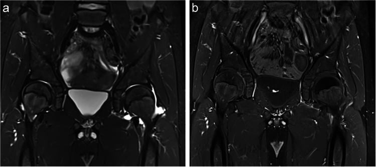
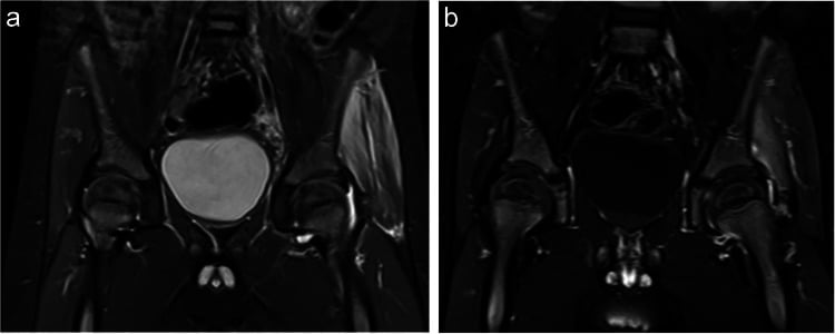
MRI. Reserved for difficult cases, where it can show bone marrow oedema (osteomyelitis) or pyomyositis. The pair below compares transient synovitis with a septic hip.
Ultrasound of the Irritable Hip
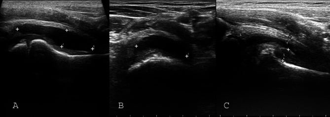
What it shows. Ultrasound is the most sensitive test for a hip effusion: an anechoic-to-hypoechoic collection distending the anterior joint recess, seen as a convex (bulging) anterior capsule lifted off the femoral neck. The usual threshold is a capsule-to-bone distance greater than about 5 mm, or a side-to-side difference of 2 mm or more compared with the asymptomatic hip.
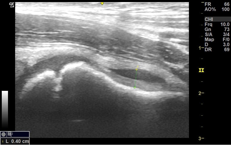
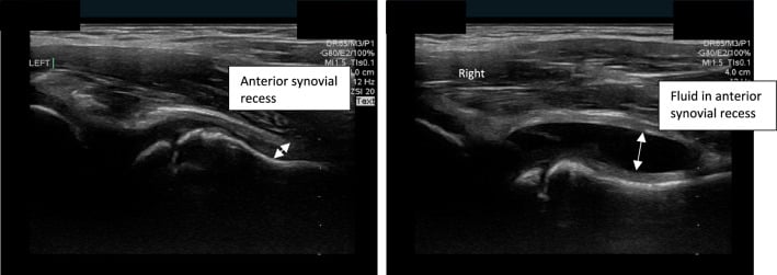
What it is for. It confirms the irritable hip, quantifies the effusion and guides aspiration. Longitudinal thickness and length plus transverse width define the anterior-recess effusion, and measurement standardises follow-up. A completely normal, symmetric scan makes a significant intra-articular hip problem unlikely.
What it cannot do. Ultrasound cannot distinguish septic arthritis from transient synovitis: both produce an effusion and the fluid looks similar. In Zamzam's series it was about 86% sensitive and 90% specific for a septic hip but was explicitly not safe to differentiate the two, and outcomes were worse when a false-negative scan delayed treatment. Only aspiration of the fluid (cell count, Gram stain, culture) tells them apart.
In practice. Use ultrasound to answer "is there an effusion, and where do I put the needle?", not "is this infected?". Never let a "just an effusion / looks like synovitis" scan reassure you out of aspirating a high-risk hip.
Detailed Differential Diagnosis
Perthes disease. Legg-Calvé-Perthes disease is the main differential for a limping child in this age group (4-8 years).
- Transient Synovitis
- Acute (days)
- Perthes Disease
- Insidious (weeks/months)
- Transient Synovitis
- Constant, antalgic
- Perthes Disease
- Activity-related, often mild
- Transient Synovitis
- Restricted in acute phase only
- Perthes Disease
- Chronic restriction (Abduction/IR)
- Transient Synovitis
- Normal / Effusion
- Perthes Disease
- Sclerosis / Fragmentation / Flattening
- Transient Synovitis
- Post-viral history
- Perthes Disease
- Well child, small stature
When it does not settle. If symptoms persist beyond 2 weeks, it is not transient synovitis. Repeat the radiograph and look for early Perthes changes (crescent sign).
The other serious causes. Maintain a high index of suspicion for each of these:
- Juvenile idiopathic arthritis - chronic (over 6 weeks), with morning stiffness, swelling and warmth; ANA may be positive, and eye examination for uveitis is required
- Slipped capital femoral epiphysis - an older age group (10-16 years), typically an obese adolescent, with an external rotation deformity and obligatory external rotation on flexion
- Osteomyelitis - in the proximal femoral metaphysis; movement is painful but not as restricted as in a septic joint, inflammatory markers are high and MRI is diagnostic
- Leukaemia - night pain, constitutional symptoms (weight loss, bruising), generalised lymphadenopathy and an abnormal FBC (blasts, cytopenia)
Management Algorithm
The sequence. Management of the irritable hip runs in four steps.
- Assess the child, not just the score. Appearance, fever history, ability to bear weight, pain severity, range, CRP trend and red flags determine pre-test concern. Escalation also weighs age, trajectory, ultrasound and the need for aspiration.
- Use ultrasound to identify an effusion. It does not determine whether the fluid is sterile.
- Aspirate when infection remains plausible or the result will change management, and send the full fluid panel (see Surgical Technique).
- Treat infection urgently when it is supported or strongly suspected, with source control plus antibiotics. Do not let a low cell count or negative routine culture override purulence, sepsis or a deteriorating course.
The confirmed typical case. Treatment is relative rest and activity modification ("cuddle therapy") with analgesia when safe: NSAIDs (ibuprofen) are the mainstay, and paracetamol is also used. Parents are educated on the red flags (high fever, refusal to walk) and given explicit return precautions, and the child is reviewed promptly if fever, worsening pain, inability to bear weight or failure to improve develops. Follow-up ensures resolution.
Antibiotics. They are not indicated for a confidently diagnosed non-bacterial transient synovitis, and are withheld only once infection has been reasonably excluded. If infection remains plausible, obtain samples promptly and start appropriate therapy without avoidable delay in a septic or deteriorating child.
The Partially-Treated Septic Arthritis Trap
The scenario. A child with an irritable hip has already received antibiotics, for another presumed infection or for the hip itself. Recent antibiotics can lower culture yield and blunt the presentation of septic arthritis, so they make a low predictor count less reassuring. Routine joint culture may be negative despite ongoing infection or undrained pus.
What to do. Reassess the full trajectory, obtain blood cultures and sample the joint urgently for cell count, Gram stain, culture and appropriate molecular testing. In a septic or deteriorating child, do not delay effective antibiotics because aspiration or theatre access is pending.
The corollary. A culture-negative aspirate is not automatically transient synovitis. Interpret it alongside prior antibiotics, organism epidemiology, fluid appearance, molecular results and the response after source control.
Q: A child with an irritable hip was started on antibiotics 3 days ago for a sore throat and now has near-normal inflammatory markers — is septic arthritis excluded? A: No. Prior antibiotics blunt fever and ESR/WCC/CRP, so the Kocher score reads falsely low. Recent antibiotic use should raise suspicion; aspirate with a low threshold and avoid giving more empirical antibiotics before sampling the joint.
Surgical Technique
No surgical role. Transient synovitis is a medical condition, and there is no role for surgery in its treatment once confirmed. Surgery's role is diagnostic: hip aspiration to exclude septic arthritis, and arthrotomy only if septic arthritis is confirmed or strongly suspected (pus on aspiration).
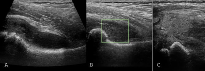
Aspiration. The approach is anterior or medial, and ultrasound guidance is standard. It is done under conscious sedation, or general anaesthesia in toddlers, with a large-bore needle. Send the fluid for:
- Cell count (WCC) and differential (PMN%)
- Gram stain and culture
- Locally appropriate molecular tests
- Crystal analysis
Reading the fluid. Turbid or purulent fluid is septic until proven otherwise, and clear or straw-coloured fluid is likely transient synovitis. Always send fluid for culture even if it appears clear, as early septic arthritis may not be frankly purulent.
No single threshold. Synovial WCC and neutrophil percentage overlap between diagnoses: elevated counts support infection, but the overlap prevents a single cutoff. Purulence and positive microbiology strongly support infection, whereas a lower count, negative Gram stain or negative routine culture does not exclude early, partially treated or Kingella infection. Interpret the fluid as a pattern, not one WCC threshold.
Complications
Missed septic arthritis. Missing septic arthritis is the critical complication. It is mitigated by interpreting the Kocher predictors in context and aspirating whenever infection remains plausible.
Recurrence. Recurrent episodes are usually milder. Parents are educated and reassured that it is benign, and each episode is treated the same way: exclude sepsis, then NSAIDs.
Perthes disease. A causal link with transient synovitis is not established. Early Perthes can initially mimic synovitis, which is why symptoms that persist are radiographed.
Coxa magna. A rare, mild enlargement of the femoral head can occur from increased blood flow (hyperaemia) due to synovitis. It is asymptomatic, usually resolves or persists without consequence, and is not Perthes disease.
Follow-Up Protocol
Transient Synovitis Follow-Up
Exclude septic arthritis. Start NSAIDs and rest.
Phone or clinical review. The child should be improving; if worse, treat as a red flag and re-evaluate for sepsis or Perthes disease.
Symptoms should largely resolve. Return to activity as tolerated.
Only needed if symptoms recur or persist. Consider X-ray to exclude Perthes (rare presentation).
Outcomes and Prognosis
Function is excellent, with 100% return to sports and activities. There is no increased risk of osteoarthritis or avascular necrosis, which differentiates transient synovitis from Perthes disease.
Guidelines, Registries & Global Practice
- Most common cause of atraumatic hip pain and limp in children worldwide
- Annual incidence estimated at roughly 0.2% of children; lifetime risk up to 3%
- Peak age 3-8 years (commonly 4-6); male predominance approximately 1.5-2:1
- Preceding viral illness (often URTI) reported in a substantial minority
- Recurrence in roughly 4-15% of children across series
- No single registry exists (non-implant, self-limiting condition)
- Kocher / Caird prediction tools are referenced internationally (AAOS, BOA, European centres)
- Universal principle: septic arthritis must be actively excluded before labelling a hip "transient synovitis"
- Antibiotic stewardship: narrow-spectrum agents (e.g. flucloxacillin / first-generation cephalosporin) reserved for proven or strongly suspected infection
- Risk Stratification
- Kocher + CRP (Caird)
- Imaging Emphasis
- Ultrasound to confirm effusion, guide aspiration
- Notable Point
- Low threshold for aspiration when 3-4 predictors
- Risk Stratification
- Clinical + inflammatory markers
- Imaging Emphasis
- Ultrasound first-line for effusion
- Notable Point
- MRI if osteomyelitis or pyomyositis suspected
- Risk Stratification
- CRP-weighted models
- Imaging Emphasis
- Ultrasound; PCR for Kingella in young children
- Notable Point
- Strong emphasis on K. kingae molecular detection
- Risk Stratification
- Clinical + basic bloods
- Imaging Emphasis
- X-ray; ultrasound where available
- Notable Point
- Aspiration and empirical cover prioritised when sepsis cannot be excluded
Where ultrasound and rapid inflammatory markers are unavailable, the safe default shifts toward aspiration and exclusion of sepsis rather than observation, because the cost of a missed septic hip is joint destruction. Conversely, in well-resourced settings serial examination, ultrasound and CRP allow more confident observation of low-risk children.
Kingella kingae is fastidious and frequently missed on standard culture. In a young child (under 4 years) with a septic-appearing but culture-negative hip, request K. kingae PCR (or inoculate aspirate into blood-culture bottles). Molecular methods have repositioned it as the leading osteoarticular pathogen in this age group.
Controversies & Areas of Uncertainty
The Kocher probabilities (under 0.2% to 99.6%) were not reproduced on external validation (Luhmann: 59% for 4/4). The tools stratify risk but should never override clinical judgment or replace aspiration when suspicion is high.
Caird's prospective data suggest CRP may be the single strongest individual predictor, yet the original four-variable rule remains the most quoted. Whether CRP should replace or supplement ESR/WCC is unsettled.
Routine early radiographs in a classic, rapidly settling case have low yield. There is no consensus on universal first-visit X-ray; most reserve it for atypical, persistent (over 2 weeks) or recurrent presentations to exclude Perthes.
A causal link between transient synovitis and later Perthes is not established; historical series report a small (around 2-3%) subsequent Perthes rate, likely reflecting early Perthes initially mislabelled rather than true progression.
MCQ Practice Points
Q: How should three Kocher predictors be interpreted? A: They raise concern substantially, but the derivation-cohort 93% is not a universal probability. Integrate age, clinical trajectory, CRP, ultrasound and aspiration/microbiology.
Q: A hip aspirate has WCC 8,000 with 45% neutrophils. Is transient synovitis proven? A: No. This lower inflammatory burden may support transient synovitis in a well, improving child, but early, partially treated and Kingella infections can have lower counts. Interpret appearance, Gram stain, culture/PCR, prior antibiotics and clinical course.
Q: What is the most common organism causing septic arthritis in the 3-8 year age group? A: Staphylococcus aureus. (Kingella kingae is increasing, especially in younger children under 4 years).
Q: What is Waldenstrom's sign? A: Widening of the medial joint space (over 2mm asymmetry) on plain X-ray. It indicates hip effusion (synovitis or septic).
Q: Does transient synovitis cause Perthes disease? A: A causal link is not established. A later Perthes diagnosis may represent early disease initially mislabelled as synovitis, which is why persistence or recurrence requires reassessment.
Exam Cheat Sheet
Diagnosis
- Age 3-8 years, acute limp
- Diagnosis of Exclusion
- Must exclude Septic Arthritis
- Exclude Perthes (X-ray)
Kocher Criteria
- History of fever
- Non-weight-bearing
- ESR at least 40
- WCC over 12,000
- Predictors support risk assessment; probabilities are cohort-specific
Management
- Analgesia and relative rest when the diagnosis is secure
- Explicit safety-net and review of clinical trajectory
- Aspirate when infection remains plausible or the result changes management
- No routine antibiotics for confirmed non-bacterial synovitis
Aspiration
- Cell count and neutrophil percentage overlap
- Purulence or positive microbiology strongly supports infection
- Negative culture does not exclude prior-antibiotic or Kingella infection
Prognosis
- Most improve over days and recover within 1-2 weeks
- Recurrence occurs and requires fresh assessment
- Persistence or atypical features require repeat imaging/work-up
- Later Perthes diagnosis may reflect an initial mimic rather than proven causation
Viva Scenarios
Practise clinical reasoning and management decisions out loud
“A 4-year-old boy presents with a limp. He had a viral URTI last week. He is afebrile and happy. Examination shows restricted internal rotation. What is your approach?”
“A 3-year-old girl presents refusing to walk. Temp 38.6. CRP 40. WCC 16. Ultrasound shows effusion. How do you manage this patient?”
“A 6-year-old boy returns 4 weeks after a diagnosis of 'transient synovitis'. He is still limping intermittently. He is afebrile. What is your differential?”
“A 7-year-old boy presents with a 4-week history of mild groin pain. He has been treated as 'transient synovitis' by his GP but is not improving. X-ray shows 'mild flattening' of the femoral head. Discuss.”
Evidence Base
Kocher Criteria — Original Prediction Algorithm
- Retrospective review of an acutely irritable hip cohort at a tertiary children's hospital (1979-1996)
- Four independent multivariate predictors: history of fever, non-weight-bearing, ESR at least 40 mm/hr, serum WCC over 12,000/mm³
- Predicted probability of septic arthritis: under 0.2% (0 predictors), 3.0% (1), 40.0% (2), 93.1% (3), 99.6% (4)
- Excellent ROC performance for combined predictors despite overlap of individual variables
External Validation of Kocher Criteria
- 165 hips (1992-2000) — applied the original Kocher algorithm at an independent institution
- With all 4 predictors present, probability of septic arthritis was only 59% (vs 99.6% in Kocher's cohort)
- Best local model used 3 variables (fever, WCC over 12,000, previous health-care visit) — 71% probability when all present
- Neither the original nor the local algorithm performed reliably outside its derivation centre
C-Reactive Protein as a Predictor (Prospective)
- Prospective study of 53 children undergoing hip aspiration for suspected septic arthritis
- CRP was the only factor strongly and independently associated with septic arthritis on multivariate analysis
- Fever (oral temperature over 38.5°C) was the single best predictor; CRP over 20 mg/L a strong independent risk factor
- Probability of septic arthritis: 83% (3 factors), 93% (4 factors), 98% (5 factors)
Transient Synovitis — Long-Term Characterisation
- 30-year retrospective review: 497 episodes in 475 children
- Femoral head measurements at 6-month follow-up showed no significant dimensional change
- Legg-Calvé-Perthes disease developed in 3 children (2.5%); recurrent synovitis (benign course) in 19
- Authors recommend radiographic assessment at 6 months after the initial episode
Ultrasound Cannot Differentiate Septic from Transient
- 154 children (81 septic arthritis, 73 transient synovitis); ultrasound correlated with final diagnosis in 127
- Ultrasound sensitivity 86.4%, specificity 89.7%, PPV 87.9% for septic hip
- Ultrasound detects minimal effusion but cannot safely distinguish septic arthritis from transient synovitis
- Worse outcomes with treatment delayed over 4 days and with false-negative scans
Kingella kingae as a Leading Cause of Osteoarticular Infection
- Prospective study; specific real-time PCR applied to culture-negative osteoarticular specimens in young children
- Culture alone identified a pathogen in 45%; adding PCR raised documentation to 66%
- Kingella kingae was the leading pathogen (45%), ahead of Staphylococcus aureus (29%)
- Standard cultures substantially under-detect K. kingae