
Article summary
How augmented reality could overlay guidance onto the surgical field, and where the technology stands today.
Educational content is reviewed for source visibility, editorial coherence, and correction readiness.
No individual clinician credential is claimed unless a named person is shown.
Verify before clinical use; this is not medical advice or a substitute for local guidance.
For decades, orthopaedic surgeons have relied on two-dimensional images and intraoperative fluoroscopy to navigate complex three-dimensional anatomy. But what if you could simply look at your patient and see the underlying bone, tumour margin, or planned screw trajectory overlaid perfectly on the surgical field? Augmented reality (AR) is rapidly evolving from a speculative concept into a practical tool that promises to fundamentally change how we operate.
The Evolution of Intraoperative Visualisation
To understand the transformative potential of augmented reality, it helps to look at the historical continuum of surgical visualisation. Orthopaedics has always been a deeply visual and spatial specialty. For decades, our intraoperative guidance relied heavily on the C-arm, a technology that revolutionised fracture fixation but comes with inherent limitations: radiation exposure for the surgical team and the patient, interrupted surgical workflow, and the cognitive burden of mentally mapping a two-dimensional, grainy image onto a three-dimensional anatomical structure.
Over recent years, navigated surgery and intraoperative three-dimensional imaging have improved our spatial awareness. These systems allow for precise implant placement, particularly in complex spinal deformity and pelvic trauma. However, traditional navigation suffers from a distinct bottleneck. It forces the surgeon to break eye contact with the patient and look away at a distant computer monitor. This "visual divide" disrupts the natural ergonomics of surgery. Augmented reality bridges this gap by bringing the navigation data directly into your line of sight, effectively merging the digital plan with the physical reality of the patient.
Defining the Technology: What Augmented Reality Actually Is
The term augmented reality is often used loosely in popular culture, but in the operating theatre, it has a highly specific definition. Unlike Virtual Reality (VR), which immerses you in a completely digital world and blocks out the physical environment, AR overlays digital information onto the real world. In the context of orthopaedic surgery, this generally means projecting virtual images of a patient's internal anatomy, pre-operative surgical plans, and real-time instrument trajectories onto your field of view.
Current AR Modalities
Currently, surgical AR is delivered through a few distinct hardware pathways, each with its own practical implications:
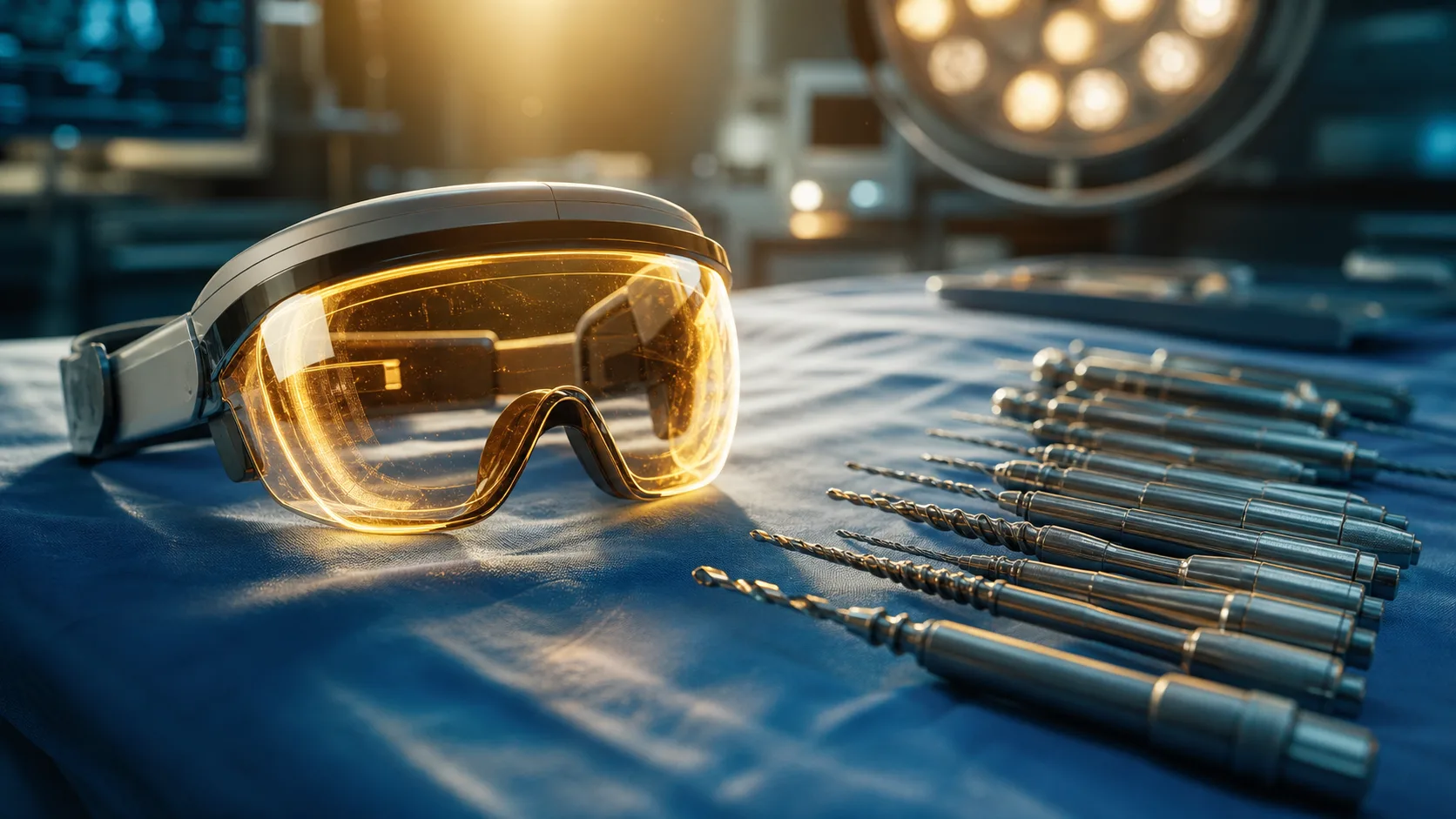
- Head-Mounted Displays (HMDs): These are perhaps the most recognisable form of AR. Surgeons wear specialised visors or smart glasses that project holographic images into their field of vision. Modern surgical HMDs are designed to be lightweight, sterilisable, and compatible with standard surgical loupes and headlights.
- Projected AR: Instead of using a headset, this technology uses high-intensity projectors to cast digital images directly onto the patient's physical skin or exposed surgical field. It is highly intuitive because it turns the patient's own anatomy into a dynamic screen.
- Tablet and Microscope-Based AR: Here, an external screen or an optical viewing system acts as the intermediary. While this still requires looking at a screen rather than the patient directly, it superimposes high-fidelity digital overlays onto the live video feed, which is particularly useful in microsurgery or for teaching audiences in the gallery.
Overlaying Guidance: The Mechanics of Superimposition
The magic of AR lies not just in the display, but in the complex mechanics of tracking and registration that make accurate superimposition possible. To safely overlay guidance, the AR system must know exactly where your instruments are in three-dimensional space and exactly how they relate to the patient's specific anatomy.
This process begins long before you enter the operating theatre. A pre-operative CT or MRI scan is segmented by advanced software, isolating the relevant bony structures, nerves, blood vessels, or tumour margins. You then plan your osteotomies or implant trajectories on this highly detailed three-dimensional model.
Once in theatre, the critical step is registration. The AR system must map this digital model perfectly to the physical patient lying on the table. Systems use various tracking methods—such as optical trackers, infrared cameras, or surface-matching algorithms—to align the digital and physical worlds. When you move your head or your hands, the system updates the holographic overlay in real time, ensuring that a virtual screw trajectory remains perfectly aligned with the physical vertebra, regardless of your viewing angle.
Practical Applications in Orthopaedic Surgery
The translation of AR from theoretical gadgetry to clinical utility is already well underway across several orthopaedic subspecialties. The ability to "see through" tissue is profoundly altering surgical approaches.
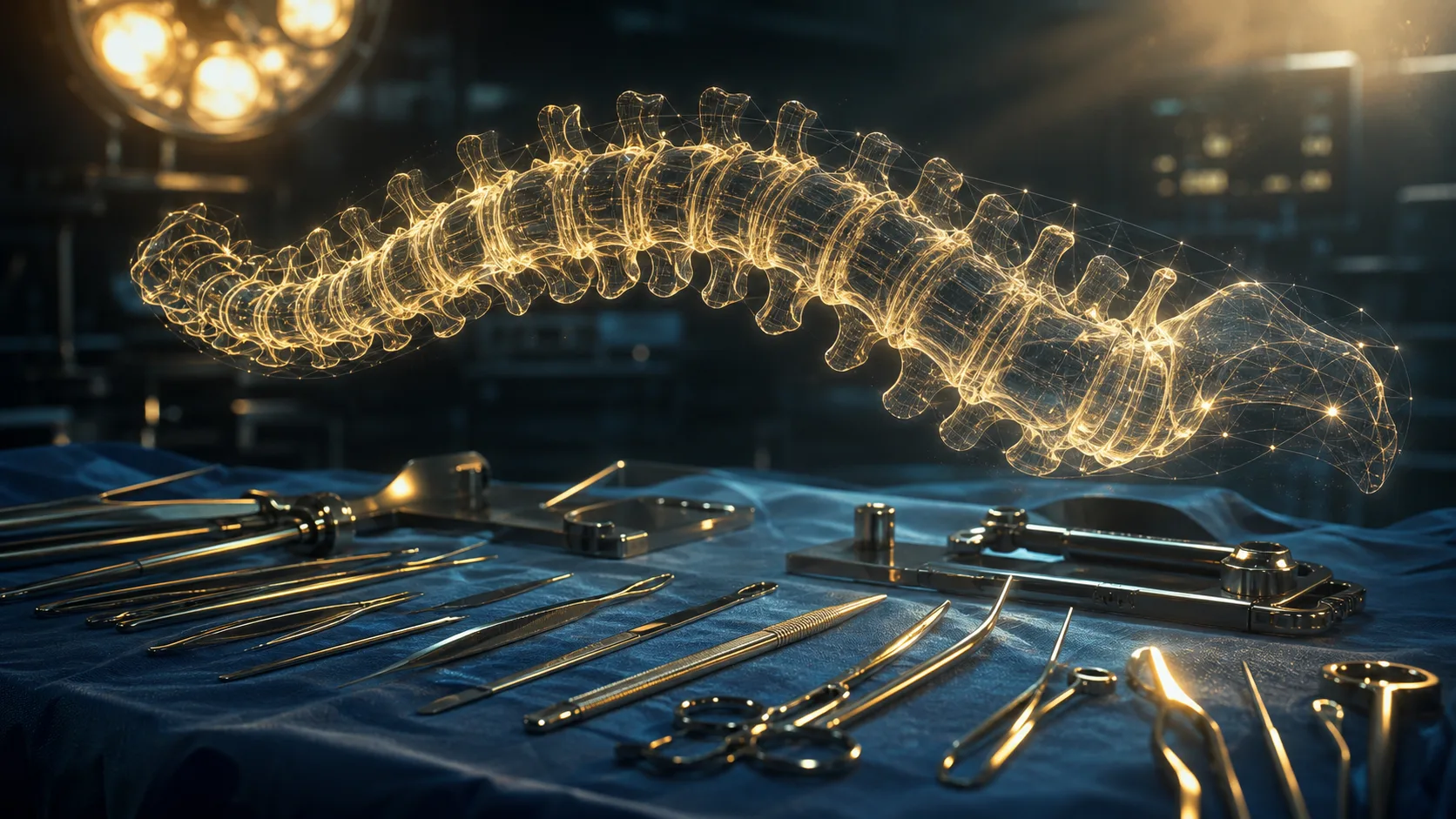
In spinal surgery, pedicle screw placement is a high-stakes procedure where spatial accuracy is paramount. AR allows you to visualise the pedicle borders, the spinal cord, and the exact planned trajectory for the screw simultaneously. By wearing an HMD, you can effectively look at the exposed posterior elements and see the anterior cortex of the vertebral body, removing the guesswork and significantly reducing the need for continuous fluoroscopic confirmation.
In orthopaedic oncology, achieving a wide en-bloc resection with clear margins while preserving as much healthy tissue as possible is a delicate balancing act. AR overlays tumour margins mapped from MRI directly onto the surgical field. This allows you to execute complex, multiplanar osteotomies with absolute precision, ensuring you are neither violating the tumour nor sacrificing unnecessary amounts of the patient's native bone.
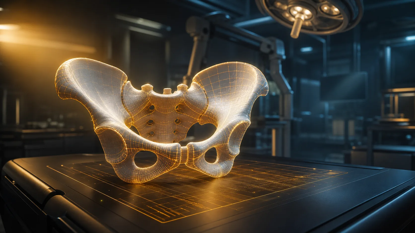
Complex trauma reconstruction also stands to benefit immensely. Pelvic and acetabular fractures, for instance, require the reduction of highly irregular, displaced fragments. AR can project a "ghosted" image of the pre-injury, uninjured contralateral pelvis over the fractured side, acting as a real-time blueprint to guide your clamps and reduction forceps to achieve perfect articular congruity.
Navigating the Limitations and Learning Curve
While the promise of AR is undeniable, it is crucial for any modern surgeon to approach the technology with a clear understanding of its current limitations. It is a powerful tool, but it is not yet infallible.
One of the most common pitfalls for early adopters is what is known as registration drift. During a long and complex case, the surgical field changes. Soft tissues retract, swelling occurs, and bony fragments move once an osteotomy is made. If the digital overlay does not dynamically update to reflect these physical changes, it can provide a false sense of security. You must maintain your standard surgical checks and balances, never blindly trusting the overlay if the intraoperative anatomy dictates otherwise.
Ergonomics and visual fatigue present additional hurdles. Wearing a head-mounted display for several hours can cause neck strain and ocular fatigue. Furthermore, the human eye sometimes struggles to focus simultaneously on a bright holographic image hovering a few centimetres away and a physical object deeper in the wound. Manufacturers are continually improving optical engineering to mitigate this, but for now, integrating AR into your practice requires patience and a willingness to adapt your physical operative posture.
Integrating AR into Your Training and Practice
For medical students, surgical trainees, and consultants looking to understand this technology, passive observation is no longer enough. The integration of AR into routine practice requires deliberate engagement with the technology's underlying principles.
If you are currently in a training pathway, your priority should be to solidify your foundational understanding of surgical anatomy and spatial awareness. Augmented reality is a tool to enhance a skilled surgeon's judgement; it cannot compensate for a lack of fundamental anatomical knowledge. Seek out simulation suites at major teaching hospitals or conference exhibition halls where AR headsets are available. Spending even a short time manipulating virtual instruments in a simulated environment will rapidly demystify the technology.
For established surgeons, introducing AR into your theatre requires a structured approach. A common mistake is attempting to use these highly complex systems on a critically ill or multiply-injured patient during a chaotic night shift. The initial learning curve should be managed in a controlled environment. Start by using AR guidance on simpler, elective cases where you are highly confident in the anatomy and the surgical plan. Build rapport with your theatre staff, as the scrub nurses and circulating staff will also need to understand the new spatial workflows. Thorough pre-operative planning is also non-negotiable; the old adage of "garbage in, garbage out" has never been more true than when programming an AR-guided resection.
The Future Horizon of Surgical Guidance
The current generation of AR is impressive, but it is merely the baseline for what is to come. As processing power increases and head-mounted displays become as lightweight as standard surgical loupes, the friction of using these systems will all but disappear.
Looking ahead, the convergence of AR with artificial intelligence and machine learning holds staggering potential. Future systems will likely offer dynamic guidance that learns and adapts in real time. Imagine an AR visor that not only shows you the planned osteotomy but uses intraoperative AI to analyse your sawing technique, warning you if your cut drifts two millimetres from the plan, or automatically adjusting the digital tumour margin overlay based on real-time changes in tissue tension and swelling.
Furthermore, as this technology becomes more globally democratised, it could act as a powerful equaliser in surgical education. A trainee could perform a complex hip resurfacing with a subtle AR safety net, preventing catastrophic errors while building muscle memory and confidence.
Augmented reality is not coming to replace your surgical skill, but it will fundamentally redefine the boundaries of what is possible in the operating theatre. By blending digital precision with human judgement, this technology offers a safer, more efficient, and ultimately more effective future for orthopaedic surgery.
Related topics
Share this article
Useful for a journal club, study list, or teaching session.