
Article summary
How simulation, digital tools and AI are reshaping the way surgeons are trained — for better and worse.
Educational content is reviewed for source visibility, editorial coherence, and correction readiness.
No individual clinician credential is claimed unless a named person is shown.
Verify before clinical use; this is not medical advice or a substitute for local guidance.
For generations, the apprenticeship model of "see one, do one, teach one" formed the bedrock of surgical training. Today, that paradigm is undergoing a profound evolution, replaced by a landscape where simulation, artificial intelligence, and digital ecosystems allow you to refine your craft long before you step up to the operating table. As orthopaedic surgeons, embracing these technological shifts is no longer optional; it is a critical component of preparing for high-stakes exams and building a resilient, forward-thinking career.
The Limitations of the Traditional Apprenticeship Model
Historically, surgical training was entirely reliant on direct patient care and hands-on operating theatre experience. While there is absolutely no substitute for the pressure and tactile reality of live surgery, relying solely on this model presents significant bottlenecks.
Firstly, working time directives and a well-deserved cultural shift towards work-life balance mean that modern trainees simply spend fewer hours in the hospital than their predecessors. Consequently, the sheer volume of primary cases you are exposed to during your training years has decreased. Secondly, the margin for error in modern orthopaedics is narrower than ever. Patient expectations are sky-high, and the ethos of modern practice dictates that it is entirely unethical for a novice to learn basic psychomotor skills on a living patient.
The traditional model also struggles with consistency. You might gain unparalleled exposure to complex polytrauma one month, only to spend the next auditing waiting lists in a clinic. Technology steps into this void, offering a standardised, deeply repeatable foundation upon which you can build your clinical judgement and manual dexterity.

Virtual Reality and High-Fidelity Simulation: The New Sandbox
When preparing for rigorous orthopaedic exams—whether you are navigating objective structured clinical examinations (OSCEs) or rigorous vivas—muscle memory and spatial awareness are paramount. Virtual reality (VR) and high-fidelity physical simulators provide a sandbox where failure is not just accepted, but encouraged as a learning tool.
Modern simulation allows you to repeatedly perform complex procedures, such as femoral canal broaching or navigating the posterior hip capsule, in a completely risk-free environment.
The Right Way to Use Simulators
The most common mistake trainees make with simulators is treating them like a video game—repeating a task until they achieve a high score, without pausing to reflect on their biomechanical errors. To genuinely extract value from these tools, you must approach a simulated procedure with the exact same gravity as a live operation.
You should perform a formal pre-operative briefing, set up your drapes precisely, and maintain the sterile field. If your simulation centre offers haptic feedback devices, pay close attention to the resistance felt when reaming a cortical bone versus cancellous bone. Utilising these platforms deliberately helps bridge the gap between theoretical anatomical knowledge and the practical, three-dimensional reality of orthopaedic surgery, ensuring you enter the theatre fully prepared for your consultant or examiner's scrutiny.
Artificial Intelligence and Surgical Analytics
Artificial intelligence (AI) is rapidly moving out of the realm of science fiction and into the daily fabric of surgical training. In orthopaedics, AI is increasingly used to analyse surgical videos and provide granular, objective feedback that a human mentor might miss.
Imagine performing an arthroscopic anterior cruciate ligament (ACL) reconstruction. By feeding a video of your procedure into an AI-driven analytics platform, the system can calculate your economy of motion, track your camera ambidexterity, and measure the exact time spent locating the anatomical footprint of the native ligament. These platforms essentially act as a tireless, objective surgical coach.
However, integrating AI into your training requires a nuanced approach. A frequent pitfall is becoming overly reliant on automated feedback to correct micro-errors, whilst ignoring the holistic picture of the surgery. AI can tell you that your instrument yaw was suboptimal during a shoulder arthroscopy, but it cannot yet fully contextualise that data against a patient's specific anatomy or tissue quality. Use AI to iron out technical inefficiencies, but always filter that data through the lens of human mentorship and established biomechanical principles.

Smart Glasses, Wearable Tech, and Remote Mentorship
The hierarchical structure of the operating theatre can sometimes be isolating for the junior trainee. You might be closing the fascia while the consultant scrubbed out to dictate the notes, leaving you to navigate a tricky anatomical plane with minimal guidance. Wearable technology and smart glasses are actively dismantling these physical barriers, enabling remote, real-time mentorship.
Equipped with smart glasses, a trainee performing a relatively routine plating of a distal radius can be virtually "shadowed" by a senior surgeon located miles away. The senior surgeon can see exactly what the trainee sees, overlaying digital arrows onto the trainee’s field of vision to guide screw trajectory or flap elevation.
This continuous connectivity is particularly revolutionary for surgeons training in remote or rural settings, where a sub-specialist hand surgeon might not be physically present in the building. For your exam preparation, recordings captured via these wearables provide an unvarnished, first-person perspective of your performance, allowing you and your educational supervisors to review your decision-making processes step-by-step during debriefs.
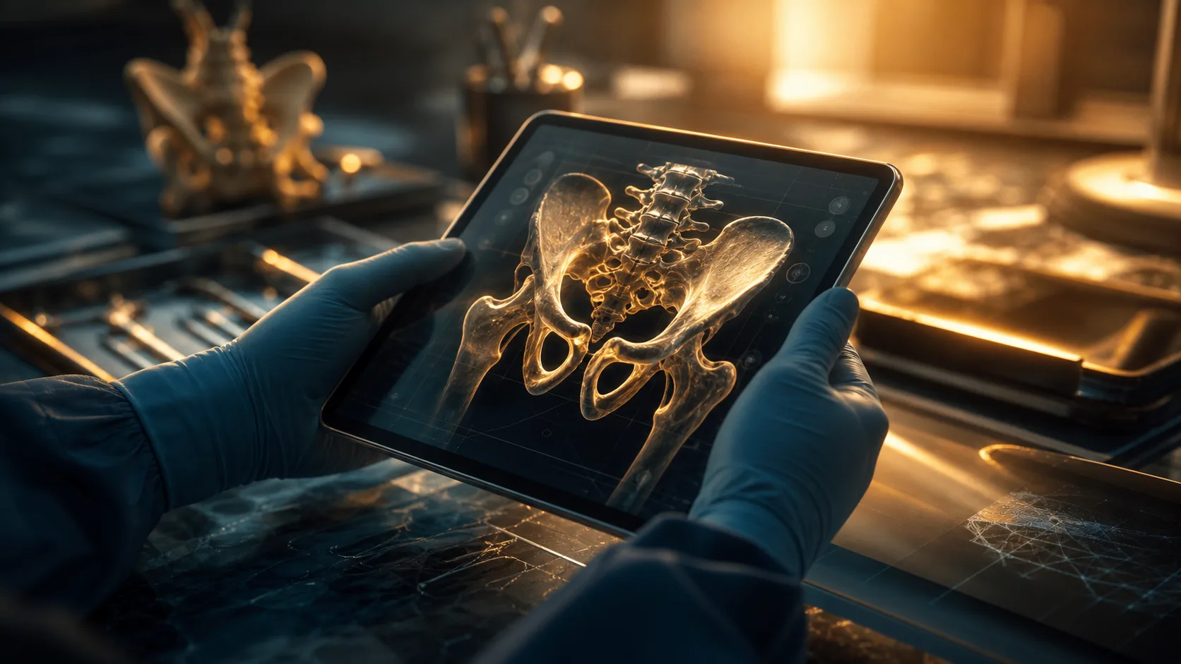
Enhanced Digital Preparation and 3D Modelling
The days of prepping for a complex joint replacement solely by reviewing two-dimensional plain radiographs are rapidly fading. Digital segmentation and 3D printing technologies have democratised pre-operative planning, making it an active, tactile part of your training.
As a trainee, you can now take a patient's computed tomography (CT) scan, segment the bony architecture using specialised software, and 3D-print an exact replica of a complex acetabular defect. Holding a physical model of the bone you are about to operate on allows you to visualise screw lengths, plan your osteotomies, and understand the deformity in a way that a screen simply cannot replicate.
If you are preparing for a major exit exam, demonstrating an understanding of digital templating and 3D modelling will mark you out as a forward-thinking, safe surgeon. A practical tip for your training is to proactively identify complex cases on your firm’s list and ask the senior surgeon if you can digitally template the operation. Presenting a comprehensive, digitally templated plan during the morning briefing not only impresses your consultant but fundamentally deepens your anatomical understanding of the procedure.
The Dark Side of Digital Dependency
While the technological revolution in surgical training is overwhelmingly positive, it carries significant risks that you must actively mitigate. The most insidious of these is the erosion of resilience and the "alert fatigue" that comes with digital overload.
Over-reliance on navigation systems and robotic assistants is a growing concern. If you train primarily with computer-navigated joint replacements, there is a genuine danger that your fundamental understanding of mechanical axes, ligament balancing, and bony landmarks will atrophy. When the power fails or the navigation camera becomes obstructed, you must possess the core surgical competence to complete the operation safely.
Furthermore, digital tools can create a false sense of competency. Scoring perfectly on a VR arthroscopy module does not equate to managing a patient who becomes suddenly haemodynamically unstable on the table. You must remember that simulation and AI are tools to augment your training, not replace the irreplaceable lessons learned from standing at the operating table for hours, managing the unanticipated complications and physiological variables of a living patient.
Integrating Tech into Exam Preparation and Career Progression
For medical students and surgical trainees reading OrthoVellum, the integration of technology must be strategic and aligned with your immediate career milestones. When preparing for high-stakes assessments, these tools can be the deciding factor between a pass and a distinction.
Building a Tech-Forward Portfolio
A common mistake among trainees is viewing digital tools merely as a crutch for their own learning, rather than a portfolio asset. Engaging actively with digital templating, contributing to departmental audits that analyse AI-assisted surgical metrics, or championing the use of simulation in your hospital’s teaching programmes demonstrates exactly the kind of leadership and innovation that training committees and consultant interview panels are looking for.
Consider utilising digital question banks and spaced-repetition algorithms not just for written exams, but to structure your viva preparation. By coupling digital fact-learning with physical model manipulation, you train your brain to recall complex management algorithms whilst simultaneously communicating your physical plan to the examiner. This dual-pronged approach ensures that when you are standing in front of the examiners, your hands and your mind are equally prepared.
Navigating the Cost and Accessibility Divide
It is impossible to discuss the technological overhaul of surgical training without addressing the stark reality of cost and accessibility. High-fidelity simulators and AI analytics suites command eye-watering budgets, and the reality is that funding and resources vary wildly across different hospital trusts and global healthcare systems.
As a trainee, you will inevitably encounter environments where the technological infrastructure lags behind the cutting edge. Rather than becoming frustrated, you must become an advocate for your own education. Seek out regional simulation networks, collaborate with university bio-engineering departments, or form trainee-led consortia to pool resources and share access to digital planning software. Furthermore, you should familiarise yourself with the Royal College of Surgeons and other international surgical bodies' recommendations on simulation, using these guidelines to lobby your programme directors for better access to modern training tools.
Technology is democratising knowledge, but the hardware remains expensive. Ensuring that you, and the cohorts of surgeons following you, have equitable access to these vital training modalities is a challenge that the current generation of trainees must actively tackle.
The surgeon of the future is not a machine operator, but a highly adaptable professional who leverages digital tools to refine their craft, prioritise patient safety, and push the boundaries of orthopaedic excellence. By thoughtfully integrating simulation, AI, and digital planning into your practice, you ensure that when the moment comes to make the critical incision, you are prepared for anything.
Share this article
Useful for a journal club, study list, or teaching session.