Article summary
Indications for tubular retractors. Reducing muscle damage without compromising visualization.
Educational content is reviewed for source visibility, editorial coherence, and correction readiness.
No individual clinician credential is claimed unless a named person is shown.
Verify before clinical use; this is not medical advice or a substitute for local guidance.
Minimally Invasive Spine Surgery (MISS) has fundamentally evolved from what was once dismissed as a niche marketing term or a passing trend into an established, evidence-based standard of care for a wide array of spinal pathologies. The underlying philosophy of MISS is emphatically not just about achieving a smaller, cosmetically appealing skin incision—often colloquially termed "keyhole surgery." Rather, the true essence of MISS lies in minimizing collateral iatrogenic damage to the stabilizing soft tissues, vascular supply, and neural elements of the posterior tension band of the spine.
Since Kevin Foley and Maurice Smitherman first described the tubular microdiscectomy technique using the METRx system in 1997, orthopaedic surgery training programs worldwide have increasingly emphasized MISS as a mandatory core competency. For trainees navigating the rigorous pathways of fellowship exam preparation—whether you are sitting the FRCS (Tr & Orth), the FRACS, or the ABOS board examinations—you must be prepared to discuss MISS comprehensively. Examiners will expect a nuanced understanding of patient selection, detailed technical pearls, the specific management of intraoperative complications, and a firm grasp of the current literature comparing MISS to traditional open techniques.
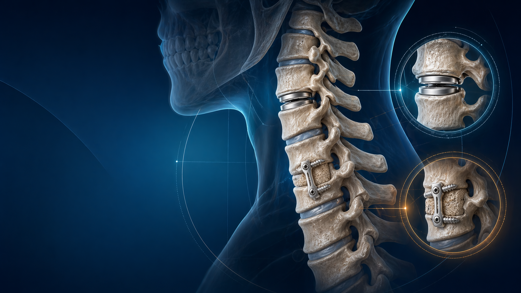
Visual Element: Cover image showing the view through a tubular retractor, highlighting the magnified visualization of the nerve root and dura.
The Muscle-Sparing Philosophy: Anatomy and Biomechanics Matter
To truly understand the value of MISS, one must first understand the devastating effects of traditional open spine surgery on the posterior paraspinal musculature. Standard open approaches necessitate the aggressive subperiosteal stripping of the paraspinal muscles from the spinous processes and the laminae to achieve adequate exposure. Prolonged retraction using self-retaining devices (like Harvey or Taylor retractors) leads to increased intramuscular pressure, resulting in ischemic necrosis.
Furthermore, this detachment creates dead space, leading to hematoma formation, subsequent fibrosis, and what is clinically recognized as "fusion disease" or postoperative failed back surgery syndrome (FBSS) driven by iatrogenic muscle denervation.
The multifidus muscle, the primary dynamic stabilizer of the lumbar spine, deserves particular anatomical respect during your orthopaedic surgery training. Its segmental innervation arises exclusively from the medial branch of the dorsal ramus. This delicate nerve enters the muscle belly posteriorly and laterally. Aggressive subperiosteal stripping and lateral retraction denervate these segments permanently. Landmark histological and MRI studies by Xu et al. and Sihvonen et al. have definitively demonstrated that open posterior approaches cause significant multifidus atrophy, selective type II muscle fiber cross-sectional area reduction, and fatty infiltration. This correlates directly with worse long-term functional outcomes and chronic postoperative axial back pain.
MISS techniques, particularly tubular microdiscectomy, utilize the Wiltse muscle-splitting approach (the paraspinal sacrospinalis-splitting plane between the multifidus and longissimus muscles). Sequential dilators are employed to gently split the muscle fibers along their natural longitudinal anatomical plane rather than cutting, stripping, or burning them with electrocautery. The static tubular retractor disperses retraction forces evenly. Once the tube is removed at the end of the procedure, the muscle fibers simply fall back into their anatomical position, preserving their structural integrity, vascular supply, and—crucially—their innervation.
Visual Element: Internal cross-section graphic comparing the approach of Open vs. MISS surgery. The open side shows muscle stripping and retraction, while the MISS side shows the tubular retractor passing through the muscle fibers.
Pre-operative Planning and Patient Positioning: Setting Up for Success
Success in MISS is entirely dependent on meticulous preoperative planning and flawless operating room setup. Before making the skin incision, study the axial, sagittal, and coronal MRI slices carefully. You must conceptually identify Kambin's triangle—the safe working zone bounded superiorly by the exiting nerve root, medially by the thecal sac and traversing root, and inferiorly by the superior endplate of the caudal vertebra.
Key planning points for orthopaedic surgery trainees:
- Level Identification: Scrutinize the radiographs and MRI for transitional anatomy (lumbarization of S1 or sacralization of L5). Counting down from the 12th rib or up from the sacrum must be correlated with sagittal MRI scout views.
- Trajectory: The tubular approach follows a "line of attack" from posterolateral to anteromedial. For a standard L4/5 paracentral disc herniation, aim toward the shoulder of the L5 pedicle. The trajectory dictates your skin incision site.
- Medialization: The tube must dock exactly on the medial pars interarticularis or the inferior laminar edge, not laterally on the facet joint. Docking too laterally risks iatrogenic facet violation, capsule destruction, and subsequent postoperative iatrogenic instability.
- Tube size and length: Standard lumbar discectomy utilizes 18mm or 22mm diameter tubes. Calculate the required tube length preoperatively by measuring the distance from the skin to the lamina on the axial MRI. Larger, more muscular patients or revision cases may require 26mm tubes.
Patient positioning is equally critical. The patient should be prone on a radiolucent Jackson table or a Wilson frame. Crucially, the abdomen must hang completely free to decrease intra-abdominal pressure. Any abdominal compression will translate to engorgement of Batson's epidural venous plexus, transforming a routine microdiscectomy into a frustrating, bloody struggle where visualization is completely lost at the base of a narrow tube.
Tubular Microdiscectomy: Step-by-Step Surgical Pearls
The tubular microdiscectomy remains the foundational workhorse procedure of minimally invasive spine surgery. Mastering this procedure is essential for any modern spinal surgeon. Here is the sequential technique enriched with clinical pearls tailored for fellowship exam preparation:
- Localization and Incision: After precise fluoroscopic localization, make a 1.5cm to 2cm vertical paramedian incision. The distance from the midline depends on patient size and target pathology (typically 1.5-2cm lateral to the midline for standard paracentral disc herniations, or 3-4cm for far-lateral/foraminal approaches). Incise the skin and subcutaneous fat.
- Fascial Incision and Dilation: Incise the lumbodorsal fascia sharply with a #15 blade. Use blunt finger dissection or a Metzenbaum scissor to palpate the bony anatomy and ensure you are entering the correct muscle plane. Pass the initial targeting guidewire onto the bony lamina or medial facet complex. Never advance the guidewire blindly into the interlaminar space or spinal canal. Pass the sequential dilators over the guidewire. You must feel the tactile change in resistance as you pass through yielding muscle versus contacting rigid bone.
- Docking and Retraction: Pass the final tubular retractor over the largest dilator and rigidly attach it to the flexible table-mounted arm. Apply slight downward pressure to prevent muscle creep under the tube. This 18mm circle is your only window to the spine—if your trajectory is incorrect, do not struggle; loosen the arm, reposition the tube, and re-dock.
- Soft Tissue Clearance: Bring in the operating microscope. The microscope provides the crucial stereoscopic vision, depth perception, and coaxial illumination essential for operating down a dark cylinder. Alternatively, 30-degree endoscopes offer superior illumination and the ability to look "around corners," though they come with a steeper learning curve. Use a long bayoneted Bovie electrocautery and pituitary rongeurs to clear the remaining soft tissue off the interlaminar window, identifying the inferior edge of the cranial lamina, the superior edge of the caudal lamina, and the medial facet.
- Bony Decompression (Hemilaminotomy): Utilize a high-speed matchstick burr (e.g., Midas Rex with a long attachment) to thin the lamina and the medial aspect of the inferior articular process. Follow this with a 2mm or 3mm Kerrison rongeur to complete the hemilaminotomy and medial facetectomy. Always keep the footplate of the Kerrison parallel to the dura.
- Flavectomy and Neural Decompression: The ligamentum flavum should be kept intact as a protective shield during the bony work. Once the bony limits are defined, use a curette to detach the flavum from the under-surface of the lamina, then resect it piecemeal with a Kerrison. This exposes the epidural fat and the underlying traversing nerve root.
- Discectomy: Gently retract the nerve root and thecal sac medially using a Penfield 4 or a specialized suction-retractor. Coagulate epidural veins with bipolar cautery. Identify the bulging annulus or extruded disc fragment. Incise the annulus with a bayoneted #11 or #15 blade, and remove the offending fragments with specialized pituitary rongeurs. Avoid overly aggressive curettage of the cartilaginous endplates in primary soft disc herniations to minimize the risk of postoperative Modic changes and discitis.
- Closure: Ensure meticulous hemostasis. Copiously irrigate the disc space and canal. Remove the tube slowly, observing for any muscular bleeding as the soft tissues fall back together. Close the lumbodorsal fascia with interrupted heavy absorbable sutures (e.g., 0 Vicryl), followed by standard dermal and skin closure.
Patient Selection: Indications and Contraindications
Choosing the right patient for the right procedure is the hallmark of a safe surgeon. During your fellowship exams, you must defend your operative plan logically.
Ideal Indications for MISS:
- Paracentral and Foraminal Disc Herniations: Particularly at L4/5 or L5/S1. The tubular approach provides a direct, magnified line of sight to the pathology.
- Far Lateral Disc Herniations: Using a Wiltse approach, docking directly on the lateral facet/transverse process junction allows direct access to the exiting nerve root without sacrificing the facet joint.
- Single-Level Degenerative Lumbar Spinal Stenosis (DLSS): For unilateral radiculopathy or neurogenic claudication without significant dynamic instability. Unilateral laminotomy for bilateral decompression (ULBD) or "over-the-top" decompression is an elegant MISS technique for central stenosis.
- Synovial Cysts: Causing lateral recess stenosis.
- Obese Patients (BMI >35): The tubular retractor provides deep, illuminated exposure without the massive, traumatic "funnel" of soft tissue dissection required by standard retractors in a thick adipose layer.
Relative Contraindications (Crucial for Fellowship Exams):
- Cauda Equina Syndrome (CES): While highly skilled MISS surgeons can perform this, the standard of care for an orthopaedic surgery trainee facing CES with profound neurological deficit is an open wide laminectomy. You need maximal speed, 360-degree visualization, and the ability to thoroughly explore the canal and lateral recesses to ensure complete decompression.
- High-Grade Spondylolisthesis or Gross Instability: These conditions generally require instrumented fusion, reduction, and wider exposure. While MIS-TLIF is an option, it is an advanced technique not suitable for primary decompression alone.
- Active Spinal Infection (Discitis/Osteomyelitis) or Malignancy: These require extensive debridement, tissue sampling, and often corpectomy and stabilization. The limited window of a tube is inadequate for source control.
- Severe Adhesive Arachnoiditis or Extensive Previous Open Surgery: Operating through dense, indistinguishable scar tissue in a revision open laminectomy case via a small tube dramatically increases the risk of catastrophic dural tears and nerve root injury.
FRCS/ABOS Viva Tip Examiners will frequently present a clinical vignette of a young patient presenting with acute urinary retention, saddle anesthesia, and bilateral dense leg weakness (Cauda Equina Syndrome). They will then ask: "You are trained in minimally invasive tubular microdiscectomy. Can you use this approach?" The correct, safe answer: "While MISS has been described in the literature for CES, the primary goal here is urgent, definitive, and unhindered neural decompression. Given the catastrophic consequences of retained compressive elements, I would perform a standard open wide laminectomy. In an emergency setting, the desire for a smaller incision must never compromise the safety and completeness of the neural decompression."
Radiation Safety: The ALARA Principle in MISS
Minimally invasive spine surgery relies heavily on intraoperative fluoroscopy for localization and hardware placement (if performing MIS fusions). This drastically increases the cumulative radiation exposure to the surgical team compared to open surgery. During your orthopaedic surgery training, you must adopt the ALARA principle (As Low As Reasonably Achievable) as a core habit:
- Distance is your best defense: The intensity of scatter radiation is inversely proportional to the square of the distance from the source (Inverse Square Law). Taking two steps back (approx. 2-3 feet) during a fluoroscopy shot reduces your exposure by over 75%.
- Shielding: Never operate without a properly fitting lead apron (0.5mm lead equivalent), a thyroid shield, and lead-lined glasses to prevent early-onset cataracts. Consider utilizing a sterile lead drape suspended from the operating table to block scatter from the patient's body.
- Technical settings: Communicate with your radiographer. Mandate the use of pulsed fluoroscopy (which delivers the lowest acceptable dose while maintaining image quality), utilize the "last-image-hold" function instead of live screening, and collimate the beam strictly to the area of interest.
- Advanced Navigation: The integration of 3D CT-guided navigation (e.g., O-arm, StealthStation) and robotic assistance (e.g., Mazor, ExcelsiusGPS) can significantly reduce radiation exposure to the surgical team, as the surgeon can leave the room during the registration spin.
Evidence-Based Outcomes: What the Literature Actually Shows
To pass your consultant exams, you cannot rely on dogma; you must quote the literature. The debate between open and MISS techniques has been extensively studied.
The current consensus, supported by rigorous systematic reviews (including landmark Cochrane reviews by Gibson et al.) and randomized controlled trials (such as those by Arts et al. and the SPORT trial subgroups), demonstrates that long-term clinical outcomes (pain relief, ODI scores, and recurrent herniation rates at 2 to 5 years) are statistically equivalent between open microdiscectomy and MISS tubular microdiscectomy.
However, MISS offers distinct, statistically significant short-term and perioperative advantages:
- Intraoperative: Significantly reduced estimated blood loss (typically <50ml in MISS vs. 100-200ml in open) and lower surgical site infection (SSI) rates due to decreased dead space and less tissue devitalization.
- Postoperative: Lower Visual Analog Scale (VAS) pain scores in the crucial first 72 hours, markedly reduced postoperative narcotic opioid consumption, and shorter hospital lengths of stay. Many centers now routinely perform tubular microdiscectomies as same-day discharge ambulatory procedures.
- Functional: Patients consistently demonstrate a faster return to work (averaging 3-4 weeks for MISS versus 6-8 weeks for open) and earlier mobilization, heavily mitigating the risks of DVT and hospital-acquired deconditioning.
The Learning Curve and Training
The primary barrier to the widespread adoption of MISS is the steep learning curve. Working through a narrow, restrictive corridor with limited degrees of freedom requires a recalibration of surgical hand-eye coordination.
For orthopaedic trainees transitioning from open to minimally invasive spine surgery, structured progression is non-negotiable:
- Cadaveric Laboratory Training: Master the spatial orientation, docking, and the handling of bayoneted instruments outside the high-stakes environment of the OR.
- Proctorship and Mentorship: Observe 10-20 cases performed by an expert. Subsequently, perform 10-20 cases under strict supervision before attempting independent practice.
- Meticulous Case Selection: For your initial independent cases, select ideal candidates: single-level, soft paracentral disc herniations at L4/5 or L5/S1 in thin, non-obese patients with clear anatomy. Strictly avoid revisions, severe multi-level stenosis, or complex deformities until you are highly proficient.
- The Asymptote of the Curve: Literature analyzing surgeon learning curves suggests that it takes approximately 30 to 50 independent cases for a surgeon's operative time, blood loss, and complication rates in MISS to plateau and match their open surgery metrics. This learning curve should be discussed transparently during the informed consent process with patients early in your fellowship or consultant practice.
Conclusion
Minimally Invasive Spine Surgery is a highly potent and refined tool in the modern spinal surgeon's armamentarium, but it is not a panacea for all spinal pathology. By fundamentally respecting the soft tissue envelope—particularly the vascularity and innervation of the multifidus—and strictly adhering to the anatomical constraints of Kambin's triangle, surgeons can achieve excellent neural decompression with dramatically less perioperative morbidity.
For the orthopaedic trainee preparing for final fellowship exams, mastering these techniques demands profound anatomical knowledge, technical patience, structured proctorship, and, above all, the maturity and clinical judgment to convert to an open procedure when patient safety or adequate decompression is ever in doubt. The ultimate goal of any spinal intervention is never simply the smallest possible incision; it is achieving the safest, most definitive clinical outcome with the least amount of iatrogenic collateral damage.
Related topics
Share this article
Useful for a journal club, study list, or teaching session.