
Article summary
Where orthopaedic implants and biomaterials are heading — smarter, more biological, and more durable.
Educational content is reviewed for source visibility, editorial coherence, and correction readiness.
No individual clinician credential is claimed unless a named person is shown.
Verify before clinical use; this is not medical advice or a substitute for local guidance.
For decades, the gold standard in orthopaedic hardware has been a relatively simple, albeit highly engineered, reality: cobalt-chrome, titanium, and ultra-high-molecular-weight polyethylene working together to provide mechanical stability. However, as our understanding of cellular biology, material science, and biomechanics deepens, we are witnessing a paradigm shift. The next generation of orthopaedic implants and biomaterials is moving away from being passive structural placeholders; they are becoming smarter, more biological, and vastly more durable.
From Inert to Bioactive: Redefining Osseointegration
Historically, the primary goal of an orthopaedic implant was to be as inert as possible. We relied on materials that would simply exist alongside bone without triggering an adverse immune response, hoping that sheer mechanical stability would lead to bone ingrowth. Today, the focus has shifted decisively toward bioactivity. We now expect our biomaterials to actively participate in the healing process, encouraging osteoinduction and osteoconduction at the cellular level.
Modern bioactive materials are designed to interact dynamically with the local biological environment. When discussing these advancements with your patients or considering them for your practice, it helps to understand the core technologies. Bioactive glasses and ceramics, for instance, are engineered to release specific ions—such as silicon, calcium, and phosphorus—as they slowly degrade. This controlled release creates a local environment that actively recruits osteoblasts and promotes angiogenesis.
A common mistake early-career surgeons make is assuming all bioactive coatings behave identically. The biological response is highly dependent on the exact composition and dissolution rate of the coating. When evaluating new implant technologies for your practice, look closely at the surface topography and chemical composition. The goal is no longer just avoiding fibrous encapsulation; it is actively directing the patient’s own cellular machinery to weave new bone directly into the implant matrix.
The Rise of Smart Implants and In Vivo Sensing
The concept of the "smart implant" is rapidly moving from theoretical engineering papers into practical surgical reality. Traditional implants are effectively blind; once the wound is closed, the surgeon relies on clinical follow-up, serial radiographs, and patient-reported outcomes to monitor success or failure. Smart implants aim to remove this guesswork by integrating micro-sensors directly into the prosthetic hardware.
These embedded sensors are designed to monitor intra-articular biomechanics in real-time. By measuring joint reaction forces, range of motion, and local temperature, the implant can gather unprecedented data on how the joint is functioning under physiological load. One of the most practical applications of this technology is the early detection of aseptic loosening or periprosthetic joint infection. A sudden change in load distribution or a localized temperature spike can alert you to micro-motion or bacterial colonization long before it becomes visible on a plain radiograph.
While highly promising, a common pitfall in interpreting this technology is underestimating the complexity of the data. As you prepare for exams or simply try to keep up with the literature, do not just memorize the fact that sensors exist. Focus on understanding the clinical utility of this data. Knowing how to translate a stream of force-vector data into actionable clinical decisions—such as altering a patient's physiotherapy protocol to prevent implant overload—will be a crucial skill set for the future orthopaedic surgeon.
Surface Engineering and the Nanoscale Frontier
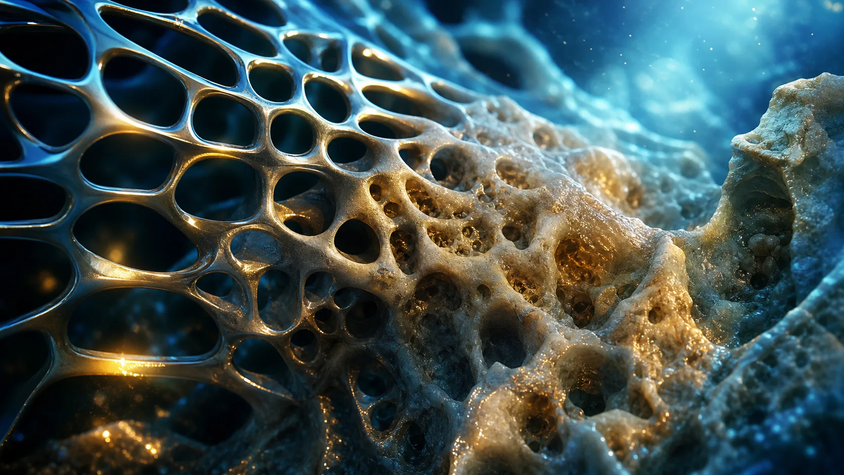
If you want to understand the future of implant durability, you have to look smaller—much smaller. Macro-level roughness, achieved through plasma spraying or bead sintering, has been the standard for achieving implant fixation. However, true biological integration happens at the cellular and protein level, which means surface engineering is increasingly shifting to the nanoscale.
By manipulating the surface of an implant at the nanometre level, we can directly influence how proteins adsorb to the metal, which in turn dictates how osteoblasts and fibroblasts attach and proliferate. Nanotubular titanium surfaces, for instance, can be engineered to promote osteoblast adhesion while simultaneously creating a hostile environment for certain strains of bacteria.
Practical Applications in Surface Engineering
When considering the practical applications of these technologies, it helps to break them down into what they actively achieve for the patient:
- Targeted Therapeutic Delivery: Nanoscale surface pores can be loaded with antibiotics or growth factors. As the bone interfaces with the implant, these substances are slowly released directly into the surgical bed, providing highly concentrated, localized therapy.
- Bacterial Adhesion Reduction: Specific nano-patterns can physically disrupt the ability of bacteria to form a protective biofilm, drastically lowering the risk of device-related infections.
- Accelerated Vasculogenesis: Nano-textured surfaces can be designed to rapidly attract endothelial cells, ensuring that a robust blood supply is established around the implant immediately after surgery.
A critical piece of advice for your surgical practice and exam preparation: understand the difference between osteoconductive and osteoinductive surfaces. Many modern proprietary coatings claim both, but the mechanism of action is vital. The surface does not just provide a scaffold; the nanoscale architecture actively triggers cellular pathways that turn precursor cells into bone-forming factories.
Additive Manufacturing and the Push for True Personalisation
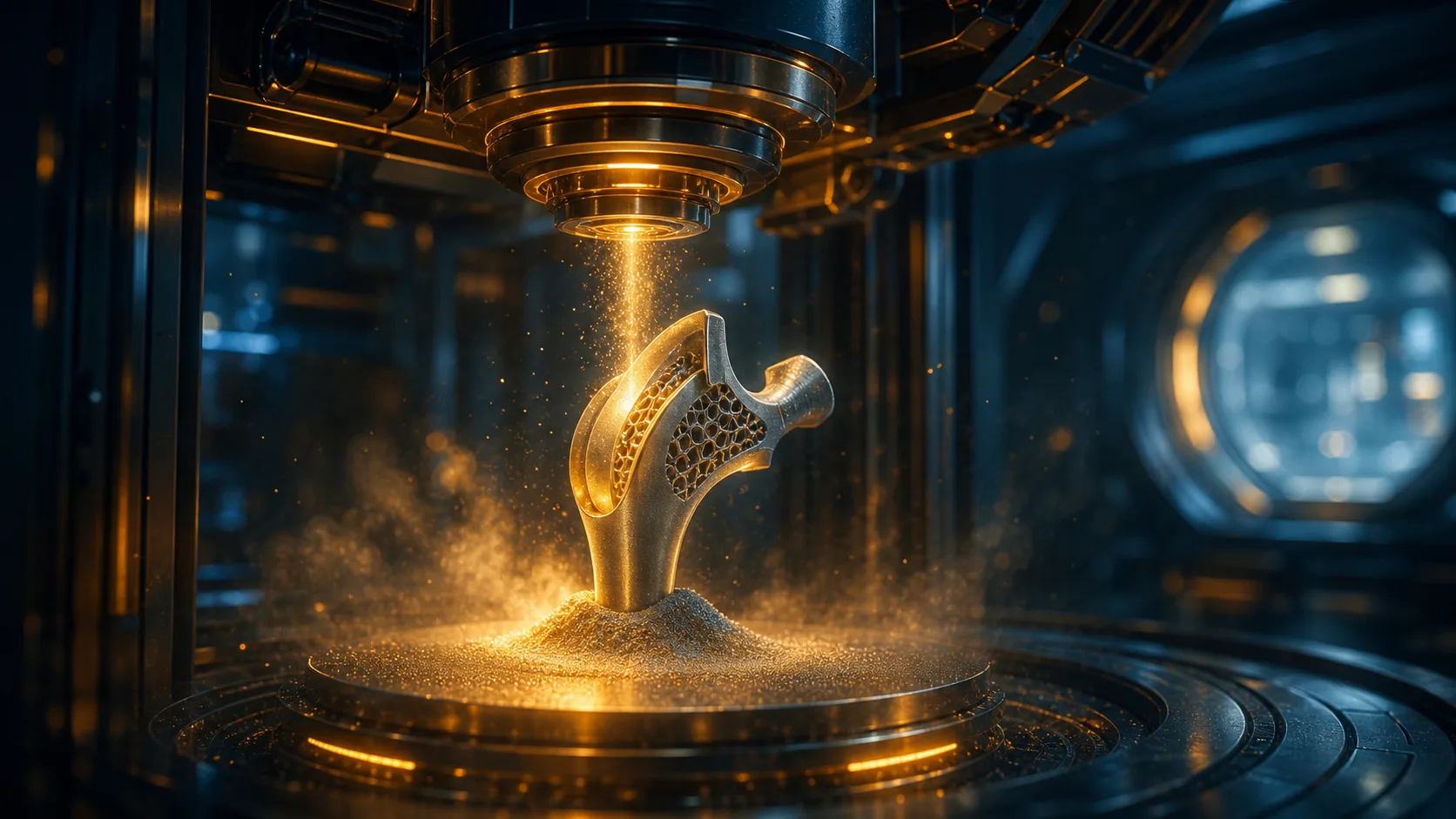
Additive manufacturing, commonly known as 3D printing, has fundamentally disrupted the orthopaedic landscape. For decades, we have been forcing human bones—which come in a vast, asymmetric variety of shapes and sizes—into standardized, off-the-shelf implants. While these mass-produced implants have served us well, the future lies in bespoke, patient-specific hardware.
Additive manufacturing allows for the creation of highly complex geometries that are simply impossible to achieve with traditional subtractive machining. We are talking about patient-specific jigs and cutting guides that perfectly mirror a patient's unique anatomy, reducing intra-operative bone cuts and minimizing soft tissue dissection. Furthermore, the implants themselves can be printed with varying densities of porous scaffolding, mimicking the gradient of human cancellous to cortical bone to reduce the risk of stress shielding.
If you are navigating your early years of surgical training or preparing for vivas, a common mistake is to focus solely on the novelty of printing a custom implant. The robustly true advantage of additive manufacturing lies in the restoration of native biomechanics. By perfectly recreating the patient's native joint geometry and center of rotation, you can theoretically reduce wear and tear on the surrounding healthy cartilage and opposing joint surfaces. This technology is not just about making things fit; it is about preserving and protecting the entire kinetic chain.
Revolutionising Polymers: Solving the Wear Debris Problem
While metals and ceramics handle the structural loads, polyethylene has long been the workhorse for the articulating surfaces in total joint arthroplasty. However, osteolysis induced by polyethylene wear debris has historically been the Achilles' heel of lower limb arthroplasty, leading to aseptic loosening and complex revision surgeries. The future of biomaterials involves a massive overhaul in how we formulate and utilize these crucial polymers.
Highly cross-linked polyethylene (HXLPE) represents a massive leap forward, but the science is not standing still. The current frontier involves the blending of Vitamin E (alpha-tocopherol) into the polymer matrix. Vitamin E acts as a powerful antioxidant, scavenging the free radicals that are generated during the sterilization and irradiation process. By neutralizing these free radicals, the material retains its structural toughness and drastically reduces the delamination and oxidative degradation that leads to the shedding of microscopic wear particles.
When you are revising for board exams or discussing implant choices in an MDT meeting, ensure you are crystal clear on the distinction between standard polyethylene, highly cross-linked variants, and antioxidant-stabilized materials. The clinical takeaway is that modern polymer formulations are not just marginally better; they fundamentally alter the long-term survival curve of the implant. By virtually eliminating the inflammatory cascade triggered by wear debris, we are giving patients a much needed biological cushion against osteolysis.
The Role of Magnesium and Novel Bio-resorbable Materials
The concept of a material that completely disappears once its job is done is one of the most exciting frontiers in orthopaedic biomaterials. Traditional hardware—such as screws and plates—often requires a second surgery for removal, particularly in paediatric populations where continued skeletal growth could be impeded by rigid, permanent metal. This is where bio-resorbable materials step in, and magnesium is currently leading the charge.
Magnesium possesses a unique and highly desirable property: its modulus of elasticity and density are remarkably similar to that of natural human cortical bone. When used for fracture fixation, a magnesium-based implant provides the necessary structural stability during the critical early phases of healing. As the bone gradually reunites and takes on more of the physiological load, the magnesium implant safely and predictably corrodes, gradually transferring the mechanical stress back to the healing bone. This prevents the stress shielding that often occurs with rigid titanium or steel plates, which can leave underlying bone weak and porotic once the hardware is removed.
However, the transition from inert metals to resorbable ones requires a distinct shift in your surgical mindset. The management of these patients differs significantly. For example, the degradation byproducts of magnesium must be safely metabolized by the body. When counseling patients or considering this technology, it is crucial to monitor the degradation rate, as a resorbable implant that loses its mechanical integrity too rapidly will result in hardware failure before the bone has adequately consolidated.
Integrating Biologics and Synthetic Scaffolds
As orthopaedic surgeons, we are increasingly realizing that mechanical stability is only half the battle; true biological healing is the ultimate goal. The future of our specialty lies in the seamless integration of orthobiologics with advanced synthetic scaffolds. We are moving beyond simple bone grafting into the realm of tissue engineering, where materials are designed to act as delivery vehicles for cellular therapies.
Imagine a scenario where a patient suffers a massive bone defect following high-energy trauma or tumour resection. Simply filling the void with cement or allograft is often insufficient for long-term biological incorporation. The next generation of biomaterials involves highly porous synthetic bone grafts impregnated with the patient's own mesenchymal stem cells or recombinant growth factors. These scaffolds provide the three-dimensional architecture necessary for vascular in-growth, while simultaneously delivering the precise molecular signals required to induce osteogenesis.
Preparing for Your Exams and Practice in a Shifting Landscape
As the biomaterials landscape evolves, so too must the approach to orthopaedic education and examination preparation. Whether you are a medical student aiming to secure a highly competitive surgical training programme, or a registrar studying for rigorous, internationally recognized board exams, demonstrating a robust understanding of these material sciences will set you apart.
Examiners are no longer satisfied with rote memorization of the periodic table or basic material names. They want to see that you understand the clinical why. If you are asked to discuss a total knee replacement, simply stating that the tibial baseplate is made of titanium is not enough. A high-scoring candidate will explain how the micro-architecture of that titanium promotes osseointegration, how the highly cross-linked polyethylene insert mitigates wear debris, and how the biomechanics of the implant design influences long-term patient mobility.
What This Means for Your Daily Surgical Practice
Adopting new technologies in the operating theatre requires a careful balance of enthusiasm for innovation and a healthy respect for evidence-based practice. While it is tempting to jump straight to the latest smart implant or bio-resorbable scaffold, a common mistake among enthusiastic surgeons is abandoning proven fundamentals for untested novelties.
Your surgical practice should evolve deliberately. Start by integrating technologies that have robust clinical backing, such as antioxidant-infused polyethylene or trabecular metal augmentations for severe bone loss. Use these cases to refine your understanding of how biomaterials interact with host tissue. Pay close attention to how the implant feels during impaction, how the bone bleeds when interacting with a bioactive surface, and how the patient recovers post-operatively. The future of orthopaedics is incredibly bright, but it requires surgeons who are as adept at understanding the microscopic behavior of biomaterials as they are at perfecting their surgical cuts.
Ultimately, the future of orthopaedic implants is not just about building a better piece of metal; it is about engineering a symbiotic relationship between man-made materials and human biology, empowering you to deliver longer-lasting, more dynamic recoveries for your patients.
Share this article
Useful for a journal club, study list, or teaching session.