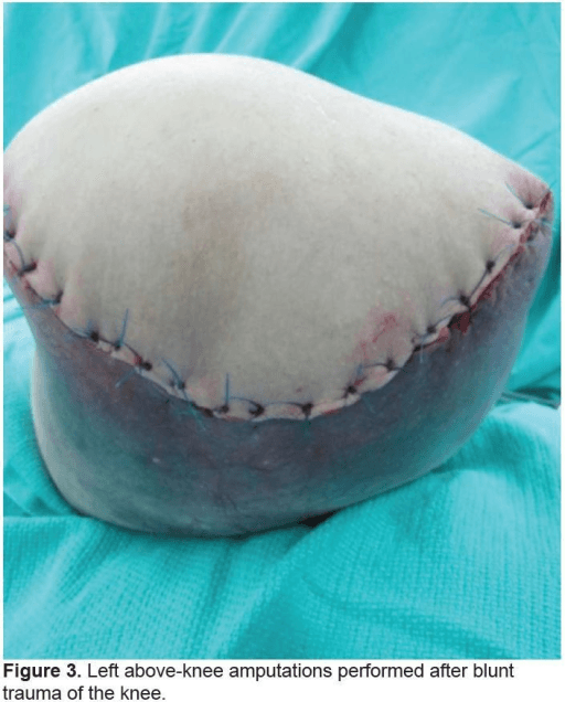
Equal anterior-posterior fish-mouth flaps with adductor magnus myodesis
- Level selection is the critical decision - preserve a functional below-knee level whenever the limb permits, because transfemoral amputation roughly doubles the metabolic cost of walking and markedly reduces independent prosthetic ambulation rates.
- Adductor magnus myodesis (suturing the adductor mass through drill holes to the lateral femur) maintains femoral adduction and prevents the abduction-flexion contracture that cripples prosthetic fitting.
- Ideal bone length is approximately 12 cm above the knee joint line to leave room for a prosthetic knee unit, with the anterior femur bevelled to avoid a sharp distal prominence.
- The major vessels (superficial femoral artery and vein) must be doubly ligated, and the sciatic nerve handled by gentle traction neurectomy with ligation of its accompanying vessel to reduce neuroma and bleeding.
When & Why
Indication. A transfemoral amputation is performed when the limb cannot be saved and a healthy, functional transtibial (below-knee) level is not achievable. The common scenarios are a dysvascular or diabetic limb with critical ischaemia or extensive tissue loss not amenable to a below-knee amputation, severe non-reconstructable trauma or failed limb salvage where the knee cannot be preserved, a life-threatening infection (necrotising soft-tissue infection or wet gangrene) extending proximal to a viable transtibial level, a tumour requiring proximal margins where limb-sparing surgery is not feasible, and a non-functional limb (fixed deformity, paralysis, intractable pain) where a transfemoral residual limb is more useful than the diseased limb. Relative indications include a failed transtibial amputation needing revision to a higher level, a severe knee flexion contracture that makes a transtibial prosthesis unworkable, and a multiply revised, chronically infected transtibial stump.
Critical ischaemia or extensive tissue loss not amenable to a healing below-knee amputation — the largest single group.
Non-reconstructable limb, failed limb salvage where the knee cannot be preserved, or a tumour needing proximal margins not amenable to limb-sparing surgery.
A necrotising soft-tissue infection or wet gangrene proximal to a viable transtibial level, or a fixed, painful, paralysed limb better replaced by a residual limb.
Preserve the knee whenever the tissue allows it. The single most important strategic decision is how proximal to amputate. Each level preserved below the knee dramatically improves function, so a transfemoral amputation is chosen only when a healed, functional transtibial stump is not achievable. Caution applies when a viable transtibial level is still possible, when the patient is a candidate for successful revascularisation (vascular assessment first), or when proximal disease will not heal at the transfemoral level and hip disarticulation may be needed. The level-selection trade-off. Losing the knee roughly doubles the metabolic cost of walking and slows comfortable walking speed. Waters' gait-laboratory work showed a stepwise rise in energy cost from transtibial to transfemoral amputation, with the dysvascular transfemoral amputee carrying the highest oxygen cost per metre and the slowest comfortable speed of all levels. In keeping with this, a substantial proportion of older dysvascular transfemoral amputees never achieve independent prosthetic ambulation, in contrast to the higher community-ambulation rates after transtibial amputation. The higher level heals more reliably in poorly perfused tissue — which is precisely why it is chosen when a below-knee amputation would fail — but that gain in healing is paid for in lost walking economy and independence. Predictors of successful prosthetic mobility include younger age, better cognition, absence of contralateral limb disease, and good cardiovascular reserve, and validated tools such as the AMPREDICT model help quantify the probability of prosthetic mobility to frame realistic counselling. Consent specifically for the realistic likelihood of household rather than community ambulation (or a wheelchair-based lifestyle), the high perioperative mortality that reflects the comorbid cardiac and diabetic population rather than the operation itself, stump and phantom limb pain, and the risk to the contralateral limb, which is often equally diseased. Setup. Supine with a sandbag or bump under the ipsilateral buttock to give access to the posterior thigh. Confirm the side and level against imaging and the vascular assessment, and mark the proposed bone level and flaps before draping. Prepare and drape for circumferential access to the thigh. A thigh tourniquet may be used in trauma or tumour but is generally avoided in dysvascular cases so that tissue bleeding and viability can be judged. Anaesthesia is general or regional (spinal or epidural), and pre-emptive multimodal analgesia (regional block, gabapentinoids) is started to reduce acute and persistent phantom limb pain.
The Operation
The goal: design equal anterior-posterior (fish-mouth) flaps, divide the thigh at the planned bone level while protecting the major vessels and the sciatic nerve, transect and bevel the femur, and restore femoral adduction with an adductor magnus myodesis before a tension-free, well-padded closure. The exposure is built into the flap design and the layered muscle division below — there is no separate incision, the flaps ARE the approach.
Operative sequence
- Supine with a bump under the ipsilateral hip to allow access to the posterior thigh; circumferential preparation of the limb.
- Confirm the side and level against the imaging and the vascular assessment before draping.
- Mark the proposed bone level (about 12 cm above the knee joint line) and the flap design on the skin.
- Plan the bone section approximately 12 cm above the knee joint line, or as dictated by tissue viability or tumour margin — about 25 to 30 cm from the greater trochanter in an average adult — leaving room for a prosthetic knee unit while preserving as long a lever arm as healing allows.
- Design equal anterior and posterior (fish-mouth) flaps whose combined length matches the thigh diameter at the level of bone section, so closure is tension-free.
- In trauma, viability rather than geometry dictates the flaps — amputate at the lowest viable level with whatever flaps the soft tissue allows.
- Incise skin and fat along the marked fish-mouth, raising the flaps as fasciocutaneous-myocutaneous units, not thin skin flaps, to preserve perfusion.
- Handle the dysvascular skin with hooks rather than crushing forceps.
- Divide the quadriceps anteriorly, bevelling the muscle distal-to-proximal so it can be folded over the bone end later.
- Identify the superficial femoral artery and vein in the adductor (Hunter's, subsartorial) canal on the medial side, where they run between the quadriceps and the adductors toward the popliteal fossa.
- Dissect the artery and vein free and doubly ligate each separately with non-absorbable suture — a transfixion ligature plus a proximal simple tie for the artery.
- Control the profunda femoris perforators and muscular branches with ties or cautery as the muscles are divided.
- Divide the hamstrings to expose the sciatic nerve deep in the posterior compartment, posterior to the adductor magnus and the femur.
- First ligate its accompanying vessel (the arteria comitans nervi ischiadici), then apply gentle distal traction and divide the nerve sharply with a fresh blade so the cut end retracts proximally into muscle, away from the bone end and the scar.
- Avoid crushing or excessive traction — both cause a painful neuroma.
- Strip a short cuff of periosteum, then transect the femur with an oscillating saw at the planned level.
- Bevel the anterior cortex and rasp all edges smooth to remove the sharp distal prominence that would tent the skin under the socket.
- Irrigate away bone debris.
- Drill holes in the lateral cortex of the distal femur.
- Bring the adductor magnus across the bone end under physiological tension with the hip held in neutral or adduction, and suture it through the drill holes.
- This myodesis restores the adduction moment and prevents the abduction-flexion contracture that follows when the adductors are left detached and the abductors and flexors pull unopposed.
- If bone fixation is unsafe (very osteoporotic or infected bone), a myoplasty suturing the adductors to the posterior and quadriceps fascia is the alternative.
- Bring the bevelled quadriceps over the distal femur and secure it to the posterior musculature and fascia, giving a stable, padded myofascial cover with the femur held in adduction.
- Avoid bulky, redundant muscle that would compromise socket fit.
- Release any tourniquet and obtain meticulous haemostasis — a haematoma is a major cause of wound breakdown in dysvascular tissue.
- Confirm the cut muscle and skin edges bleed (viability) in non-tourniquet cases.
- Place a closed suction drain deep to the muscle layer, brought out away from the suture line (remove at 24 to 48 hours depending on output).
- Close the deep fascia and muscle envelope, then subcutaneous tissue and skin without tension, so the scar lies away from the weight-bearing distal end.
- Apply a rigid plaster dressing (which shapes the stump, controls oedema, protects from trauma and may shorten time to prosthetic casting) or a soft compressive dressing with stump bandaging, depending on local practice and the ability to monitor the wound.
- Position the hip in neutral extension and adduction, and avoid pillows under the stump.
Before dividing the femur, identify and doubly ligate the superficial femoral artery and vein separately in the adductor canal — inadequate ligation causes reactionary haemorrhage and a stump haematoma that compromises healing in dysvascular tissue. Handle the sciatic nerve by first ligating its accompanying arteria comitans, then applying gentle distal traction and dividing it sharply so it retracts into muscle away from the bone end; crushing or crude traction causes a painful neuroma.
When you drill the lateral femoral cortex for the adductor magnus myodesis, hold the hip in adduction and tension the adductor across the bone end before securing it through the drill holes. A correctly tensioned myodesis holds the femur in adduction and neutralises the unopposed abductor and flexor pull — the difference between a prosthetically fittable stump and one that drifts into abduction.
Detaching the adductors and hamstrings leaves the gluteus medius and minimus and the iliopsoas relatively unopposed, driving the residual femur into abduction and flexion. Gottschalk's biomechanical work showed that failing to reattach the adductor magnus shortens the adductor lever arm and lets the femur drift. The surgical answer is adductor magnus myodesis; the nursing and therapy answer is a neutral hip, no pillow under the stump, and daily prone lying.
Aftercare & Complications
Rehabilitation | Phase | Timing | Focus | Positioning and therapy | |-------|--------|-------|--------------------------| | Immediate | 0 to 2 weeks | Wound protection, oedema control | Rigid or compressive dressing; hip in neutral extension and adduction; no pillow under the stump; drain out at 24 to 48 hours; sutures or staples out at about 2 to 3 weeks | | Intermediate | 2 to 6 weeks | Contracture prevention, strengthening | Daily prone lying; strengthen hip extensors and the remaining adductors; stump shrinker or compression once the wound has healed | | Rehabilitation | 6 weeks onward | Prosthetic fitting | Cast the socket once the stump has healed and matured (volume stabilised); ischial-containment or sub-ischial socket, a prosthetic knee unit and a foot or ankle matched to mobility goals | | Long-term | Months onward | Maintenance | Serial stump-volume and skin review, socket fit and gait; protect the contralateral limb; ongoing review for phantom or neuroma pain | Set realistic expectations from the outset. Many older dysvascular transfemoral amputees achieve household rather than community ambulation, and a frail patient may reasonably choose a wheelchair-based lifestyle; the energy cost of walking is substantially higher than with a transtibial prosthesis. Mobility, walking distance, prosthetic use and quality-of-life tools frame outcome assessment, and the contralateral limb — often equally at vascular and diabetic risk and now bearing extra load — needs active surveillance and protection (foot care, glycaemic and vascular control) to avoid bilateral amputation. Review for warning signs: a non-healing or breaking-down wound, increasing pain or discharge, a developing fixed hip posture, and new rest pain or tissue change in the contralateral limb. Complications
- Recognition
- Marginal flap dusky or black, dehiscence with serous or purulent discharge, non-healing skin edge over the bone end
- Prevention
- Choose a level with adequate perfusion, tension-free fasciocutaneous flaps, atraumatic handling, meticulous haemostasis, rigid or compressive dressing
- Management
- Local wound care for minor necrosis, debride non-viable tissue, re-assess perfusion, revision to a higher level if extensive
- Recognition
- Tense swollen stump, expanding ecchymosis, soakage of dressing, dropping haemoglobin, increasing early post-op pain
- Prevention
- Doubly ligate the femoral artery and vein separately, ligate the sciatic accompanying vessel, control profunda perforators, suction drain
- Management
- Small: observe with compression; large or expanding: return to theatre for evacuation and haemostasis; resuscitate and correct coagulopathy
- Recognition
- Erythema, warmth, discharge, systemic sepsis, wound dehiscence with pus, raised inflammatory markers
- Prevention
- Prophylactic antibiotics, control source infection pre-op, debride necrotic tissue at the correct level, glycaemic optimisation, drain dead space
- Management
- Antibiotics guided by culture, drainage and debridement of collections, open management or delayed closure if heavily contaminated
- Recognition
- Femur held abducted and flexed at rest, limited passive hip extension and adduction, difficulty aligning the prosthesis, poor gait
- Prevention
- Adductor magnus myodesis to restore adduction, neutral hip positioning, no pillow under the stump, daily prone lying, early physiotherapy
- Management
- Aggressive physiotherapy and stretching for early contracture, prone-lying programme, surgical release for established fixed contracture
- Recognition
- Focal exquisite point tenderness with a positive Tinel sign, pain reproduced by socket pressure, sharp or electric pain in the nerve distribution
- Prevention
- Sharp division of the nerve under gentle traction high in the wound, allow retraction into muscle away from scar and bone, ligate accompanying vessel, avoid crushing
- Management
- Desensitisation, socket modification, injection, neuropathic agents; refractory: neuroma excision with proximal relocation into muscle
- Recognition
- Cramping, burning or shooting pain in the missing limb, phantom sensation distinct from stump pain, onset early and may persist long-term
- Prevention
- Perioperative multimodal and regional analgesia, good acute pain control to reduce central sensitisation, early prosthetic use and mirror therapy
- Management
- Multimodal analgesia (gabapentinoids, TCAs, opioids if needed), mirror therapy, desensitisation, TENS, pain-team referral for refractory cases
Viva & Exam Focus
FLAPSFLAPS — key technical goals
STUMPSTUMP — contracture and stump care
Clinical Decision Scenarios
Practise clinical reasoning and management decisions out loud
“A 68-year-old man with type 2 diabetes and peripheral vascular disease has wet gangrene of the forefoot extending into the leg. The vascular team report unreconstructable distal disease and a non-viable posterior calf. You are deciding between a below-knee and an above-knee amputation. How do you choose the level, and what do you tell the patient about the consequences of an above-knee amputation?”
“Describe the key technical steps of a transfemoral amputation. In particular, how do you manage the femur, the major vessels, and the sciatic nerve, and what is a myodesis?”
“A patient is six weeks post transfemoral amputation. The stump has healed but the physiotherapist reports the hip is held flexed and abducted and prosthetic casting is being delayed. Separately, the patient describes severe burning pain in the foot that is no longer there. How do you explain and manage these two problems?”
Indications
- Dysvascular or diabetic limb with critical ischaemia or tissue loss not amenable to a healing below-knee amputation
- Severe non-reconstructable trauma or failed limb salvage where the knee cannot be preserved
- Life-threatening infection, necrotising soft-tissue infection or gangrene proximal to a viable transtibial level
- Tumour needing proximal margins not amenable to limb salvage, or a non-functional (painful, paralysed or deformed) limb
Level selection — energy and mobility trade-off
- Save the knee whenever tissue allows — transtibial amputees have far better walking economy and ambulation rates
- Transfemoral amputation roughly doubles the metabolic (oxygen) cost of walking and slows comfortable speed (Waters)
- Dysvascular transfemoral amputees have the highest energy cost and lowest independent prosthetic ambulation rates
- The higher level heals more reliably in poor perfusion — the gain in healing is paid for in lost function
- Use predictors and tools (e.g. AMPREDICT) and comorbidity to counsel on realistic prosthetic mobility
Critical anatomy
- Femur: section about 12 cm above the knee joint line to fit a prosthetic knee unit, bevel the anterior cortex
- Superficial femoral artery and vein: in the adductor (Hunter's) canal medially — the vessels to doubly ligate
- Sciatic nerve: deep in the posterior compartment with the arteria comitans nervi ischiadici (ligate before division)
- Adductor magnus (obturator nerve): the key muscle for myodesis to maintain femoral adduction
- Muscle balance: detaching adductors and hamstrings leaves abductors and flexors unopposed — drives the abduction-flexion contracture
Critical operative steps
- Equal anterior-posterior or fish-mouth flaps, combined length about the thigh diameter for tension-free closure
- Raise fasciocutaneous-myocutaneous flaps; handle dysvascular skin atraumatically with hooks
- Identify and doubly ligate the femoral artery and vein separately in the adductor canal
- Sciatic nerve: ligate the accompanying vessel, gentle traction, sharp high division to allow proximal retraction
- Transect the femur about 12 cm above the knee, bevel the anterior cortex, rasp the edges smooth
- Adductor magnus myodesis through lateral femoral drill holes with the hip in adduction
- Cover with bevelled quadriceps, meticulous haemostasis, suction drain, tension-free closure, rigid or compressive dressing
Myodesis vs myoplasty
- Myodesis = muscle sutured directly to bone through drill holes — stable, dynamic stump, maintains adduction (preferred)
- Adductor magnus myodesis is the key step to prevent the abduction-flexion contracture (Gottschalk)
- Myoplasty = opposing muscle groups sutured to each other over the bone end — simpler but less securely anchored
- Choose myoplasty when bone fixation is unsafe (very osteoporotic or infected bone)
Danger zones
- Femoral vessels — inadequate ligation causes reactionary haemorrhage and stump haematoma
- Sciatic nerve — crude handling causes a painful neuroma; ligate its accompanying vessel to avoid bleeding
- Dysvascular soft-tissue envelope — tension or haematoma causes flap necrosis and conversion to a higher level
- Hip muscle imbalance — missed adductor myodesis or poor positioning causes the abduction-flexion contracture
- Cut nerve endings — source of phantom pain and neuroma; pre-emptive analgesia and good technique reduce the risk
Major complications — prevention and management
- Wound breakdown or flap necrosis: correct level, tension-free flaps, haemostasis; debride or revise if extensive
- Haematoma or haemorrhage: double vessel ligation and a drain; return to theatre if expanding
- Abduction-flexion contracture: adductor myodesis, prone lying, no pillow under the stump; surgical release if fixed
- Neuroma: sharp high nerve division into muscle; excise and relocate if refractory
- Phantom limb pain: perioperative multimodal or regional analgesia; gabapentinoids, mirror therapy, TENS
Aftercare and rehab
- Positioning: hip in neutral extension and adduction, no pillow under the stump, daily prone lying
- Dressing: rigid plaster (shapes the stump, controls oedema, protects) or soft compressive bandaging
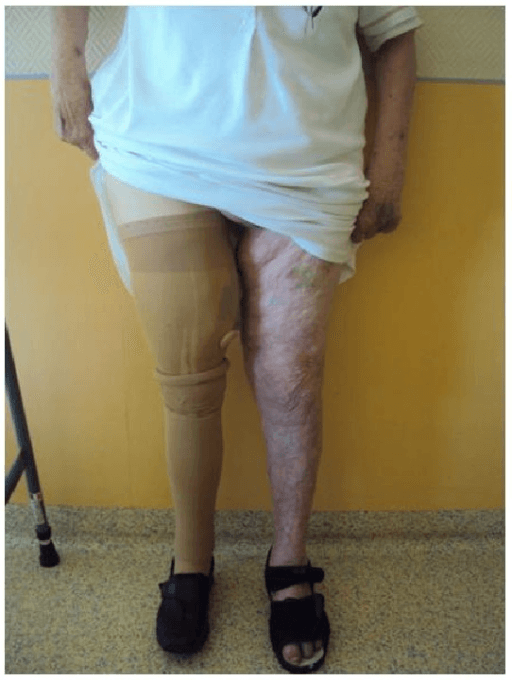
- Prosthetics: cast once the stump has matured; the short femur leaves room for a prosthetic knee unit
- Expectations: many older dysvascular transfemoral amputees become household rather than community ambulators
- Protect the contralateral limb — high risk in diabetic and vascular disease; foot care and risk-factor control
Background & Evidence
Epidemiology. Dysvascular disease (peripheral arterial disease and diabetes) is the dominant cause of major lower-limb amputation, so the amputee population is older and heavily comorbid with cardiac and diabetic disease. This comorbidity — not the operation itself — drives the high perioperative mortality after major dysvascular lower-limb amputation, and it must frame every pre-operative conversation. Cross-sectional anatomy of the thigh. - Anterior compartment (quadriceps) — rectus femoris and the three vasti, femoral nerve — provides the anterior myofascial flap folded over the bone end.
- Medial compartment (adductors) — adductor longus, brevis, magnus and gracilis, obturator nerve. Adductor magnus is the key muscle for myodesis to maintain femoral adduction.
- Posterior compartment (hamstrings) — biceps femoris, semitendinosus and semimembranosus, sciatic (tibial division) nerve — contributes to the posterior flap and hip extension.
- Femur — the shaft is transected at about 12 cm above the knee joint line; a longer femur improves the lever arm and prosthetic control, so length is preserved as much as healing permits while leaving room for the knee unit, and the anterior cortex is bevelled.
- Neurovascular — the superficial femoral artery and vein run in the adductor (Hunter's, subsartorial) canal on the medial side toward the popliteal fossa and are the principal vessels to ligate; the profunda femoris gives muscular perforators that must be controlled; the sciatic nerve lies deep in the posterior compartment with its arteria comitans; and cutaneous nerves (posterior femoral cutaneous, saphenous branches) cross the flaps and contribute to neuroma or scar sensitivity if handled roughly.
Muscle sutured directly to bone through drill holes — a stable, dynamic stump that maintains muscle tension and femoral adduction. The evidence-based default for the adductor magnus.
Opposing muscle groups sutured to each other over the bone end — technically simpler, but the envelope is less securely anchored and may retract, giving weaker adduction control.
Reserved for when bone fixation is unsafe — very osteoporotic or infected bone — where a drill-hole myodesis would not hold.
Hip muscle balance — why myodesis matters. Detaching the adductors and hamstrings leaves the abductors (gluteus medius and minimus) and flexors (iliopsoas, rectus femoris) relatively unopposed. Their pull drives the residual femur into abduction and flexion — the basis of the dreaded abduction-flexion contracture. Gottschalk's biomechanical work showed that losing the distal attachment of the adductor magnus strips away most of its adduction moment arm; re-anchoring it with a myodesis is the surgical antidote, restoring the adduction moment and a more neutral femoral position for prosthetic alignment. Energy expenditure and prosthetic mobility. Walking after amputation costs more oxygen and is slower at each more proximal level. Waters' gait-laboratory comparison established the stepwise rise in energy cost from transtibial to transfemoral amputation, with the dysvascular transfemoral amputee carrying the highest oxygen cost per metre and the slowest comfortable walking speed, and vascular amputees performing worse than traumatic amputees at every level. A substantial proportion of dysvascular transfemoral amputees never achieve independent prosthetic ambulation. Predictors of successful mobility include younger age, better cognition, the absence of contralateral limb disease and good cardiovascular reserve, and validated multivariable models such as AMPREDICT quantify the probability of prosthetic mobility to guide prescription and counselling. Healing and outcomes. Transfemoral amputation has a higher primary healing rate than transtibial amputation in poorly perfused limbs — precisely why it is chosen when a below-knee amputation would fail — but this gain in healing is offset by a markedly inferior functional outcome. Mortality after major dysvascular lower-limb amputation is high, reflecting the comorbid cardiac and diabetic population rather than the operation itself, and the contralateral limb is frequently also at risk.
References
Energy cost of walking of amputees: the influence of level of amputation
- Gait-laboratory comparison of 70 unilateral traumatic and vascular amputees against 40 normal controls
- Walking performance was significantly better the lower the amputation level in both traumatic and vascular groups
- Transfemoral amputees walked slower and at substantially higher oxygen cost per metre than transtibial or Syme amputees
- Vascular amputees performed worse than traumatic amputees at every level
Transfemoral amputation. Biomechanics and surgery
- Defines the muscle-stabilised transfemoral technique built on biomechanical principles
- Preservation and myodesis of the adductor magnus to the residual femur keeps the femoral shaft axis close to normal
- Patients are easier to fit with a prosthesis and more likely to remain ambulatory
- Associated with fewer stump problems and improved stump strength
The biomechanics of trans-femoral amputation
- Cadaveric study quantifying the moment arms of the three adductor muscles in the thigh
- Adductor magnus has the dominant mechanical advantage for holding the thigh in its normal axis
- Loss of the distal third of adductor magnus attachment produces a 70% loss of its effective moment arm
- This loss of the adductor lever explains the abducted residual femur seen after standard transfemoral amputation
Multidisciplinary preoperative assessment and late function in dysvascular amputees
Demonstrates the value of preoperative assessment and the functional limitations of dysvascular amputees.
Controversies in lower-extremity amputation
Instructional review of level selection, technique, and rehabilitation controversies in lower-limb amputation.
AMPREDICT PROsthetics - Predicting Prosthesis Mobility to Aid in Prosthetic Prescription and Rehabilitation Planning
- Multivariable prediction model developed and validated in 357 veterans after incident dysvascular transtibial or transfemoral amputation
- Predicts 12-month mobility category (wheelchair, household, basic community, advanced community) at the time of prosthetic prescription
- Final model uses 23 routinely available clinical predictors and discriminates community from household ambulation
- Model performance was weaker at separating wheelchair from household mobility
Medium-term outcomes following limb salvage for severe open tibia fracture are similar to trans-tibial amputation
Comparative trauma outcome data informing the salvage-versus-amputation decision.
Removable Rigid Dressings for Postoperative Management of Transtibial Amputations: A Review of Published Evidence
Review supporting rigid dressings for oedema control, stump shaping, and faster prosthetic readiness (principles applicable to transfemoral care).
Limb amputation and limb deficiency: epidemiology and recent trends in the United States
Population-level epidemiology confirming dysvascular disease as the dominant cause of major lower-limb amputation.
Phantom limb pain
- Authoritative review of the epidemiology, mechanisms and management of phantom limb pain
- Phantom limb pain is common after limb amputation and may persist long-term
- Peripheral and central sensitisation underpin the pain, distinct from residual-limb (stump) pain
- Identifies perioperative pain control and multimodal or neuropathic strategies as management cornerstones