Forequarter, hindquarter and rotationplasty for musculoskeletal malignancy when limb salvage is not possible | advanced
- Limb salvage is now the standard of care for extremity sarcoma - amputation is required in only about 5 to 10 percent of cases, reserved for tumours where a wide margin cannot otherwise be achieved with an acceptable functional limb.
- The amputation level is dictated by ONCOLOGICAL clearance - tumour extent, compartment involvement and skip lesions on whole-bone MRI - NOT by classic prosthetic or anatomical levels. The bone marrow margin must be confirmed (frozen section or imaging) to be tumour-free.
- Amputation does NOT improve survival over limb salvage with adequate margins - it is a local control and function decision, not a cure-rate decision. State this early in any viva.
- Forequarter amputation (Berger) removes the entire upper limb with the scapula and clavicle; hindquarter amputation (external hemipelvectomy) removes the entire lower limb with the hemipelvis. Internal hemipelvectomy is the limb-SPARING pelvic resection equivalent.
- Van Nes rotationplasty converts a distal femoral resection into a functional below-knee-equivalent limb by rotating the distal segment 180 degrees so the ankle acts as a knee - durable, active and ideal for the growing child.
When & Why
The shift to limb salvage. Historically amputation was the primary treatment for extremity sarcoma. Following the landmark work of Rosen and the introduction of neoadjuvant chemotherapy, plus the limb-salvage comparison studies of the 1980s, limb salvage has become the standard of care. - Limb salvage is now performed in roughly 85 to 90 percent of extremity sarcomas
- Amputation is required in only about 5 to 10 percent of cases
- Survival is equivalent between amputation and limb salvage when a wide margin is achievable - amputation does NOT improve cure rates
- Local recurrence rates are slightly higher with limb salvage than amputation, but with no survival penalty when recurrence is detected and managed early
"Amputation for tumour is a decision about local control and function, not about survival. I would never tell a patient that an amputation gives them a better chance of cure than a properly executed limb salvage with a wide margin."
Indications for amputation. The decision is reserved for the tumour, the limb or the patient in whom a wide margin and a useful limb cannot otherwise be achieved: - Major neurovascular encasement - tumour circumferentially involving the main artery, vein and nerve such that a wide margin cannot be achieved without sacrificing limb viability or function
- Extensive joint or multi-compartment involvement - disease crossing fascial planes into several compartments or into a joint
- Large recurrent tumour - particularly after previous surgery and radiotherapy where re-excision margins are inadequate
- Fungating or infected tumour - for local control, hygiene and palliation of symptoms
- Pathological fracture - when the fracture haematoma contaminates compartments beyond the resectable field
- Failed limb salvage - chronic deep infection of a megaprosthesis, failed reconstruction, or a non-functional salvaged limb
- Distal tumours where salvage is non-functional - reconstruction would leave a limb worse than a well-fitted prosthesis
- Informed patient preference - some patients choose a single definitive procedure over prolonged, multi-operation salvage
- Palliation - in advanced disease for a fungating, painful or haemorrhaging tumour, even in the presence of metastases, to improve quality of life Oncological principles of the amputation. Every amputation for tumour is planned and executed to oncological rules, not prosthetic convenience: - Plan from staging imaging - whole-bone MRI defines the proximal tumour edge and detects skip lesions (discontinuous intramedullary deposits). A bone cut must be made a safe distance proximal to all disease
- Wide margin - a continuous cuff of normal tissue around the tumour in every plane, including the bone marrow margin
- Marrow margin confirmation - frozen section of the divided bone end confirms tumour-free marrow before closure
- Excise the biopsy tract - the tract is contaminated and must be removed en bloc within the specimen
- Plan flaps from uninvolved tissue - use atypical or non-standard flaps if needed to keep the tumour margin wide. Oncology trumps the textbook flap
- MDT and neoadjuvant therapy - decisions are made within the sarcoma MDT; chemotherapy and radiotherapy status influence timing, flap choice and wound healing The decision in one table. Whether to amputate turns on whether a wide margin and a useful limb can be achieved another way:
- Favours Limb Salvage
- Free or only displaced - wide margin achievable
- Favours Amputation
- Encased - cannot clear without losing limb viability
- Favours Limb Salvage
- Single compartment, clear planes
- Favours Amputation
- Multi-compartment or joint contamination
- Favours Limb Salvage
- Undisplaced, contained
- Favours Amputation
- Displaced, haematoma contaminating compartments
- Favours Limb Salvage
- First presentation, clear margins possible
- Favours Amputation
- Large recurrent disease, prior radiotherapy
- Favours Limb Salvage
- Reconstruction gives useful limb
- Favours Amputation
- Salvaged limb would be non-functional or chronically infected
- Favours Limb Salvage
- Equivalent to amputation
- Favours Amputation
- Equivalent - no survival advantage
Consent and preparation. Counsel specifically on the amputation level, phantom pain, the prosthetic plan and psychological support. For hindquarter and forequarter amputations carry major haemorrhage risk, so cross-match generously, plan for cell salvage and large-bore access, and involve vascular and general or urology surgery as needed for the proximal dissection. Re-mark the biopsy tract for en bloc excision and plan flaps from uninvolved tissue.
The Operation
The goal is to remove the tumour-bearing segment with a continuous wide margin while achieving proximal vascular control before division, preserving a tumour-free marrow margin, excising the biopsy tract en bloc, and closing with viable soft tissue fashioned from uninvolved, non-irradiated tissue. The exposure and vascular control are the heart of each operation and are laid out step by step below.


Operative sequence
- Confirm the sarcoma MDT decision, neoadjuvant chemotherapy/radiotherapy status and staging - whole-bone MRI, chest CT, bone scan or PET.
- Counsel specifically on level, phantom pain, the prosthetic plan and psychological support.
- Hindquarter and forequarter cases carry major haemorrhage risk: cross-match generously, plan cell salvage and large-bore access, and involve vascular and general or urology surgery.
- Re-mark the biopsy tract for en bloc excision and plan flaps from uninvolved tissue.
- Define the proximal tumour edge and exclude skip lesions on whole-bone MRI before choosing the bone cut.
- Set the bone cut a defined safe distance proximal to all disease, with a wide margin in every plane.
- Confirm a tumour-free marrow margin by frozen section of the divided bone end.
- Excise the biopsy tract en bloc within the specimen.
- Fashion flaps from uninvolved, non-irradiated tissue - oncological clearance takes priority over the textbook flap; standard flaps may pass through tumour-bearing tissue.
- Lateral or semi-lateral position.
- The incision encircles the shoulder girdle - an anterior limb over the clavicle and a posterior limb around the scapula - planned to excise the biopsy tract and keep the tumour widely covered.
- Anterior (Berger) approach begins with division of the clavicle and control of the subclavian/axillary vessels; posterior (Littlewood) approach begins posteriorly, freeing the scapula before tackling the vessels - useful when anterior tumour bulk obstructs anterior access.
- Expose and divide the clavicle in its medial third.
- Identify, doubly ligate with transfixion sutures, and divide the subclavian/axillary artery and vein before they retract.
- Ligate the vessels FIRST to control blood loss.
- Divide the brachial plexus roots or trunks under traction and allow them to retract (reduces neuroma symptoms).
- Divide the muscles attaching the shoulder girdle to the chest wall and spine - pectoralis major and minor, trapezius, rhomboids, latissimus dorsi, serratus anterior and levator scapulae.
- Remove the entire upper limb with the scapula and clavicle en bloc.
- Confirm haemostasis.
- Close using a chest-wall (posterior) flap or a fillet flap over a drain, shaping a soft shoulder contour for prosthetic and cosmetic fitting.
- Lateral decubitus with the affected side up and the limb draped free.
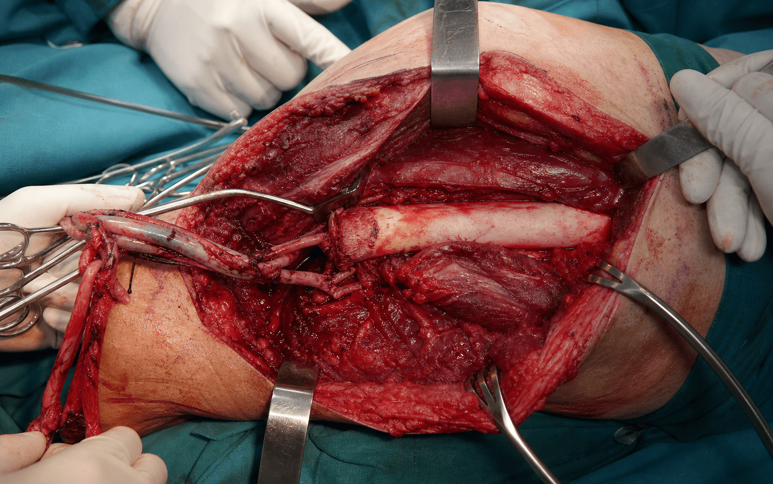
- The classic incision runs anteriorly along the inguinal ligament and posteriorly around the buttock, designed around the tumour and biopsy tract.
- Raise a posterior gluteal myocutaneous flap for closure wherever the posterior compartment is uninvolved.
- Through the anterior approach, identify and protect the ureter.
- Gain proximal control of the common iliac artery and vein (or the external iliac, depending on level).
- Ligate and divide the vessels with transfixion sutures, and divide the femoral nerve.
- Divide the pubic symphysis anteriorly.
- Disarticulate or osteotomise through the sacroiliac joint posteriorly, extending into the sacrum if disease demands (extended hindquarter).
- Divide the remaining pelvic floor and gluteal muscles.
- Remove the limb and hemipelvis en bloc and achieve meticulous haemostasis.
- Close with the posterior gluteal flap (preferred, robust blood supply) or an anterior thigh fillet flap if the posterior compartment is involved.
- Drain widely and anticipate a large dead space and seroma.
- Perform a wide resection of the distal femur (the tumour segment) together with the knee.
- Preserve the sciatic/tibial and peroneal nerves and the popliteal vessels in continuity.
- Rotate the distal limb 180 degrees and reattach it.
- The vessels are preserved and must not kink on rotation - the vascular pedicle is the limiting structure (vessels may be divided and re-anastomosed in some techniques).
- Confirm the foot is well perfused and there is no kinking before fixation.
- Achieve bony fixation between the proximal femur and distal tibia with a plate or an intramedullary device, aiming for solid union.
- Set the final rotation so the foot points posteriorly and the ankle's plantarflexion (extension of the new knee) and dorsiflexion (flexion) drive the new "knee".
A divided subclavian vessel can retract into the thorax causing catastrophic, difficult-to-control haemorrhage - so gain proximal control of the subclavian/axillary vessels (supraclavicular approach) and ligate them securely with transfixion sutures BEFORE division. Watch also for pneumothorax during the root-of-neck dissection; inadequate ligation leads to massive blood loss.
The procedure divides the common or internal iliac vessels and massive blood loss is expected - secure proximal control of the common iliac artery and vein with transfixion ligatures before division. Identify and protect the ureter early, and beware the bladder and rectum during the medial pubic and symphyseal dissection.
"After the 180-degree rotation I confirm there is no kinking and that the foot is well perfused before fixation. Preserving the tibial nerve gives a sensate, functional ankle-knee. Bony union must be solid before prosthetic loading."
Aftercare & Complications
Energy expenditure rises with each more proximal level. A central principle is that the energy cost of walking rises - and prosthetic function falls - with each more proximal amputation level. This drives the surgical preference to preserve length and joints where oncologically safe, and underpins the case for rotationplasty in children.
- Relative energy cost of walking
- Modestly increased
- Functional outlook
- Good - most return to independent walking
- Relative energy cost of walking
- Substantially increased
- Functional outlook
- Reduced - knee-unit dependence, higher fatigue
- Relative energy cost of walking
- Greatly increased
- Functional outlook
- Poor - many become wheelchair-dependent
- Relative energy cost of walking
- Approaches below-knee level
- Functional outlook
- Excellent - active, sporting children
Rehabilitation pathway. Recovery is structured around stump maturation, pain control and prosthetic fitting, tailored to the level: - Early - stump oedema control (compression), pain management (phantom and stump pain), wound care, early mobilisation and transfers
- Prosthetic phase - a preparatory prosthesis once the wound and stump volume stabilise, progressing to a definitive socket
- Upper limb (forequarter) - prosthetics are often largely cosmetic; many patients function well as one-handed with a lightweight cosmetic shoulder prosthesis. Functional myoelectric options exist but uptake at this level is limited
- Lower limb proximal (hindquarter) - a complex socket with hip and knee units; sitting balance and skin care over the resection are key challenges
- Rotationplasty - a custom prosthesis harnesses ankle motion as knee motion, and intensive physiotherapy teaches the patient to drive the new knee with ankle plantar and dorsiflexion Osseointegration. A transcutaneous bone-anchored implant is an emerging option for selected amputees, particularly transfemoral, who cannot tolerate a socket. Advantages are direct skeletal load transfer, osseoperception and no socket-related skin problems. In oncology it requires a disease-free status and intact host bone - infection at the skin-implant interface (stoma) is the principal concern, so selection and surveillance are essential. Psychological and survivorship support. Amputation for tumour combines limb loss with a cancer diagnosis, so structured psychological support, peer mentoring (including meeting prior rotationplasty patients and families) and long-term survivorship follow-up are integral, not optional. Complications. Recognise, prevent and manage the major complications of oncological amputation:
- Incidence / setting
- Hindquarter and forequarter - high
- Recognition
- Rapid blood loss on dividing iliac or subclavian vessels; haemodynamic instability
- Prevention and management
- Prevention: proximal vascular control before division, transfixion ligatures, cross-match, cell salvage. Management: rapid transfusion, direct vessel control, vascular surgery support
- Incidence / setting
- Elevated after chemotherapy and radiotherapy
- Recognition
- Wound dehiscence, marginal flap necrosis, delayed healing
- Prevention and management
- Prevention: design flaps from non-irradiated, uninvolved tissue; meticulous haemostasis; avoid tension. Management: debridement, negative-pressure dressing, secondary closure or further flap
- Incidence / setting
- Common with large dead space (hindquarter)
- Recognition
- Fever, collection, purulent drainage, raised inflammatory markers
- Prevention and management
- Prevention: wide drainage, dead-space management, perioperative antibiotics. Management: drainage, antibiotics, washout; control before prosthetic fitting
- Incidence / setting
- Very common (majority of amputees)
- Recognition
- Perceived pain or sensation in the absent limb
- Prevention and management
- Prevention: perioperative/pre-emptive analgesia, regional blocks, traction neurectomy. Management: multimodal analgesia, gabapentinoids, amitriptyline, mirror therapy, pain team
- Incidence / setting
- Common at divided nerve ends
- Recognition
- Focal tender nodule, Tinel sign, localised stump pain on contact
- Prevention and management
- Prevention: divide nerves under traction and allow retraction into soft tissue; consider TMR or RPNI. Management: revision, traction neurectomy, targeted muscle reinnervation
- Incidence / setting
- Higher if margin inadequate
- Recognition
- New mass or nodularity in the stump or scar; imaging change
- Prevention and management
- Prevention: wide margin, marrow frozen section, excise biopsy tract. Management: restaging, MDT, re-excision or higher amputation, oncology referral
- Incidence / setting
- Common, especially proximal/cosmetic amputations
- Recognition
- Depression, body-image distress, poor prosthetic adaptation
- Prevention and management
- Prevention: pre-operative counselling, peer support, psychology input. Management: ongoing psychological support, rehabilitation, peer mentoring
- Incidence / setting
- Proximal levels (forequarter, hindquarter) hardest
- Recognition
- Poor socket fit, inability to bear load, stump volume change
- Prevention and management
- Prevention: surgeon-prosthetist planning of stump shape and length. Management: custom sockets, osseointegration in selected cases, prosthetist review
- Incidence / setting
- Rises with more proximal level
- Recognition
- Fatigue, low walking distance, prosthetic non-use
- Prevention and management
- Prevention: preserve length where oncologically safe; consider rotationplasty in children. Management: gait training, lighter componentry, energy-storing feet
Prosthetic and rehabilitation priorities by level:
- Prosthetic goal
- Cosmetic shoulder; optional myoelectric
- Main challenge
- Acceptance, limited functional gain
- Rehab focus
- One-handed function training, body image
- Prosthetic goal
- Energy-storing foot, good socket
- Main challenge
- Stump volume change
- Rehab focus
- Early walking, gait symmetry
- Prosthetic goal
- Microprocessor knee where available
- Main challenge
- High energy cost, socket fit
- Rehab focus
- Knee control, falls prevention; consider osseointegration
- Prosthetic goal
- Hip-knee-ankle prosthesis
- Main challenge
- Sitting balance, skin over resection
- Rehab focus
- Transfers, wheelchair skills, selective ambulation
- Prosthetic goal
- Custom ankle-as-knee prosthesis
- Main challenge
- Cosmetic acceptance
- Rehab focus
- Driving new knee via ankle, sport reintegration
Viva & Exam Focus
AMPUTATEAMPUTATE — Indications for amputation in tumour
MARGINSMARGINS — Oncological amputation principles
The trap: Choosing the amputation level by classic prosthetic landmarks (for example a mid-thigh transfemoral level) rather than by tumour extent. The fix: Review whole-bone MRI for the proximal tumour edge AND skip lesions. The bone cut must be a defined distance proximal to disease with a tumour-free marrow margin confirmed on frozen section. A higher amputation is chosen if oncology demands it.
Principle: Any prior biopsy tract is considered contaminated and MUST be excised en bloc within the amputation specimen. The fix: A poorly planned biopsy (wrong plane, transverse incision, off the definitive surgical axis) can force a higher amputation. The biopsy should always be performed by, or in discussion with, the operating sarcoma unit.
Principle: Standard amputation flaps may pass through tumour-bearing or contaminated tissue. The fix: Use atypical flaps fashioned from uninvolved tissue (for example posterior or fillet flaps). Oncological clearance takes priority over the textbook flap - viability and coverage are then solved with the tissue that remains.
Danger: The subclavian/axillary vessels and brachial plexus are ligated and divided at the root of the neck during forequarter amputation - uncontrolled retraction of a divided subclavian vessel into the chest is catastrophic. The fix: Gain proximal vascular control of the subclavian vessels first (supraclavicular approach), ligate securely with transfixion sutures before division.
Danger: External hemipelvectomy divides the common or internal iliac vessels - massive blood loss is expected. The ureter, bladder, rectum and iliac vessels are all at risk. The fix: Proximal control of the common iliac artery and vein before division, identify and protect the ureter, cross-match generously and anticipate large-volume transfusion.
The trap: Believing amputation guarantees a better margin or cure than limb salvage. Reality: With modern chemotherapy and wide local excision, limb salvage gives equivalent survival and local control in suitable tumours. Amputation is chosen when salvage CANNOT achieve a wide margin or an acceptable limb - not to improve survival.
Clinical Decision Scenarios
Practise clinical reasoning and management decisions out loud
“A 16-year-old presents with a high-grade osteosarcoma of the distal femur. Imaging shows the tumour encasing the popliteal vessels and extending across multiple compartments, with a skip lesion in the proximal femoral diaphysis. The family asks whether amputation will give a better chance of cure than limb salvage. How do you counsel and manage?”
“You are planning a hindquarter amputation (external hemipelvectomy) for a large recurrent pelvic chondrosarcoma. What are the principal intra-operative dangers and how do you mitigate them?”
“A patient who had a transfemoral amputation for soft tissue sarcoma 18 months ago presents with severe phantom limb pain and a tender stump nodule with a positive Tinel sign. How do you assess and manage this?”
Core principles
- Limb salvage is standard - amputation needed in only about 5 to 10 percent of extremity sarcomas
- Amputation does NOT improve survival versus wide-margin limb salvage - it is a local control and function decision
- Level is set by ONCOLOGY (tumour extent, skip lesions on whole-bone MRI), NOT by classic prosthetic levels
- Confirm a tumour-free bone marrow margin (frozen section)
- Excise the biopsy tract en bloc - it is contaminated
Indications for amputation
- Major neurovascular encasement preventing a wide margin
- Extensive joint or multi-compartment involvement
- Large recurrent tumour (often previously irradiated)
- Fungating or infected tumour (local control or palliation)
- Pathological fracture contaminating compartments
- Failed limb salvage; non-functional salvageable limb; informed patient preference
Specific amputations
- Forequarter (Berger): whole upper limb plus scapula and clavicle - proximal humerus / shoulder-girdle tumour
- Tikhoff-Linberg: limb-sparing proximal humerus plus scapula resection, preserves hand and forearm
- Hindquarter (external hemipelvectomy): whole lower limb plus hemipelvis - pelvic or proximal thigh tumour
- Internal hemipelvectomy: limb-SPARING pelvic resection (Enneking types I to III)
- Rotationplasty (Van Nes): rotate distal limb 180 degrees, ankle becomes knee - paediatric distal femur
Forequarter — key steps and dangers
- Anterior (Berger) or posterior (Littlewood) approach
- Divide the clavicle; gain proximal control of the subclavian/axillary vessels FIRST
- Ligate vessels with transfixion sutures before division - retraction into the chest is catastrophic
- Divide the brachial plexus under traction to reduce neuroma; close with a chest-wall or fillet flap
Hindquarter — key steps and dangers
- Multidisciplinary case (vascular, general, urology); major haemorrhage expected
- Proximal control of the common or internal iliac vessels before division
- Identify and protect the ureter; beware the bladder and rectum medially
- Divide the pubic symphysis and sacroiliac joint; close with a posterior gluteal myocutaneous flap
Rotationplasty — why and how
- Resect the distal femur, rotate the distal limb 180 degrees, the ankle acts as the knee
- Preserve and avoid kinking the popliteal vessels (the limiting structure); preserve the tibial nerve
- Grows with the child, no implant to revise, durable and active
- Energy cost approaches below-knee level; the main barrier is cosmetic acceptance
Complications
- Massive haemorrhage (forequarter or hindquarter) - proximal vascular control
- Wound failure or flap necrosis - higher after chemo or radiotherapy; use uninvolved-tissue flaps
- Phantom limb pain and symptomatic neuroma - multimodal analgesia, TMR or RPNI
- Deep infection or seroma (large dead space); local recurrence - exclude with imaging and MDT
- Psychological morbidity and prosthetic fitting difficulty - support and prosthetist planning
Prosthetics and rehabilitation
- Energy cost of walking rises with a more proximal level - preserve length where oncologically safe
- Transtibial good; transfemoral reduced; hindquarter often wheelchair-dependent
- Forequarter prosthesis often largely cosmetic; myoelectric optional
- Osseointegration emerging for selected (transfemoral) cases - needs disease-free status and infection surveillance
- Structured psychological support and survivorship follow-up are integral
Background & Evidence
Epidemiology and the shift to limb salvage. Limb salvage is now the standard of care for extremity sarcoma, performed in roughly 85 to 90 percent of cases, with amputation reserved for about 5 to 10 percent. Following the landmark work of Rosen on neoadjuvant chemotherapy and the limb-salvage comparison studies of the 1980s, survival has been shown to be equivalent between amputation and limb salvage when a wide margin is achievable. Local recurrence rates are slightly higher with limb salvage, but with no survival penalty when recurrence is detected and managed early. Definitions and classification of the specific amputations. The level and extent of each operation is defined by what is removed and by whether a functional limb can be preserved: - Forequarter amputation (Berger) removes the entire upper limb together with the scapula and clavicle at the level of the root of the neck. Indicated for high-grade tumours of the proximal humerus, shoulder girdle or scapula not amenable to limb salvage, and for large or recurrent shoulder-girdle sarcomas.
- Tikhoff-Linberg resection is the limb-sparing en bloc removal of the proximal humerus, scapula and lateral clavicle, preserving a functional hand and forearm - the shoulder-girdle equivalent of internal hemipelvectomy.
- Hindquarter amputation (external hemipelvectomy) removes the entire lower limb together with the hemipelvis (innominate bone). The standard hindquarter divides through or near the sacroiliac joint and pubic symphysis; the extended hindquarter includes part of the sacrum for more medial disease; the conservative (modified) hindquarter preserves a portion of the iliac wing when oncologically safe, improving sitting balance. Indicated for high-grade sarcomas of the pelvis, hip or proximal thigh that cannot be cleared by internal hemipelvectomy.
- Internal hemipelvectomy is the limb-SPARING resection of part of the pelvis without amputating the lower limb (Enneking pelvic resection types I to III: ilium, periacetabular, ischiopubic).
- Transfemoral and transtibial amputation are used for thigh and leg tumours where a more distal limb-sparing option is not possible - again the level is set by tumour clearance and marrow margin, NOT by the classic prosthetic level. Van Nes rotationplasty — concept and why it is ideal in children. For paediatric distal femoral (or proximal tibial) sarcoma, the tumour-bearing segment is resected and the distal limb is rotated 180 degrees and reattached so that the ankle now functions as a knee joint - plantarflexion of the ankle produces extension of the new knee, and dorsiflexion produces flexion. Functionally it converts an above-knee-level resection into a below-knee-equivalent prosthetic limb. It is ideal in children because it grows with the child (retaining the proximal tibial physis, so there is no leg-length crisis), has no implant to revise, is durable and active (children can run, cycle and play sport, with energy expenditure approaching that of a below-knee amputee), and its main barrier is cosmetic and psychological acceptance - thorough counselling with families and prior rotationplasty patients is essential.
- What is removed
- Whole upper limb plus scapula and clavicle
- Typical indication
- Unsalvageable proximal humerus / shoulder-girdle tumour
- Key hazard
- Subclavian/axillary vessels, brachial plexus
- What is removed
- Proximal humerus plus scapula (limb-sparing)
- Typical indication
- Shoulder-girdle tumour, salvageable hand and forearm
- Key hazard
- Preserve the neurovascular bundle to the forearm
- What is removed
- Whole lower limb plus hemipelvis
- Typical indication
- Pelvic or proximal thigh tumour beyond internal resection
- Key hazard
- Iliac vessels, ureter, bladder, rectum
- What is removed
- Part of pelvis (limb-sparing)
- Typical indication
- Pelvic tumour with a salvageable limb
- Key hazard
- Reconstruction stability, vascular injury
- What is removed
- Limb at thigh or leg level
- Typical indication
- Thigh or leg tumour, level set by clearance
- Key hazard
- Inadequate marrow margin or skip lesion
- What is removed
- Distal femur; rotate distal limb 180 degrees
- Typical indication
- Paediatric distal femoral osteosarcoma
- Key hazard
- Vascular kinking on rotation, non-union, acceptance
References
Chemotherapy, en bloc resection and prosthetic bone replacement in the treatment of osteogenic sarcoma
- 20 patients with primary osteogenic sarcoma of the distal femur (15) or proximal tibia (5) given intensive pre-operative high-dose methotrexate with citrovorum-factor rescue plus doxorubicin
- 17 of 18 measurable tumours regressed on chemotherapy, allowing en bloc resection with prosthetic replacement instead of amputation - all 15 operated patients had grossly and microscopically tumour-free margins
- Established the principle that effective neoadjuvant chemotherapy can convert an amputation-only disease into one treatable by limb salvage with adequate margins
Limb-salvage treatment versus amputation for osteosarcoma of the distal end of the femur
- Retrospective multi-institutional study of 227 patients comparing limb-sparing surgery, above-knee amputation and hip disarticulation
- No significant difference between the three groups in disease-free survival or overall survival (Mantel-Cox p = 0.8) at a median 5.5-year follow-up
- Local recurrence in the limb-salvage group was 8 of 73; 18 limb-salvage patients ultimately required amputation, but this did not compromise long-term survival
Resection and reconstruction for primary neoplasms involving the innominate bone
- Defined the three anatomical zones of pelvic resection - Type I (iliac wing), Type II (periacetabular) and Type III (pubis or ischiopubis) - the basis of internal hemipelvectomy
- Of more than 200 patients evaluated, 32 underwent limb-sparing pelvic resection rather than hindquarter amputation, demonstrating that limb-sparing surgery is feasible in selected pelvic tumours
- Recurrence was 4 percent after an oncologically adequate procedure but 100 percent when the procedure was compromised by poorly planned biopsy, occult extension or surgical error
Rotationplasty
- Comprehensive description of the rotationplasty classification (types AI to BIIIb) covering femoral, proximal tibial and pelvic variants, with operative technique and prosthetic care
- Main indication is as an alternative to amputation for malignant tumours of the femur or tibia, particularly in very young children where growth-dependent complications limit endoprosthetic reconstruction
- The rotated ankle functions as a knee, giving a durable, active, sensate limb that grows with the child and avoids lifelong implant revision
Hindquarter amputation: is it still needed and what are the outcomes?
- 157 hindquarter amputations over 30 years (13 percent of all pelvic bone sarcomas) - 140 curative and 17 palliative
- Peri-operative mortality 1.3 percent, but major wound-healing or infection complications in 45 percent; survival with curative intent 45 percent at 5 years and 38 percent at 10 years
- Local recurrence 15 percent; phantom pain was a major problem and only 20 percent used their prosthesis regularly, with a mean functional score of 57 percent
Osteosarcoma of the proximal humerus: long-term results with limb-sparing surgery
- 23 patients with proximal humeral osteosarcoma treated by extra-articular resection and cemented endoprosthetic reconstruction (the limb-sparing alternative to forequarter amputation)
- At a median 10-year follow-up, 65 percent were alive without disease, with NO local recurrences and 100 percent prosthetic survival in survivors
- MSTS upper-limb functional scores of 80 to 90 percent with stable, pain-free shoulders and preserved elbow and hand function; the commonest complication was transient neurapraxia
Long-term follow-up of Van Nes rotationplasty in proximal focal femoral deficiency
- Long-term (mean 21.5 years) follow-up reporting durable function and quality of life after Van Nes rotationplasty
- Supports rotationplasty as giving a long-lasting, functional, sensate limb that performs well over decades of use
Targeted muscle reinnervation treats neuroma and phantom pain in major limb amputees: a randomized clinical trial
- The first surgical RCT for post-amputation pain - 28 amputees randomised to targeted muscle reinnervation (TMR) versus standard neuroma excision and muscle burying
- TMR significantly improved phantom limb pain in longitudinal mixed-model analysis (mean difference 3.5 on the numerical rating scale, p = 0.03) and trended toward reduced residual limb pain
- Supports primary or secondary TMR at the time of amputation or neuroma revision to reduce neuroma-related and phantom pain