Reamed antegrade intramedullary nailing of the femoral shaft and subtrochanteric femur | advanced
- Entry point determines reduction. The PIRIFORMIS fossa is collinear with the femoral canal, so a straight nail reduces the shaft anatomically, but it is technically harder, passes through the abductor insertion, and its start point lies close to the deep branch of the medial femoral circumflex artery — the dominant supply to the femoral head. The GREATER TROCHANTER tip entry is easier and abductor-sparing but mandates a nail with a proximal valgus bend of about 4 to 6 degrees, or the shaft is driven into varus with medial cortex blow-out.
- NEVER use a piriformis entry in the skeletally immature or adolescent femur — the start point can injure the medial femoral circumflex artery (its lateral epiphyseal branches supply the capital femoral epiphysis) and cause avascular necrosis of the femoral head, a catastrophic complication. Use a lateral trochanteric entry rigid nail, flexible (elastic) nails or a sub-muscular plate in children.
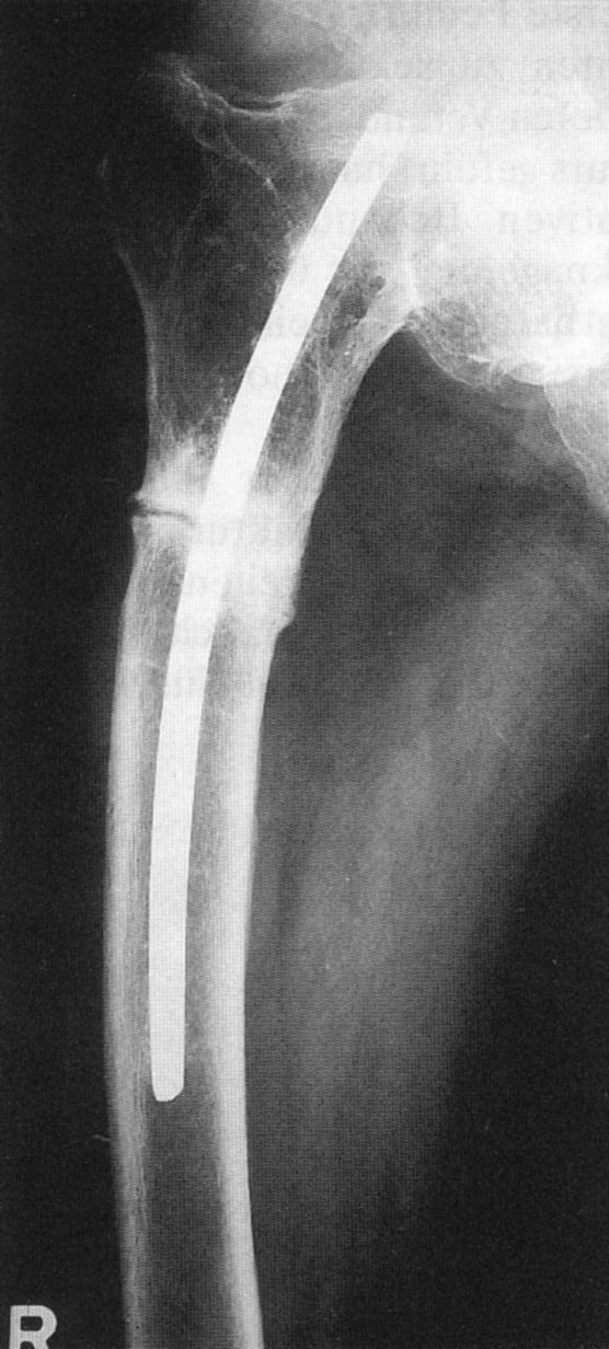
- Subtrochanteric fractures malreduce into VARUS and PROCURVATUM (apex anterior) because the proximal fragment is FLEXED, ABDUCTED and EXTERNALLY ROTATED by iliopsoas, the abductors and the short external rotators. Anticipate this — reduce before reaming, use a trochanteric-entry nail, and accept a low threshold for open or assisted reduction (clamp, blocking screws, cerclage, unicortical plate).
- Malrotation is the most common and most frequently MISSED complication — up to 20 to 30 percent have a clinically relevant rotational difference. Set rotation against the contralateral limb intra-operatively using the lesser trochanter profile sign and the cortical step-sign, and confirm length before final distal locking.
When & Why
Indication. Reamed antegrade intramedullary nailing is the GOLD STANDARD for the adult femoral shaft (diaphyseal) fracture. It is also the workhorse for subtrochanteric fractures (with a trochanteric-entry nail), for ipsilateral femoral shaft and neck fractures (after the neck is prioritised), and for pathological or impending pathological fractures of the diaphysis. Standard indications
- Femoral shaft (diaphyseal) fractures — reamed antegrade nailing is the benchmark
- Subtrochanteric femoral fractures — with a trochanteric-entry (cephalomedullary or long reconstruction) nail
- Ipsilateral femoral shaft and neck fractures — the femoral NECK takes priority; the shaft is then managed with a nail or a retrograde or plate strategy depending on neck fixation
- Pathological or impending pathological fractures of the diaphysis (prophylactic stabilisation) Relative indications — segmental fractures (a length-stable construct restoring alignment), selected distal-third shaft fractures (retrograde nailing or plating may be preferable nearer the knee), and femoral shaft nonunion (exchange reamed nailing). Contraindications
- Absolute — active infection at the entry site or along the canal (without staged management); and piriformis entry in the skeletally immature or adolescent femur (AVN risk via the medial femoral circumflex artery).
- Relative — unstable or borderline polytrauma physiology (favours damage control orthopaedics), pre-existing femoral deformity or retained hardware preventing canal passage, and very distal or very proximal fracture extension where nail fixation is biomechanically poor. The one decision that matters — entry point. Every antegrade femoral nail begins with the same choice of start point, and the start point dictates both the reduction and the implant:
Collinear with the medullary canal, so a straight nail reduces a diaphyseal shaft fracture anatomically with no built-in bend. It is technically harder to localise (especially in the muscular or obese patient), passes through the abductor insertion and deep external rotators, and the start point lies close to the deep branch of the medial femoral circumflex artery. Never used in the skeletally immature.
Easier and faster to localise, more abductor-sparing, less fluoroscopy, lower medial femoral circumflex artery risk — the routine adult choice. It is off-axis to the canal, so it mandates a nail manufactured with a proximal mediolateral (valgus) bend of about 4 to 6 degrees. A mismatched straight nail through this entry drives the shaft into varus and blows out the medial cortex.
Timing — match the intervention to the physiology. The decision between early definitive nailing and damage control is driven by the patient's response to resuscitation (lactate clearance, base deficit) rather than a fixed clock:
Definitive reamed nailing within the first 24 hours benefits the stable, resuscitated patient — earlier mobilisation, fewer pulmonary complications, shorter ICU and hospital stay.
Temporary spanning external fixation for the unstable or borderline patient (haemodynamic instability, severe chest or head injury, coagulopathy, hypothermia, lactate not clearing) limits the reaming-driven second hit of fat and marrow emboli. Definitive nailing is staged until physiology is restored.
Special situations
- Skeletally immature or adolescent femur — avoid piriformis entry (medial femoral circumflex artery injury, femoral head AVN). Choose by age and weight: flexible (elastic) nails for most school-age children with a length-stable shaft fracture; a lateral-entry rigid (trochanteric) nail or sub-muscular bridge plating for older, heavier adolescents and length-unstable patterns. Respect the proximal femoral physis and the trochanteric apophysis.
- Bilateral femoral shaft fractures — a higher-energy injury carrying greater systemic insult and mortality; have a lower threshold for damage control (bilateral external fixation) in the borderline patient, with staged definitive nailing once resuscitated, and vigilance for fat embolism syndrome and ARDS.
- Ipsilateral femoral neck and shaft fracture — the femoral NECK is the priority and is easily missed; scrutinise the hip on the trauma series, stabilise the neck reliably, and choose a shaft strategy (retrograde nail, plate, or a construct compatible with neck fixation) around it. Consent specifically for malrotation and malreduction, varus (subtrochanteric), nonunion or delayed union, AVN of the femoral head (immature femur, piriformis entry), fat embolism and ARDS, infection, nerve injury (pudendal from the perineal post; rarely sciatic or peroneal from positioning), heterotopic ossification, hardware failure, and the possible need for dynamisation or revision. Setup. Two common positions — a fracture (traction) table (supine or lateral, longitudinal traction for length and reduction, mandating a well-padded perineal post and careful traction limits) or a free-leg (flat radiolucent) table (faster set-up, easier rotational control and access for assisted reduction, manual traction; preferred by many for subtrochanteric patterns). Image intensify with a clear view of the hip (entry, neck) and the knee (length, distal locking, distal alignment), and drape the contralateral limb accessible if rotation comparison on the table is planned.
The Operation
The goal: choose the correct entry point, achieve and hold reduction before reaming, deliver a nail matched to the entry across the reduced fracture, and lock it in correct length and rotation. The exposure is the proximal entry — laid out in full as the opening steps below — and the whole sequence turns on reducing the fracture before the nail commits the alignment.
Operative sequence
- Fracture (traction) table or free-leg radiolucent table; ensure resuscitation is complete in the trauma patient before a reamed definitive nail.
- Image intensifier with clear hip (entry, neck) and knee (length, distal locking) views; drape the contralateral limb accessible for rotation comparison.
- General or regional anaesthesia; consent confirmed for malrotation, varus, nonunion, AVN, fat embolism, infection and nerve injury.
- Make a proximal incision in line with the femoral shaft above the greater trochanter; palpate the trochanter.
- Trochanteric entry — start point at the tip of the greater trochanter, slightly medial on the AP and central on the lateral, in line with the canal. Use a trochanteric-entry nail with the matching proximal bend.
- Piriformis entry — piriformis fossa, just medial to the trochanteric tip, collinear with the canal. Use a straight (piriformis-design) nail.
- Confirm the start point on a TRUE AP and a TRUE lateral before opening the cortex; a start point too lateral creates varus and risks medial cortex blow-out, and too medial threatens the medial femoral circumflex artery (especially in a child).
- Achieve and provisionally hold reduction before reaming — once the nail is in, the deforming forces are fixed.
- Use traction (table or manual), and for difficult patterns add a percutaneous clamp, a ball-spike pusher, blocking (Poller) screws, cerclage wire (for a spiral subtrochanteric component) or a small unicortical reduction plate.
- In subtrochanteric fractures anticipate varus and procurvatum (the proximal fragment is flexed, abducted and externally rotated) and over-correct accordingly.
- Open the proximal cortex with the entry reamer or awl over the guidewire, protecting soft tissue with a sleeve.
- Pass the ball-tipped guidewire across the reduced fracture into the centre of the distal fragment on both AP and lateral — confirm it sits central distally, not eccentric and not out through a fracture gap.
- Measure nail length over the guidewire (against the contralateral femur if needed).
- Ream sequentially with sharp reamers, advancing slowly to limit intramedullary pressure and the embolic load; ream typically 1 to 1.5 mm beyond the chosen nail diameter.
- Keep the fracture reduced while reaming so the reamer does not chatter eccentrically across the fracture.
- Pass the nail over the guidewire across the reduced fracture by hand — hammering suggests under-reaming or malreduction.
- Seat the nail to the correct depth proximally (trochanteric tip for a trochanteric nail); avoid distraction at the fracture as the nail advances.
- Lock proximally through the jig.
- Before final distal locking, SET ROTATION against the contralateral limb — rotate the uninjured hip under fluoroscopy to capture the lesser trochanter profile in the patella-forward position, then match the injured side; cross-check with the cortical step-sign and canal-diameter comparison.
- Lock distally with a free-hand perfect-circles technique (or jig).
- Confirm length is correct (not distracted, not shortened) and rotation is set before drilling the distal interlocks.
- STATIC locking (both ends locked) for comminuted, segmental or length-unstable patterns — the default for most femoral shaft and all subtrochanteric fractures.
- DYNAMIC locking (one end only, or a dynamic slot) only for stable transverse or short-oblique mid-shaft fractures with cortical contact, to allow controlled axial loading.
- Later DYNAMISATION (removing a locking bolt) is a salvage option for delayed union of a stable pattern.
- Confirm on AP and lateral: reduction (alignment, no varus or procurvatum), nail seating, all locking bolts engaged, length and rotation.
- Wash out reaming debris (reduces heterotopic ossification) and close in layers.
Reduce before you ream — a reamer or nail follows an unreduced fracture and locks in varus and procurvatum. Never use a piriformis entry in the skeletally immature femur (medial femoral circumflex artery injury and femoral head AVN). On a traction table use a well-padded perineal post and the minimum effective traction, releasing it periodically, to avoid a pudendal nerve palsy.
For a subtrochanteric fracture I reduce before I ream, because once the nail is in the deforming forces are fixed. I anticipate that the proximal fragment is flexed, abducted and externally rotated — so I expect varus and procurvatum. I have a low threshold for assisted reduction: a clamp, blocking (Poller) screws placed to push the nail away from the apex of the deformity, cerclage for a spiral component, or a unicortical plate to hold length and alignment while I pass the nail.
The entry must match the nail design — a straight (piriformis-design) nail to the piriformis fossa, a bent (valgus) nail to the trochanteric tip. A straight nail forced down a trochanteric entry levers the shaft into varus and blows out the medial cortex of the proximal fragment. Confirm a correct start point on a true AP and a true lateral before reaming.
Aftercare & Complications
Immediate (Day 0 to 2)
- Neurovascular check — confirm distal pulses and sensation; specifically check perineal sensation if a traction table and post were used (pudendal nerve).
- Respiratory monitoring — vigilance for fat embolism syndrome (hypoxia, confusion, petechiae) in the first 24 to 72 hours, particularly with a chest injury or bilateral femoral fractures.
- Multimodal analgesia and chemical plus mechanical thromboprophylaxis — a femoral fracture is high VTE risk.
- Radiographs — AP and lateral of the full femur including hip and knee to document reduction, alignment, length and locking. Weight-bearing
- Length-stable patterns (transverse, short oblique, stable comminution) — often early weight-bearing as tolerated.
- Length-unstable, highly comminuted or subtrochanteric patterns — protected or partial weight-bearing initially, advancing as callus appears; individualise to construct stability.
- Early mobilisation reduces pulmonary and thromboembolic complications. Rehabilitation — early knee and hip range of motion; quadriceps and abductor strengthening (abductor function may be affected by the entry, especially piriformis); gait re-education and any Trendelenburg from abductor compromise; progressive return to function over 3 to 6 months with union typically at 3 to 6 months on serial radiographs. Complications
- Incidence
- up to 20 to 30 percent greater than 15 degrees
- Recognition
- Rotational asymmetry on clinical hip rotation; gait disturbance; confirmed on CT rotational profile versus the contralateral femur
- Prevention and management
- Prevention: set rotation intra-operatively against the contralateral limb (lesser trochanter profile, cortical step-sign) before final distal locking. Management: CT to quantify; if symptomatic and greater than 15 degrees, de-rotation osteotomy or nail removal and re-locking
- Incidence
- common without anticipation
- Recognition
- Apex-anterior and varus angulation on intra-op or post-op AP or lateral; proximal fragment flexed and abducted
- Prevention and management
- Prevention: reduce before reaming; trochanteric-entry nail; blocking screws, clamp, cerclage or unicortical plate to control the proximal fragment. Management: revise reduction if recognised early; established symptomatic malunion may need corrective osteotomy
- Incidence
- reported with piriformis entry in the immature femur
- Recognition
- Hip pain, limp, collapse; AVN changes on MRI or radiograph months later
- Prevention and management
- Prevention: never use piriformis entry in the skeletally immature or adolescent — use lateral trochanteric, flexible nail or sub-muscular plate; protect the medial femoral circumflex artery. Management: established AVN — joint preservation versus salvage by severity and age
- Incidence
- clinically significant in a minority; higher with chest injury
- Recognition
- Hypoxia, tachycardia, confusion, petechiae 24 to 72 hours post-injury or surgery; bilateral pulmonary infiltrates
- Prevention and management
- Prevention: damage control (external fixation) in the borderline, unstable or chest-injured patient; sharp reamers, slow reaming, canal venting to lower intramedullary pressure. Management: supportive — oxygen, ventilatory support, ICU care
- Incidence
- 1 to 10 percent depending on pattern and host
- Recognition
- Persistent pain and tenderness at the fracture; no bridging callus at 6 months; hardware loosening or failure
- Prevention and management
- Prevention: avoid distraction; reamed nailing; restore alignment; address biology (smoking, NSAIDs, diabetes). Management: dynamisation for a stable pattern; exchange reamed nailing; augmentation plating with bone graft for atrophic or recalcitrant nonunion
- Incidence
- uncommon, usually transient
- Recognition
- Perineal or genital numbness, erectile dysfunction post-op after fracture-table use
- Prevention and management
- Prevention: well-padded perineal post; minimum effective traction; release traction periodically. Management: usually resolves spontaneously over weeks; reassure and monitor
- Incidence
- common radiographically, usually asymptomatic
- Recognition
- New bone around the trochanteric entry on follow-up films; occasional abductor pain or stiffness
- Prevention and management
- Prevention: gentle abductor handling; thorough lavage of reaming debris. Management: usually observe; excision only if symptomatic and mature
- Incidence
- low; higher with nonunion
- Recognition
- Broken interlocking bolt or nail on radiograph, often with recurrent pain — a sign of underlying nonunion
- Prevention and management
- Prevention: appropriate nail diameter and working length; achieve union. Management: treat the nonunion (the usual root cause); exchange nailing or revision fixation; remove broken hardware
- Incidence
- uncommon
- Recognition
- New fracture lines on insertion; distal anterior cortex perforation from radius-of-curvature mismatch
- Prevention and management
- Prevention: adequate reaming; insert by hand not hammer; match the nail radius of curvature (especially the bowed elderly femur). Management: additional fixation or locking; revise the nail if mal-seated
Viva & Exam Focus
ENTRYENTRY — Choosing and Making the Start Point
Clinical Decision Scenarios
Practise clinical reasoning and management decisions out loud
“A 28-year-old man sustains an isolated closed left subtrochanteric femoral fracture in a motorbike crash. He is haemodynamically stable. You plan antegrade intramedullary nailing. Talk me through your entry point choice and how you will avoid malreduction.”
“A 12-year-old child has a femoral shaft fracture. A trainee suggests a standard adult piriformis-entry reamed nail. Why is this the wrong choice, and what would you do instead?”
“A 35-year-old polytrauma patient has bilateral femoral shaft fractures, a flail chest with pulmonary contusions, and is hypotensive with a rising lactate. The trauma team asks whether you will nail both femurs tonight. What is your approach?”
Entry point — core concept
- Piriformis fossa = collinear with the canal, so a straight nail reduces the shaft anatomically; technically harder, abductor damage, near the medial femoral circumflex artery
- Greater trochanter tip = off-axis, so it needs a nail with a proximal valgus bend of about 4 to 6 degrees; easier, abductor-sparing, lower MFCA risk
- A mismatched straight nail through a trochanteric entry causes varus and medial cortex blow-out
- Trochanteric entry is the routine adult choice; piriformis remains the most collinear for the shaft
- Confirm a correct start point on a TRUE AP and lateral before reaming
Paediatric or adolescent rule
- NEVER use piriformis entry in the skeletally immature or adolescent — AVN of the femoral head (medial femoral circumflex artery)
- Also protect the trochanteric apophysis and the proximal physis
- Flexible (elastic) nails for most length-stable school-age shaft fractures
- Lateral trochanteric rigid nail or sub-muscular plate for older, heavier or length-unstable patterns
- Children remodel and unite fast — but rotation does not remodel, so still set it
Subtrochanteric deforming forces
- Iliopsoas — proximal fragment FLEXION
- Gluteus medius and minimus — proximal fragment ABDUCTION
- Short external rotators — proximal fragment EXTERNAL ROTATION
- Adductors — distal fragment medial pull (varus)
- Net deformity = VARUS plus PROCURVATUM (apex anterior) — reduce before reaming
Timing — ETC versus DCO
- Stable, resuscitated patient — early total care (early definitive reamed nailing)
- Borderline or unstable, chest injury, head injury, coagulopathy — damage control (external fixation first)
- Reaming is a second hit (intramedullary pressure, fat and marrow emboli) — ARDS risk with lung injury
- Definitive surgery timing is driven by response to resuscitation (lactate, base deficit), not a fixed clock
- Bilateral femurs — higher systemic insult and mortality; lower threshold for DCO
Operative technique — key steps
- 1. REDUCE FIRST — clamp, blocking (Poller) screws, cerclage or unicortical plate as needed
- 2. Correct start point matched to nail design; confirm on true AP and lateral
- 3. Central guidewire across the reduced fracture on both views
- 4. Ream gently with sharp reamers (limit intramedullary pressure and fat embolism)
- 5. Insert the nail by hand (hammering equals under-reaming or malreduction)
- 6. Set ROTATION against the contralateral limb (lesser trochanter profile, cortical step-sign) before the distal lock
- 7. Static lock unstable, comminuted or subtrochanteric patterns; dynamic only for a stable transverse fracture
- 8. Lavage reaming debris to reduce heterotopic ossification
Danger zones
- Medial femoral circumflex artery (piriformis entry) — femoral head AVN, especially in children
- Abductor insertion (piriformis approach) — weakness, Trendelenburg, heterotopic ossification
- Perineal post (traction table) — pudendal nerve palsy
- Reaming plus chest injury — fat embolism or ARDS
- Lateral start with a straight nail — varus and medial cortex blow-out
Complications
- Malrotation — up to 20 to 30 percent greater than 15 degrees; the commonest MISSED complication; set versus the contralateral limb, CT to confirm
- Varus or procurvatum (subtrochanteric) — anticipate and reduce before reaming
- AVN (paediatric piriformis) — avoid piriformis entry in the immature femur
- Fat embolism or ARDS — damage control in the borderline or chest-injured patient
- Nonunion — dynamise a stable pattern or exchange reamed nail; hardware failure usually signals nonunion
Special cases
- Skeletally immature — flexible nail or lateral-entry rigid nail or sub-muscular plate; never piriformis
- Bilateral femurs — high mortality; fat embolism syndrome or ARDS vigilance; consider DCO
- Ipsilateral neck plus shaft — the NECK is the priority and is easily missed; scrutinise the hip series
- Reamed versus unreamed — reamed favoured for union; consider unreamed only when the pulmonary embolic load is a genuine concern
Background & Evidence
Proximal femoral entry anatomy. The two start points define the operation. The piriformis fossa lies just medial to the tip of the greater trochanter, at the base of the femoral neck; it is collinear with the medullary canal, so a straight nail introduced here lies in line with the shaft and reduces a diaphyseal fracture anatomically — but the start point is close to the deep branch of the medial femoral circumflex artery posteriorly, passes through the abductor insertion and deep external rotators, and is technically demanding to localise, particularly in the muscular or obese patient. The greater trochanter tip start point (slightly medial to the very tip on the AP, central on the lateral) is off-axis to the canal; a straight nail would drive the shaft into varus, so trochanteric-entry nails are manufactured with a proximal mediolateral bend of about 4 to 6 degrees. Medial femoral circumflex artery — the femoral head supply. The medial femoral circumflex artery, a branch of the profunda femoris, gives off the deep branch that ascends posterior to the femoral neck and terminates as the lateral epiphyseal (retinacular) arteries — the dominant supply to both the adult and the paediatric femoral head. A medially placed proximal femoral start point can injure this vessel; this is the anatomical basis for avoiding piriformis entry in children and adolescents, where AVN of the femoral head is a recognised, devastating complication. Femoral canal and isthmus. The canal narrows at the isthmus (mid-diaphysis), where the nail engages cortical bone and where the reaming diameter is set. The femur has a natural anterior bow (radius of curvature about 120 cm); a nail with a mismatched radius of curvature risks anterior cortical perforation distally, especially in the older patient with a more bowed femur. Femoral neck anteversion of about 10 to 15 degrees is the reference for setting rotation — match the contralateral side. Subtrochanteric deforming forces. The subtrochanteric region (from the lesser trochanter to about 5 cm distal) is dominated by powerful muscle attachments that deform the proximal fragment into the classic varus and procurvatum malreduction:
- Attachment
- Lesser trochanter
- Deforming effect
- FLEXION of the proximal fragment
- Attachment
- Greater trochanter
- Deforming effect
- ABDUCTION of the proximal fragment
- Attachment
- Trochanteric region
- Deforming effect
- EXTERNAL ROTATION of the proximal fragment
- Attachment
- Distal medial shaft
- Deforming effect
- Medial pull on the distal fragment (varus)
References
Trochanteric versus piriformis entry portal for antegrade femoral nailing
Comparative study of trochanteric versus piriformis entry antegrade femoral nails. Trochanteric entry was associated with shorter operative time and less fluoroscopy than piriformis entry, with equivalent alignment and union when an entry-appropriate (correctly bent) nail design was used. Trochanteric entry is a reliable, often easier alternative to piriformis entry provided the nail is designed for a trochanteric start point.
Damage control orthopaedics versus early total care in polytrauma with femoral fracture (EPOFF)
Borderline or unstable polytrauma patients had a higher inflammatory response and complication burden with immediate reamed nailing; initial external fixation (damage control) followed by staged definitive nailing reduced the early systemic insult. Match the intervention to the patient's physiology — early definitive nailing for the stable patient, damage control external fixation for the borderline or unstable patient to avoid the reaming second hit.
Reamed versus unreamed intramedullary nailing of the femur
Randomised comparison of reamed versus unreamed intramedullary nailing in femoral shaft fractures. Reamed nailing showed a lower rate of nonunion and reoperation than unreamed nailing in the femur; reaming concerns relate chiefly to the pulmonary insult in the severely injured chest, not to femoral union. Reamed nailing is preferred for femoral shaft union; reserve consideration of unreamed technique for the patient in whom the pulmonary embolic load of reaming is a genuine concern.
Malrotation after antegrade femoral nailing — incidence and detection
Clinically relevant rotational malalignment (greater than 15 degrees) occurred in a substantial proportion of antegrade femoral nails. Malrotation is frequently missed intra-operatively and on plain radiographs; CT is the reference standard, and comparison with the contralateral femur (lesser trochanter profile, cortical step-sign) reduces malrotation. Set and confirm rotation against the uninjured limb intra-operatively, with a low threshold for a CT rotational profile if clinically suspected.
Subtrochanteric femoral fractures — deforming forces and reduction principles
The proximal fragment flexes (iliopsoas), abducts (gluteus medius and minimus) and externally rotates (short external rotators), producing the characteristic varus and procurvatum (apex-anterior) malreduction with intramedullary nailing. Adequate reduction (open or assisted) before or while nailing is critical to avoid malunion and nonunion; subtrochanteric fractures behave differently from diaphyseal fractures, so anticipate the deformity, use a trochanteric-entry nail and accept assisted reduction.