Long posterior myocutaneous flap (Burgess) or skew flap (Robinson) | level ~12-15cm below tibial tuberosity | intermediate
- Preserving the knee is the whole point - a transtibial amputee walks with roughly a 25% increase in energy cost versus about 65% for a transfemoral amputee, so a BKA is strongly preferred over an AKA whenever the limb will heal at that level. A failed BKA that converts to an AKA is a worse outcome than a primary AKA, so level selection is the critical judgement.
- Aim for about 12-15cm of tibia (a hand's breadth) below the tibial tuberosity. Bevel the anterior tibia 45-60 degrees, smooth every edge, and divide the fibula 1-2cm shorter so the stump is conical and prosthesis-friendly.
- Two flap designs dominate: the Burgess long posterior myocutaneous flap (robust gastrocnemius-based blood supply, the workhorse for dysvascular limbs) and the Robinson skew flap (equal sagittal flaps on the cutaneous perforator supply). The multicentre RCT (Ruckley) found no significant difference in primary healing between them.
- Ligate every major pedicle individually - the anterior tibial, posterior tibial and peroneal vessels - and specifically ligate the artery that accompanies the tibial nerve. A missed vessel here is the classic cause of a stump haematoma that threatens the flap.
- Healing is governed by perfusion and nutrition: select the level on transcutaneous oxygen pressure (TcPO2 greater than 30-40 mmHg predicts primary healing), ankle-brachial index, serum albumin and lymphocyte count - not on cosmetic length.
When & Why
The operation in one line. A below-knee (transtibial) amputation divides the tibia and fibula roughly a hand's breadth below the knee, covers the bone end with a cushioned gastrocnemius-based muscle flap, and preserves the knee joint - the single most important determinant of efficient prosthetic walking. The Big Four indications ("Dead, Dangerous, Damaged, Damn nuisance").
Critical limb ischaemia with unreconstructable disease or failed revascularisation, and diabetic foot sepsis or gangrene. Peripheral vascular disease and diabetes account for the large majority of lower-limb amputations in older patients.
A non-salvageable mangled lower leg after failed limb salvage, prolonged warm ischaemia, or segmental soft-tissue and bone loss. The decision rests on clinical judgement and shared decision-making rather than any single score in isolation.
Life-threatening sepsis - necrotising fasciitis, gas gangrene, uncontrolled diabetic foot infection, or chronic osteomyelitis unresponsive to debridement and antibiotics.
When limb-sparing resection is not feasible or would leave a non-functional limb; also a chronically painful or useless deformed limb that is a "damn nuisance" to the patient.
Why preserving the knee matters. Whenever the limb can heal at a transtibial level, a BKA is preferred over an AKA: - Energy cost of gait - transtibial amputees ambulate with roughly a 25% increase in energy expenditure above normal, whereas transfemoral amputees require around 65% more. The knee joint is the single biggest determinant of efficient prosthetic walking.
- Prosthetic rehabilitation - preserving the anatomical knee dramatically improves the proportion of patients who become independent prosthetic walkers, particularly the frail, elderly and dysvascular in whom every extra metabolic demand matters.
- Functional independence - lower fall risk, better balance, and a higher likelihood of returning to community ambulation than with an above-knee amputation.
- The trade-off - this benefit only holds if the BKA level will heal. A non-healing BKA that converts to an AKA is worse than a primary AKA, so level selection is the critical judgement. Level selection. The level is dictated by where the flap will heal, not by cosmetic length - but a well-shaped stump is the aim wherever perfusion allows. - Ideal length - about 12-15cm of tibia from the tibial tuberosity, classically a hand's breadth below the tuberosity (a rough guide is about 2.5cm of bone per 30cm of body height).
- Minimum functional length - a stump shorter than about 8cm of tibia below the joint line gives a poor lever arm, is hard to fit and is prone to flexion contracture; an extremely short stump may be no better functionally than a knee disarticulation.
- Maximum length - avoid an over-long distal stump, because soft-tissue cover and perfusion are poorer in the lower third of the leg and the muscle bulk for padding is thinner.
- Perfusion overrides geometry - in the dysvascular limb the level is ultimately set by where the flap will heal. Contraindications to a transtibial level. - Inadequate perfusion at the proposed level (very low TcPO2, absent popliteal pulse with unreconstructable disease).
- A knee flexion contracture or a non-functional knee that would preclude prosthetic use.
- Extensive proximal soft-tissue loss, sepsis or tumour reaching the upper calf.
- A non-ambulatory patient in whom a higher, more reliably healing level may be more pragmatic (individualised). Pre-operative optimisation. Wound healing in the dysvascular and diabetic limb is governed by perfusion and nutrition, so optimise before committing to a level: - Vascular assessment and intervention - confirm inflow and decide whether revascularisation could lower the amputation level or improve healing.
- Sepsis control - in wet gangrene or ascending infection, consider a staged guillotine amputation first to control sepsis before definitive flap-based closure.
- Metabolic and nutritional optimisation - tight glycaemic control, correct anaemia, and address malnutrition (serum albumin and total lymphocyte count are practical, validated healing predictors).
- Medical and anaesthetic work-up - these patients are frequently elderly with significant cardiac, renal and respiratory comorbidity; multidisciplinary peri-operative care reduces mortality. Consent specifically for wound failure and possible conversion to a higher level, stump or phantom limb pain, neuroma, infection, knee flexion contracture, and the realistic prosthetic and mobility outlook given the patient's comorbidity and the contralateral limb.
The Operation
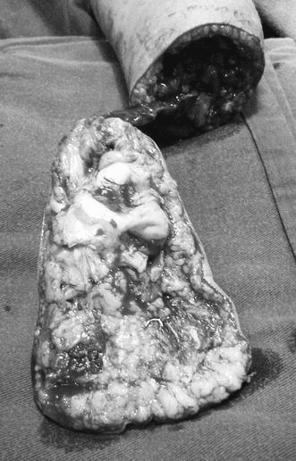
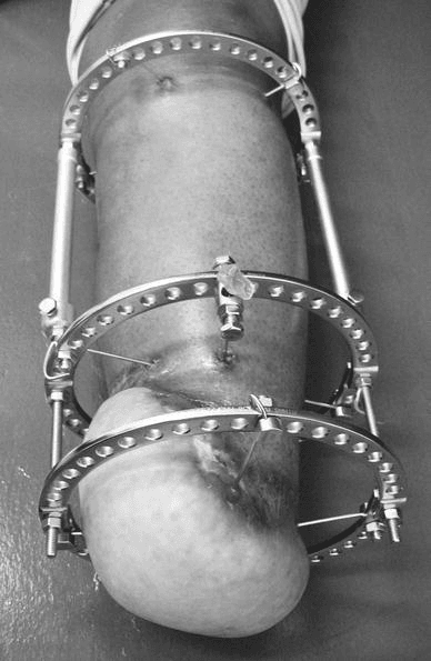
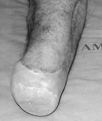
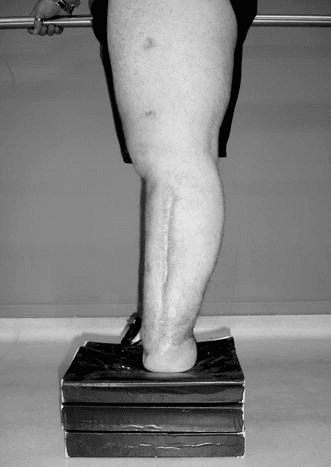
The goal is to divide the tibia and fibula at a level that will heal, cover the bevelled bone end with a cushioned, stable gastrocnemius-based flap, handle each nerve and vessel deliberately, and close without tension so the scar lies off the weight-bearing bone-end - then protect it all in a rigid dressing with the knee in extension. The exposure is laid out as the opening steps below. The images below illustrate one specialised transtibial technique - a sensate calcaneal (plantar) flap transferred onto the tibial end to permit terminal weight-bearing - shown intra-operatively and through to long-term follow-up.
Operative sequence - Burgess long posterior flap
- Supine, the leg prepped free and draped for circumferential access; a sandbag under the ipsilateral buttock helps neutralise external rotation. Pad the contralateral heel and bony prominences.
- Mark the landmarks: the tibial tuberosity (the reference for level measurement), the subcutaneous anterior tibial crest (the line to bevel), and the fibular head and neck (the common peroneal nerve winds round it). Mark flaps with the limb in neutral.
- Tourniquet is optional and debated in dysvascular limbs (see below). In a non-ischaemic limb (trauma, tumour) it gives a bloodless field; in critical ischaemia many surgeons omit it.
- Identify the bone-division level about 12-15cm (a hand's breadth) below the tibial tuberosity. Measure the leg circumference at that level.
- The anterior incision sits at the level of bone section.
- Mark the long posterior myocutaneous flap to a length of roughly the antero-posterior diameter of the leg at that level plus about 1cm, so it folds anteriorly over the bone end without tension. The medial and lateral limbs of the incision descend from the anterior mark to meet the posterior flap.
- Incise skin, subcutaneous fat and deep fascia along the marked lines.
- Anteriorly, divide the anterior compartment muscles (tibialis anterior, EHL, EDL) at the level of bone section.
- Identify, doubly ligate and divide the anterior tibial artery and venae comitantes, and perform a traction neurectomy of the deep peroneal nerve.
- Divide the fibularis (peroneal) muscles.
- Perform a traction neurectomy of the superficial peroneal nerve.
- Expose the fibula; it will be divided 1-2cm proximal to the planned tibial cut.
- Strip periosteum minimally at the cut site.
- Saw the tibia at the chosen level and create a 45-60 degree anterior bevel; round and smooth every edge with a rasp so no spike remains.
- Divide the fibula 1-2cm shorter than the tibia and bevel its lateral edge so it cannot become a distal prominence.
- With the bones divided, retract them and divide the deep posterior compartment.
- Identify and doubly ligate and divide the posterior tibial vessels and the peroneal vessels individually under direct vision.
- Perform a traction neurectomy of the tibial nerve, and specifically ligate the accompanying artery to the tibial nerve before dividing it to prevent a stump haematoma.
- Take the sural nerve in the posterior flap with traction neurectomy.
- The posterior flap is skin, fascia, soleus and gastrocnemius.
- Tailor (debulk) the soleus and bevel the muscle so the flap is not too bulky and folds easily over the bone end - excessive bulk impairs prosthetic fitting and may compromise distal perfusion.
- Bring the gastrocnemius (and tailored soleus) anteriorly to cover the bevelled tibial end.
- Perform a myoplasty - suture the posterior muscle flap to the anterior compartment fascia and periosteum (or a drill-hole myodesis to bone) - so muscle is anchored over the bone end, giving a padded, stable, non-mobile soft-tissue envelope.
- Release the tourniquet if used and secure meticulous haemostasis - re-check each pedicle.
- Place a suction drain deep to the muscle layer, brought out away from the suture line.
- Trim redundant flap and any dubiously perfused margin.
- Close fascia then skin so the scar lies anteriorly across the distal stump, off the weight-bearing bone-end, with durable posterior calf skin facing the prosthesis. Avoid "dog-ears" and tension; re-palpate the bone end through the closed flap to confirm no spike remains.
- Apply a well-padded rigid (plaster) dressing or rigid removable dressing with the knee in full extension to control oedema, protect the stump and prevent a knee flexion contracture - the foundation of early/immediate postsurgical prosthetic progression.
Identify, doubly ligate and divide the anterior tibial, posterior tibial and peroneal pedicles individually under direct vision before transecting muscle, and specifically ligate the artery that accompanies the tibial nerve. A slipped ligature or an unligated vasa nervorum is the classic cause of an expanding stump haematoma that compresses and kills the flap. Release the tourniquet before closure and re-check every pedicle, then leave a suction drain.
A smooth tibia with a 45-60 degree anterior bevel and a fibula divided 1-2cm shorter and bevelled is what makes a stump tolerable in a prosthesis. A square or spiked tibial end, or a long fibula, tents the flap, causes skin breakdown over the bone and prevents socket fit. Re-palpate the bone end through the closed flap before you leave theatre.
The skew flap uses two equal-length sagittal skin flaps rotated about 25 degrees from antero-posterior so the apices sit antero-medially and postero-laterally, based on the cutaneous perforator/angiosomal supply; a gastrocnemius myoplasty still pads the bone end. The aim is a more cylindrical, conical stump with the scar away from bony prominences. The multicentre RCT (Ruckley) showed no significant difference in primary healing versus the long posterior flap, so choose on familiarity, the limb's perfusion pattern and the soft tissue available.
A prospective comparative study (Wolthuis, 89 patients) showed a pneumatic tourniquet reduced blood loss (haemoglobin fall 5.6% versus 14.8%) and roughly halved the revision rate (14.3% versus 38.3%) without increasing wound-healing failure - but it was a non-randomised cohort, not an RCT, so the evidence is suggestive rather than definitive. Many surgeons still avoid it in critical ischaemia for fear of compromising marginal flap perfusion. Be ready to argue both sides in a viva.
Myoplasty (muscle-to-fascia) is quicker and standard; myodesis (muscle-to-bone through drill holes) gives a more stable envelope and may suit active patients but is avoided where perfusion is marginal. If the flap will not reach without tension, the level is too distal or the flap too short - shorten the bone within functional limits rather than close tightly.
Aftercare & Complications
Immediate post-operative phase (0-2 weeks) - Rigid dressing (or rigid removable dressing) with the knee in full extension to control oedema, protect the stump and prevent flexion contracture.
- Elevate the residual limb early but avoid a pillow under the knee (it promotes contracture).
- Remove the suction drain at about 24-48 hours; monitor the flap for perfusion and haematoma.
- Multimodal analgesia with attention to phantom and residual limb pain: regional catheters, gabapentinoids or amitriptyline, paracetamol or NSAIDs and short-term opioids; introduce mirror therapy early.
- Encourage knee extension and prone-lying periods; begin gentle hip and knee range of motion and bed mobility. Intermediate phase (2-6 weeks) - Suture or clip removal at about 2-3 weeks (later in dysvascular and diabetic patients who heal more slowly).
- Progress to a stump shrinker or elastic compression once the wound is sound, to shape the stump for prosthetic casting; continue oedema control and desensitisation.
- Early walking aids (such as a pneumatic post-amputation mobility aid) allow trial weight-bearing and gait re-education before the definitive prosthesis; Burgess-style early or immediate postsurgical prosthetic fitting accelerates rehabilitation in suitable patients.
- Multidisciplinary care: vascular and diabetic optimisation, podiatry, protection of the contralateral limb (high risk of subsequent amputation), wound care and dietitian input. Rehabilitation (6 weeks to 6-12 months) - Cast for a definitive socket once the stump is mature and stable; progressive prosthetic gait training toward independent community ambulation, leveraging the preserved knee and the favourable ~25% energy cost of transtibial gait.
- Counsel realistically - outcomes depend on age, cardiorespiratory reserve, comorbidity and the contralateral limb; set expectations honestly and individualise goals (community versus household ambulation versus wheelchair independence). Mortality after major lower-limb amputation for vascular disease is substantial.
- Reinforce knee extension at every stage - the flexion contracture is the most preventable cause of a functionally useless transtibial stump. Watch also for late neuroma, stump breakdown over a prominent bone end, and the contralateral limb. Warning signs requiring urgent review: increasing pain, swelling, erythema or discharge (infection or haematoma); new flap discolouration or dehiscence; a developing knee flexion contracture compromising prosthetic fitting.
- Recognition
- Dusky or black flap margin, non-healing edges, dehiscence, serous or purulent discharge; most evident in the first 1-2 weeks
- Prevention
- Perfusion-based level selection (TcPO2 greater than 30-40 mmHg, ABI, Doppler); atraumatic flap handling with skin hooks; tension-free closure; excise dubious margins; optimise glycaemia and nutrition
- Management
- Minor margin necrosis: dressings, debridement, secondary healing. Significant necrosis or dehiscence: debridement; if non-salvageable, revision or conversion to AKA
- Recognition
- Tense, swelling stump, increasing pain, ooze through the dressing, falling haemoglobin
- Prevention
- Individual double ligation of the posterior tibial, peroneal and anterior tibial pedicles; ligate the artery accompanying the tibial nerve; release the tourniquet and re-check; suction drain
- Management
- Small and stable: observe with a compressive rigid dressing. Expanding or large with falling Hb: return to theatre for evacuation and haemostasis to protect the flap
- Recognition
- Spreading erythema, fever, purulent discharge, raised inflammatory markers; deep collection on imaging
- Prevention
- Control sepsis before definitive closure; prophylactic antibiotics; debride non-viable tissue; staged guillotine first if grossly infected
- Management
- Antibiotics per culture; drain collections; debride. Deep or persistent infection may require proximal revision
- Recognition
- Focal Tinel-positive tender nodule, sharp shooting pain, intolerance of the socket at that point
- Prevention
- Traction neurectomy with sharp division high, so the cut end retracts into muscle away from bone and scar; consider TMR or a regenerative peripheral nerve interface in selected patients
- Management
- Desensitisation, socket modification, injections. Refractory: neuroma excision and burial, or targeted muscle reinnervation
- Recognition
- Painful sensation referred to the absent foot or leg (phantom) versus pain localised to the stump (residual)
- Prevention
- Perioperative multimodal and pre-emptive analgesia, regional or peripheral nerve blocks, good nerve handling
- Management
- Multimodal analgesia (gabapentinoids, amitriptyline), mirror therapy, graded motor imagery, psychological support; treat any neuroma or socket cause of residual pain
- Recognition
- Fixed flexion at the knee, inability to achieve full extension for socket alignment
- Prevention
- Avoid prolonged knee-flexed positioning and a pillow under the knee; rigid dressing in extension; early physiotherapy and prone lying
- Management
- Aggressive physiotherapy, serial splinting or casting; an established severe contracture may compromise prosthetic use
The non-healing or failing stump. Re-assess perfusion (consider angiography or revascularisation if not already optimised), nutrition and infection. Minor distal margin necrosis can be managed conservatively, but a substantially non-viable flap is best dealt with by timely revision rather than repeated futile debridements. Conversion to an AKA is a salvage decision - explain that the rehabilitation trade-off (higher energy cost) is the price of a reliably healing wound. Staged ("guillotine") amputation. In severe ascending infection or wet gangrene, a rapid open guillotine amputation through healthy tissue controls sepsis; definitive flap-based BKA closure is performed once infection is controlled, reducing the risk of closing over infected tissue.
Viva & Exam Focus
STUMPSTUMP - principles of a good transtibial stump
HEALHEAL - wound-healing predictors at the chosen level
Clinical Decision Scenarios
Practise clinical reasoning and management decisions out loud
“A 68-year-old diabetic with critical limb ischaemia has unreconstructable disease and dry gangrene of the forefoot extending to the mid-foot. The popliteal pulse is palpable. How do you decide between a below-knee and an above-knee amputation, and how do you select the level?”
“Describe the long posterior flap (Burgess) technique for a below-knee amputation. How does it differ from the skew flap, and what does the evidence say about which to use?”
“What are the main complications of a below-knee amputation and how do you prevent them? A diabetic patient's stump becomes tense and increasingly painful 12 hours after surgery with ooze through the dressing - what has happened and what do you do?”
Key indications (the Big Four)
- Dysvascular or diabetic limb (commonest): critical limb ischaemia unreconstructable or failed revascularisation; diabetic foot sepsis or gangrene
- Trauma: non-salvageable mangled lower leg after failed limb salvage
- Infection: necrotising fasciitis, gas gangrene, uncontrolled diabetic foot infection, chronic osteomyelitis
- Tumour: when limb-sparing resection is not feasible or leaves a non-functional limb
Why BKA over AKA
- Energy cost: transtibial gait ~25% above normal vs transfemoral ~65% - the knee is the key determinant of efficient walking
- Far more BKA patients become independent prosthetic walkers, especially the elderly and dysvascular
- Caveat: only if the BKA level will heal - a failed BKA converted to AKA is worse than a primary AKA
- Level selection is the critical judgement and is driven by perfusion, not cosmesis
Level selection
- Ideal: ~12-15cm of tibia (a hand's breadth) below the tibial tuberosity
- Minimum: about 8cm below the joint line for a functional lever arm and to avoid contracture
- Avoid an over-long distal stump - poorer perfusion and padding in the lower third
- Healing predictors (HEAL): ABI and toe pressures, TcPO2 greater than 30-40 mmHg, albumin and lymphocytes, local viability plus glycaemia
Flap options
- Burgess long posterior myocutaneous flap: gastrocnemius-based robust blood supply, workhorse for dysvascular limbs, with myoplasty and immediate or early prosthetic fitting
- Robinson (Kingsley Robinson) skew flap: equal sagittal flaps rotated ~25 degrees on cutaneous perforators for a cylindrical stump
- Evidence: Ruckley multicentre RCT - no significant difference in primary healing between skew and long posterior flap
- Choice rests on surgeon familiarity, limb perfusion pattern and available soft tissue
Critical operative steps
- Mark the anterior incision at bone level; long posterior flap length ~ AP diameter of the leg at that level
- Divide the tibia with a 45-60 degree anterior bevel, smooth edges; fibula 1-2cm shorter and bevelled
- Individually doubly ligate the anterior tibial, posterior tibial and peroneal pedicles; ligate the artery accompanying the tibial nerve
- Traction neurectomy of the tibial, common, superficial and deep peroneal, sural and saphenous nerves
- Tailor the soleus; gastrocnemius myoplasty over the bone end; close so the scar lies anteriorly off the bone-end, tension-free
Tourniquet controversy
- Comparative study (Wolthuis): pneumatic tourniquet reduced blood loss and transfusion and roughly halved the revision rate without increasing wound failure (prospective non-randomised, not an RCT)
- Many surgeons still avoid it in critical ischaemia for fear of compromising marginal flap perfusion
- Reasonable to use in non-ischaemic limbs (trauma, tumour) for a bloodless field
- Be ready to argue both sides in a viva
Major complications
- Flap necrosis or wound failure: prevent with perfusion-based level selection, atraumatic handling, tension-free closure; may need revision or AKA
- Stump haematoma: individual pedicle ligation plus ligate the tibial nerve's artery plus drain; evacuate if expanding or with falling Hb
- Infection: control sepsis pre-closure; staged guillotine if grossly infected
- Neuroma and phantom or residual pain: traction neurectomy into muscle, pre-emptive multimodal analgesia, mirror therapy
- Knee flexion contracture: rigid dressing in extension, no pillow under the knee, early physiotherapy and prone lying
Post-op and rehabilitation pearls
- Rigid dressing (Burgess) in knee extension: controls oedema, protects the stump, prevents contracture, enables early or immediate prosthetic progression
- Early walking aids for gait re-education before the definitive prosthesis; stump shrinker once the wound is sound
- Protect the contralateral limb (high risk of further amputation) and optimise diabetes and vascular risk factors
- Definitive prosthesis once the stump is mature; goal is independent community ambulation via the preserved knee
Background & Evidence
Surgical anatomy that governs the operation. At the level of section the leg holds four compartments, and each must be addressed deliberately: - Anterior compartment - tibialis anterior, EHL, EDL and fibularis tertius, carrying the anterior tibial artery and deep peroneal nerve on the interosseous membrane; these are divided and ligated as the anterior flap and muscles are taken.
- Lateral compartment - fibularis longus and brevis, with the superficial peroneal nerve that emerges through the deep fascia in the lower leg.
- Superficial posterior compartment - gastrocnemius and soleus, the muscle bulk that forms the durable long posterior myocutaneous flap with its robust overlying calf skin.
- Deep posterior compartment - tibialis posterior, FDL and FHL, carrying the posterior tibial artery and nerve and the peroneal artery, the principal pedicles to ligate. The bones. The tibia is triangular with a subcutaneous antero-medial surface; the sharp anterior crest must be bevelled 45-60 degrees with all edges smoothed. The fibula is divided 1-2cm shorter than the tibia so it does not become a distal prominence, and its lateral edge is bevelled. Nerves to address. The tibial nerve is the largest; it lies deep posteriorly with an accompanying artery (a sizeable epineurial vessel) that must be ligated to prevent a stump haematoma, and is treated with gentle traction neurectomy. The common peroneal nerve is addressed proximally - vulnerable as it winds round the fibular neck, so the traction neurectomy is kept high to keep the neuroma away from the lateral stump. The sural nerve runs subcutaneous in the posterior flap with the short saphenous vein, and the saphenous nerve medially with the long saphenous vein. Blood supply relevant to the flaps. The long posterior flap is perfused chiefly by the posterior tibial and peroneal arteries via the sural arteries to gastrocnemius, giving it reliable perfusion even in dysvascular limbs. The skew flap is based on the perforating or angiosomal supply to skin, with the sagittal flaps positioned to optimise cutaneous perfusion. The angiosome concept - matching the retained flap to the dominant patent inflow (for example the posterior tibial) - supports healing in ischaemic limbs. Surface anatomy and landmarks for marking. The tibial tuberosity is the primary reference for level (the cut sits a hand's breadth distal to it). The anterior tibial crest is subcutaneous and palpable - the line to bevel, and where skin breakdown occurs if a spike is left. The fibular head and neck mark the common peroneal nerve proximally. The posterior calf bulk - the gastrocnemius-soleus mass - becomes the durable posterior flap; mark its length against the AP diameter of the leg at the level of section. Functional anatomy for the prosthesis. The patellar tendon, tibial flares and gastrocnemius pad are the weight-tolerant areas a patellar-tendon-bearing or total-surface-bearing socket loads, so preserving padded, mobile-free soft tissue over the bone end is essential. A flexion contracture at the knee changes socket alignment and the moment arm, which is why maintaining full passive extension is both an anatomical and a rehabilitation priority. Wound-healing predictors - why level selection fails or succeeds. Arterial perfusion (a palpable popliteal pulse, ankle-brachial index, toe pressures) reflects inflow to the flap; the long posterior flap's gastrocnemius supply makes it relatively forgiving in ischaemia. Transcutaneous oxygen pressure measured at the proposed level - values above about 30-40 mmHg predict primary healing, while very low values predict failure. Nutrition and immunocompetence matter: low serum albumin and a depressed total lymphocyte count are associated with non-healing and amputation in diabetic foot disease. Uncontrolled diabetes and active infection both impair healing and should be addressed before definitive closure.
- Burgess long posterior flap
- Single long posterior myocutaneous flap
- Robinson skew flap
- Two equal-length sagittal (skew) skin flaps rotated ~25 degrees
- Burgess long posterior flap
- Gastrocnemius-based supply via sural arteries - robust in ischaemia
- Robinson skew flap
- Cutaneous perforator or angiosomal supply to the skin flaps
- Burgess long posterior flap
- Gastrocnemius-soleus myoplasty over the bone end
- Robinson skew flap
- Gastrocnemius myoplasty over the bone end (as for Burgess)
- Burgess long posterior flap
- Classic posterior-flap stump
- Robinson skew flap
- More cylindrical, conical stump; scar away from bone
- Burgess long posterior flap
- No significant difference in primary healing versus skew flap
- Robinson skew flap
- No significant difference in primary healing versus long posterior flap
Rigid dressings and early rehabilitation. Burgess popularised the immediate postoperative rigid dressing and immediate or early postsurgical prosthetic fitting, allowing earlier mobilisation and oedema control. Rigid removable dressings protect the stump, control oedema, may speed healing and reduce knee flexion contracture compared with soft dressings, and facilitate progression to weight-bearing. The evidence base. Burgess (1971) established the long posterior myocutaneous flap with myoplasty for peripheral vascular insufficiency, the workhorse for dysvascular limbs; its gastrocnemius-based blood supply underpins its reliability in critical ischaemia. Robinson (1982) introduced the skew flap based on the cutaneous perforator supply to produce a cylindrical, well-perfused stump. The multicentre randomised trial by Ruckley (1991) randomised 191 patients across 11 centres and found no significant difference in primary healing between the skew and long posterior flaps. Wolthuis (2006) challenged the dogma that a tourniquet is contraindicated in dysvascular amputation, but as a non-randomised cohort the evidence is suggestive rather than definitive. Waters (1976) established the objective basis for preserving the knee: walking performance and oxygen cost were significantly better the lower (more distal) the amputation level.
References
Long posterior myocutaneous flap for the dysvascular leg (Burgess)
- Original description of the long posterior myocutaneous flap transtibial amputation with myoplasty for peripheral vascular insufficiency
- The posterior gastrocnemius-soleus flap carries a robust blood supply, making it forgiving in the ischaemic and diabetic limb
- Established the principles of bone-end management, muscle stabilisation and durable posterior calf skin over the weight-bearing stump
Skew-flap myoplastic below-knee amputation (Robinson)
- Introduced equal-length sagittal (skew) skin flaps based on the cutaneous perforator or angiosomal supply rather than a single long posterior flap
- Combined with a gastrocnemius myoplasty to produce a more cylindrical, conical stump with the scar away from bony prominences
- Designed to optimise cutaneous perfusion and ease prosthetic socket fitting
Skew flap versus long posterior flap: multicentre randomised trial (Ruckley)
- 191 patients with end-stage occlusive vascular disease randomised across 11 centres (98 skew flap, 93 long posterior flap)
- Primary healing at one week was 60% in both groups, with no significant difference in same-level revision (7% vs 8%) or revision to a higher level (10% vs 8%)
- Prosthetic fitting (84% vs 77%) and walking (78% vs 71%) at 6 months also did not differ significantly
Pneumatic tourniquet in trans-tibial amputation for vascular disease (Wolthuis)
- Prospective non-randomised comparison of 89 patients (42 tourniquet, 47 no tourniquet) undergoing trans-tibial amputation
- Haemoglobin fall was 5.6% with a tourniquet versus 14.8% without, with a lower transfusion requirement
- Revision rate was roughly halved (14.3% with tourniquet vs 38.3% without) with similar mortality and no increase in wound-healing failure
Energy cost of prosthetic walking by amputation level (Waters)
- Compared gait and energy cost in 70 unilateral traumatic and vascular amputees (above-knee, below-knee, Syme) against 40 normal controls
- Walking performance and oxygen cost were significantly better the lower (more distal) the amputation level in both aetiology groups
- Quantified the metabolic penalty that escalates sharply once the knee is sacrificed
Further reading and source references 1. Burgess EM, Romano RL, Zettl JH, Schrock RD Jr. Amputations of the leg for peripheral vascular insufficiency. J Bone Joint Surg Am. 1971;53(5):874-890. Classic description of the long posterior flap transtibial amputation with myoplasty in dysvascular limbs. 2. Burgess EM. Amputations below the knee. Artif Limbs. 1969;13(1):1-12. Foundational account of below-knee amputation principles and stump management. 3. Romano RL, Burgess EM. Level selection in lower extremity amputations. Clin Orthop Relat Res. 1971;74:177-184. Principles of choosing the amputation level to balance healing and prosthetic function. 4. Robinson KP, Hoile R, Coddington T. Skew flap myoplastic below-knee amputation: a preliminary report. Br J Surg. 1982;69(9):554-557. Original description of the skew-flap technique based on cutaneous perforator supply. 5. Robinson KP. Skew-flap below-knee amputation. Ann R Coll Surg Engl. 1991;73(3):155-157. Refinement and rationale of the skew-flap design for a cylindrical, well-perfused stump. 6. Ruckley CV, Stonebridge PA, Prescott RJ. Skewflap versus long posterior flap in below-knee amputations: multicenter trial. J Vasc Surg. 1991;13(3):423-427. Multicentre RCT showing no significant difference in primary stump healing between the two flaps. 7. Wolthuis AM, Whitehead E, Ridler BM, Cowan AR, Campbell WB, Thompson JF. Use of a pneumatic tourniquet improves outcome following trans-tibial amputation. Eur J Vasc Endovasc Surg. 2006;31(6):642-645. Reduced blood loss and transfusion with tourniquet, without increased wound failure. 8. Burgess EM, Romano RL. The management of lower extremity amputees using immediate postsurgical prostheses. Clin Orthop Relat Res. 1968;57:137-146. Introduction of the rigid dressing and immediate postsurgical prosthetic fitting concept. 9. Holloway GA Jr, Burgess EM. Cutaneous blood flow and its relation to healing of below knee amputation. Surg Gynecol Obstet. 1978;146(5):750-756. Early evidence linking skin perfusion to transtibial wound healing and level selection. 10. Brookes JDL, Jaya JS, Tran H, et al. Broad-Ranging Nutritional Deficiencies Predict Amputation in Diabetic Foot Ulcers. Int J Low Extrem Wounds. 2020;19(1):27-33. Demonstrates nutritional status (including albumin) as a predictor of amputation and healing. 11. Waters RL, Perry J, Antonelli D, Hislop H. Energy cost of walking of amputees: the influence of level of amputation. J Bone Joint Surg Am. 1976;58(1):42-46. Prosthetic walking performance and oxygen cost are progressively better the lower (more distal) the amputation level. 12. Ameli FM, Byrne P, Provan JL. Selection of amputation level and prediction of healing using transcutaneous tissue oxygen tension (PtcO2). J Cardiovasc Surg (Torino). 1989;30(2):220-224. Prospective study supporting transcutaneous oxygen tension as a valid predictor of primary amputation healing.