Core needle, open incisional and excisional biopsy of bone and soft-tissue tumours, with tract planning at the sarcoma centre | advanced
- The biopsy is the single most important and most commonly mishandled step in sarcoma care — it must be performed by, or in direct discussion with, the surgeon who will deliver the definitive resection at the treating sarcoma centre.
- The biopsy is done LAST, after all staging imaging is complete — needle tracts, haematoma and oedema distort MRI and can render subsequent imaging uninterpretable for resection planning.
- The incision is LONGITUDINAL and placed directly in line with the planned definitive resection incision, so that the entire biopsy tract can be excised en bloc with the tumour. Transverse incisions are forbidden.
- Mankin's landmark studies showed errors, complications and changes in the course/outcome were 2 to 12 times more common (p less than 0.001) when biopsy was performed at the referring rather than the treating institution — including unnecessary amputation.
- “A poorly-placed biopsy can convert a limb-salvageable sarcoma into an amputation — the tract is treated as contaminated tumour and must be excised with the specimen.
- “Do NOT cross uninvolved compartments and never violate a neurovascular bundle — contamination of an NV bundle may force its sacrifice and convert a salvageable limb to amputation.
- “Meticulous haemostasis is mandatory: haematoma carries tumour cells along tissue planes. Avoid drains, or if essential bring the drain out in line with and immediately adjacent to the incision so the drain track can also be excised.
- “Sample VIABLE representative tissue from the tumour periphery — the necrotic centre is non-diagnostic. Send tissue for histology AND microbiology, because infection is the great mimic of malignancy.
When & Why
The Central Principle The biopsy is the most important and most commonly mishandled step in the management of a musculoskeletal tumour. A technically perfect resection cannot rescue a patient from a badly-placed biopsy that has contaminated tissue planes, an uninvolved compartment, or a neurovascular bundle. The governing rule is that the biopsy must be performed by, or in direct discussion with, the surgeon who will perform the definitive resection, at the specialist sarcoma / multidisciplinary team (MDT) centre. ### Mankin's Evidence — Why the Centre Matters The landmark studies by Mankin and colleagues remain the cornerstone evidence: - Mankin et al. (1982) PMID 7130225 — original Musculoskeletal Tumor Society survey of 329 biopsies: troubling rates of error in diagnosis and technique, with complications that adversely affected patient care, occurring far more often when biopsy was done at the referring rather than the treating institution.
- Mankin et al. (1996) PMID 8642021 — repeat study (597 patients from 21 institutions) showed the problem had not improved: the overall diagnostic error rate was 17.8%; a biopsy problem forced a different, often more complex operation in 19.3%; the course/outcome changed (more complex resection, disability, local recurrence or death) in 10.1%; and 18 patients had an unnecessary amputation attributable to the biopsy. Errors, complications and changes in course/outcome were 2 to 12 times greater (p less than 0.001) when the biopsy was done in a referring institution rather than a treatment centre. The conclusion of both studies: refer the patient with the lesion intact; biopsy at the centre that will treat the patient. ## Indications for Biopsy A biopsy is performed to obtain a tissue diagnosis that will determine definitive treatment, once clinical assessment and staging imaging raise concern for a primary bone or soft-tissue tumour. ### When Biopsy is Indicated - Any soft-tissue mass that is deep to fascia, greater than 5 cm, enlarging, or symptomatic — treat as sarcoma until proven otherwise
- An aggressive or indeterminate bone lesion on imaging (cortical destruction, periosteal reaction, soft-tissue extension, indeterminate matrix)
- Suspected primary bone sarcoma (osteosarcoma, Ewing sarcoma, chondrosarcoma)
- A solitary destructive bone lesion in an adult where metastasis or myeloma is possible but a tissue diagnosis is required (after screening for a known primary)
- Confirmation of metastatic disease where it would change management and the primary is unknown or in doubt ### When Biopsy May Be Deferred or Avoided - Classic benign latent lesions with pathognomonic imaging (non-ossifying fibroma, classic osteochondroma, simple bone cyst, classic enchondroma) — observe; biopsy is not required
- Lesions where the diagnosis is established radiologically by the MDT (e.g. typical osteoid osteoma treated directly)
- A known primary carcinoma with multiple typical bone metastases — biopsy of the most accessible lesion only if it will alter management ### Sequence — Biopsy is the LAST Step The biopsy must follow, not precede, the staging work-up: 1. History, examination, plain radiographs
- Staging imaging completed first — whole-bone MRI with contrast (local stage, skip lesions), CT chest (pulmonary metastases), bone scan / FDG-PET as indicated
- MDT discussion of imaging and a definitive surgical plan
- Biopsy performed last, with the tract chosen to fit the planned resection Performing the biopsy before staging imaging produces haematoma and oedema that distort the MRI, can over-stage the lesion, and may render the imaging uninterpretable for resection planning. ## Staging Imaging — Why It Must Precede the Biopsy All staging imaging must be complete and reviewed before the biopsy, for three reasons: 1. Avoid distortion — a biopsy creates haematoma, oedema and a needle tract that contaminate the MRI signal and obscure true tumour margins
- Target the biopsy — contrast MRI and PET identify the viable, enhancing periphery (where representative tissue lies) and the necrotic centre (to avoid)
- Plan the tract — cross-sectional imaging defines compartmental anatomy and the neurovascular bundle, allowing a safe corridor to be chosen
- Purpose
- First-line for bone lesions; matrix, margin, periosteal reaction
- Key point
- Lodwick grading of aggressiveness; often the single most useful film
- Purpose
- Local stage; soft-tissue extent; marrow involvement; SKIP LESIONS
- Key point
- Image the WHOLE bone and both adjacent joints to detect skip metastases
- Purpose
- Pulmonary metastases (commonest site for sarcoma spread)
- Key point
- Mandatory staging for any confirmed or suspected sarcoma
- Purpose
- Polyostotic disease, skip lesions, distant bone metastases
- Key point
- Screens the whole skeleton
- Purpose
- Metabolic staging; identifies most-avid (viable) region to biopsy
- Key point
- Helps target the biopsy and assess chemotherapy response
- Best use
- First-line for most bone and soft-tissue tumours
- Diagnostic yield
- High (pooled about 84%, up to 90-95% in specialist series)
- Contamination / morbidity
- Low — small tract, easily excised
- Key caveat
- Plan the needle corridor with the resection team; tract still must be excisable
- Best use
- Confirming recurrence or metastasis where cytology suffices
- Diagnostic yield
- Good for benign vs malignant (sens about 96%, spec about 97%) but only about 76% for a definitive subtype
- Contamination / morbidity
- Minimal
- Key caveat
- Cannot reliably grade or subtype a primary sarcoma — not first-line for a new diagnosis
- Best use
- When core biopsy is non-diagnostic or insufficient tissue
- Diagnostic yield
- Highest — abundant tissue, architecture preserved
- Contamination / morbidity
- Higher — larger wound and contamination field
- Key caveat
- Longitudinal incision in line with resection; meticulous haemostasis; no drains
- Best use
- Small (less than 3-5 cm), superficial, clearly benign or low-grade lesions only
- Diagnostic yield
- Definitive (whole lesion)
- Contamination / morbidity
- High if used on a sarcoma — contaminates the whole bed
- Key caveat
- NEVER excise a suspected sarcoma as a biopsy — turns the whole bed into the tract
Fine needle aspiration (FNA) yields cytology only — no tissue architecture, limited grading and subtyping. It reliably triages benign versus malignant and is useful for confirming recurrence or metastasis when the diagnosis is already known, but it is not adequate as the primary diagnostic biopsy of a new suspected sarcoma. Excisional biopsy is appropriate only for small, superficial, clearly benign or low-grade lesions; removing a sarcoma whole as a "biopsy" contaminates the entire bed and is forbidden for a suspected high-grade or deep sarcoma.
The Operation
The goal of the operation is to obtain a representative, diagnostic tissue sample with the lowest possible contamination and morbidity, with every step — sequence, site, orientation, technique, haemostasis, specimen handling and documentation — made to protect the option of limb salvage and an accurate diagnosis. The exposure is the biopsy tract itself: it is planned jointly with the definitive surgeon, laid out before any tissue is taken, and treated throughout as a contaminated extension of the tumour that will be excised en bloc.
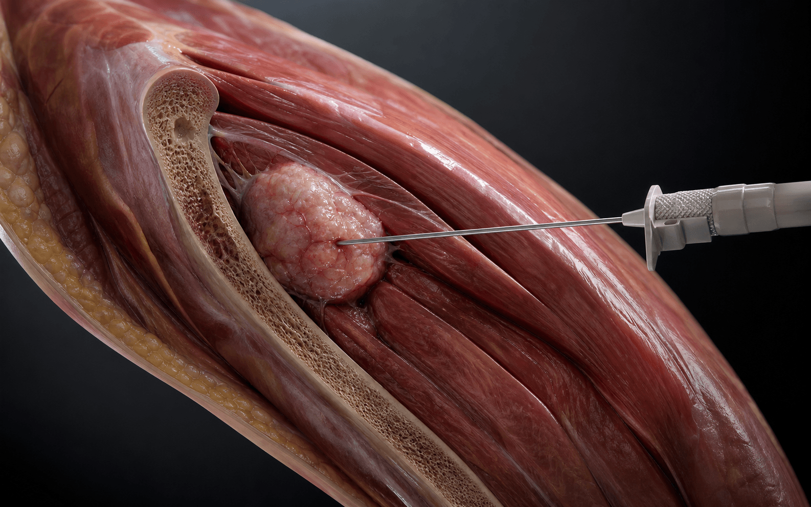


Operative sequence
- Before any procedure, the tract is agreed with the surgeon who will perform the resection, so that it can be excised en bloc with the specimen.
- Longitudinal orientation, aligned with the planned resection incision; a transverse incision is forbidden because it contaminates the whole limb width and cannot be excised.
- Shortest direct route from skin to the most representative (viable) part of the tumour.
- Single compartment — never cross an uninvolved compartment, joint, or neurovascular interval.
- Clear of the neurovascular bundle — image-guidance lets the radiologist choose a corridor that avoids vessels and nerves.
- Marked / tattooed so the tract is unmistakably identified and excised at definitive surgery.
- Confirm all staging imaging is complete and MDT-reviewed, and that the biopsy is genuinely the last step.
- Perform the biopsy under image guidance — ultrasound for soft-tissue lesions, CT for deep bone lesions — so the corridor and target are precisely controlled.
- Use a coaxial system so multiple cores pass through a single skin entry, minimising the contaminated tract.
- Target the viable enhancing periphery identified on contrast MRI / PET-avid regions; avoid the necrotic centre.
- For a core biopsy, a single skin puncture placed directly over the most superficial part of the tumour, in line with and excisable by the definitive resection.
- For an open biopsy, a small longitudinal incision directly over the tumour, in line with the resection incision.
- Go straight down through one compartment to the tumour — do NOT raise flaps, do NOT cross compartments, do NOT violate the neurovascular bundle.
- Pass the coaxial needle to the viable enhancing periphery and obtain multiple cores.
- Take enough tissue for histology, immunohistochemistry and molecular testing.
- Send tissue for histology AND microbiology — infection is the principal mimic of a bone tumour.
- The skin entry point and tract remain those agreed with the definitive surgeon and marked for later excision.
- Reserved for cases where core biopsy is non-diagnostic or where larger tissue volume is required.
- Through the longitudinal incision, take a representative sample of viable peripheral tumour under direct vision; avoid the necrotic centre.
- Confirm diagnostic lesional tissue on frozen section before closing.
- For a bone lesion, if a cortical window is unavoidable make it small, round/oval and stress-riser sparing, and fill it (bone wax / sealant) to limit haematoma and fracture.
- Achieve meticulous haemostasis — any haematoma carries tumour cells along tissue planes and expands the contaminated field.
- Apply bone wax or sealant to a cortical window.
- No drains; if drainage is genuinely unavoidable, bring the drain out in line with and immediately adjacent to the incision so the drain track lies within the future resection field.
- Close in layers to seal the tumour; avoid wide undermining.
- Frozen section (where used) confirms diagnostic lesional tissue was obtained before the patient leaves theatre — it does not give the final diagnosis.
- Document and mark the tract (skin tattoo / clear operative note, with a photograph of the entry point) so the definitive surgeon excises it en bloc with the specimen.
- Send fresh tissue promptly to pathology — the specialist sarcoma pathologist may need fresh material for cytogenetics / molecular studies (e.g. translocation studies for Ewing sarcoma and synovial sarcoma).
- Ensure adequate tissue for immunohistochemistry and molecular testing — multiple cores or a generous incisional sample.
- Longitudinal incision only — a transverse incision contaminates the whole limb width and cannot be excised.
- Do not cross uninvolved compartments or violate the neurovascular bundle — contamination of a major NV bundle may require its sacrifice and convert a salvageable limb to an amputation.
- Meticulous haemostasis — haematoma spreads tumour along tissue planes; no drain, or a drain brought out in line with the incision.
- Sample viable peripheral tissue and confirm diagnostic yield on frozen section before closing.
Before I touch the patient, the staging imaging is complete and the case has been through the sarcoma MDT. I plan the biopsy tract with the surgeon who will do the resection — it is longitudinal, in line with the resection incision, takes the shortest route to viable tumour, stays in one compartment, and never crosses a neurovascular bundle. My first-line is image-guided core needle biopsy; I sample the viable enhancing periphery, take multiple cores, send histology AND microbiology, achieve absolute haemostasis, avoid a drain, and clearly mark and document the tract so it is excised en bloc.
Aftercare & Complications
After the Biopsy — Specimen, Documentation & MDT Handling the specimen. Send fresh tissue promptly to pathology — the specialist sarcoma pathologist may need fresh material for cytogenetics and molecular studies (for example translocation studies in Ewing sarcoma and synovial sarcoma). Send tissue for histology AND microbiology in every case, because infection is the principal differential of a destructive bone lesion. Frozen section intra-operatively confirms that diagnostic lesional tissue has been obtained before the patient leaves theatre (it does not give the final diagnosis). Ensure adequate tissue for immunohistochemistry and molecular testing. Documentation and marking. Document and mark the tract (skin tattoo and a clear operative note) so the definitive surgeon excises it en bloc with the specimen. Photograph and record the entry point — a forgotten or undocumented tract may be left behind and act as a focus for recurrence. Multidisciplinary team (MDT) review. The biopsy result is interpreted in the specialist sarcoma MDT, where the radiologist, pathologist and surgeon correlate imaging, histology and clinical findings — discordance triggers re-review or re-biopsy. The definitive plan is set: neoadjuvant chemotherapy (e.g. osteosarcoma, Ewing sarcoma), radiotherapy, and the type and timing of resection. The margin required is defined (intralesional / marginal / wide / radical per Enneking) and the resection includes the biopsy tract. Radiology–pathology discordance. If the histology does not fit the imaging (for example, benign-looking histology from an aggressive-looking lesion), the result is treated as non-diagnostic / sampling error, not reassurance. The MDT re-reviews and the patient is re-biopsied rather than discharged. ## Complications of a Poorly-Planned Biopsy The complications of biopsy are overwhelmingly the consequence of poor planning and technique — and many are catastrophic for the patient's oncological outcome.
- Cause
- Poorly-placed, transverse, or cross-compartment tract; haematoma spread
- Consequence
- Forces a much wider resection or converts limb-salvage to amputation
- Prevention
- Longitudinal tract in line with resection, single compartment, meticulous haemostasis, excise the tract en bloc
- Cause
- Tract directed across or adjacent to a major NV bundle; tumour spillage
- Consequence
- May require sacrifice of the vessel/nerve at resection — limb loss or major morbidity
- Prevention
- Image-guided corridor that avoids the NV bundle; plan the tract with the definitive surgeon
- Cause
- Sampling necrotic centre or reactive rim rather than viable tumour
- Consequence
- False-negative or non-diagnostic result; delayed diagnosis; repeat biopsy
- Prevention
- Target enhancing viable periphery on MRI/PET; multiple cores; frozen section to confirm yield
- Cause
- Large biopsy window or hole in a bone already weakened by tumour
- Consequence
- Fracture haematoma contaminates surrounding tissues widely; may force amputation
- Prevention
- Use core needle where possible; if a cortical window is needed make it small, round/oval (stress-riser sparing) and fill it
- Cause
- Inadequate haemostasis after biopsy
- Consequence
- Spreads tumour cells along tissue planes; expands the contaminated field requiring excision
- Prevention
- Meticulous haemostasis; bone wax / sealant for the bone window; no drain or drain in line with the incision
- Cause
- Wide flaps, poor closure, biopsy at a non-specialist centre
- Consequence
- Delays neoadjuvant therapy and definitive surgery; further contamination
- Prevention
- Minimal-access technique, layered closure; send tissue for microbiology to distinguish infection from tumour
- Cause
- Cumulative effect of the above, classically when biopsy is done outside the treating centre
- Consequence
- Loss of limb salvage, worse oncological and functional outcome
- Prevention
- Refer intact to the sarcoma centre; biopsy by/with the definitive surgeon (Mankin principle)
- Transverse incision — contaminates the whole limb width; longitudinal only.
- Crossing compartments / NV bundles — converts salvage to sacrifice.
- Biopsy before staging imaging — distorts the MRI and ruins resection planning.
- Excisional biopsy of a sarcoma — turns the whole bed into a contaminated tract.
- Failure to send microbiology — infection is the great mimic; always culture as well as biopsy.
- Sampling the necrotic centre — non-diagnostic; target the viable periphery. The biopsy is part of a continuum that begins with referral to the sarcoma centre and ends with en-bloc excision of the tract at definitive surgery. Every decision is made to protect the option of limb salvage and an accurate diagnosis.
Viva & Exam Focus
BIOPSYBIOPSY — Principles of musculoskeletal tumour biopsy
TRACTTRACT — Planning the biopsy tract
How to plan a biopsy tract that does not compromise definitive surgery
The trap: Performing a biopsy of a suspected sarcoma at the local hospital before referral. Mankin (1982, repeated 1996) showed errors, complications and changes in course/outcome were 2 to 12 times more frequent (p less than 0.001) when biopsy is done at the referring rather than the treating centre. The fix: Refer ALL suspected bone or soft-tissue sarcomas to a specialist sarcoma centre BEFORE any biopsy. The biopsy is performed by, or in discussion with, the definitive treating surgeon.
The trap: A transverse biopsy incision contaminates the whole width of the limb and cannot be excised within a longitudinal resection ellipse — it commits the patient to a much larger resection or amputation. The fix: Always use a LONGITUDINAL incision aligned with the planned definitive resection. Transverse incisions are absolutely forbidden in extremity tumour biopsy.
The trap: Biopsy performed before staging MRI. The resulting haematoma, oedema and tract distort the tumour margins on subsequent imaging and can make resection planning impossible. The fix: Complete all staging imaging (whole-bone MRI, CT chest, bone scan / PET as indicated) BEFORE the biopsy. Biopsy is the LAST step in the staging pathway.
The trap: A tract that crosses from one muscular compartment into an uninvolved compartment contaminates that compartment, forcing it to be included in the resection. The fix: Plan the shortest tract through a single compartment, directly over the tumour. Never traverse an uninvolved compartment, joint, or neurovascular interval to reach the lesion.
Why critical: Contamination of a major neurovascular bundle by tumour spillage or a misdirected tract may require its sacrifice at resection, converting a salvageable limb to an amputation. The fix: Plan the tract to stay well clear of major vessels and nerves. Image-guided core biopsy lets the radiologist choose a safe corridor that avoids the NV bundle entirely.
The trap: Sampling the necrotic, haemorrhagic or reactive centre of a tumour yields non-diagnostic tissue and a false-negative result. The fix: Target VIABLE tissue at the enhancing periphery (guided by contrast MRI / PET-avid regions). Take multiple cores. Use frozen section to confirm lesional, diagnostic tissue is present before leaving theatre.
Clinical Decision Scenarios
Practise clinical reasoning and management decisions out loud
“A 22-year-old man is referred to your sarcoma unit with a 9 cm aggressive distal femoral lesion. The referring orthopaedic surgeon has already performed an open biopsy through a transverse incision over the medial thigh at their local hospital. What are your concerns and how does this affect his management?”
“You are planning the biopsy of a suspected soft-tissue sarcoma in the posterior compartment of the thigh. Talk me through your principles for planning and performing this biopsy.”
“Why is it so important to complete staging imaging before a biopsy, and which imaging would you obtain for a suspected primary bone sarcoma of the proximal tibia?”
The governing principles
- The biopsy is the most important and most commonly mishandled step in sarcoma care
- Performed by, or in discussion with, the definitive treating surgeon at the sarcoma / MDT centre
- Refer the lesion INTACT — do NOT biopsy a suspected sarcoma at the local hospital first
- Biopsy is the LAST step, after all staging imaging is complete and MDT-reviewed
- Mankin (1982, 1996): errors, plan changes and unnecessary amputations 2 to 12 times more common (p less than 0.001) when biopsy done at referring vs treating centre; overall diagnostic error 17.8%
Tract planning rules
- LONGITUDINAL incision, in line with the planned definitive resection — transverse is forbidden
- Tract must be excisable en bloc with the tumour — treat it as contaminated tumour
- Shortest direct route to viable tumour; stay within a single compartment
- NEVER cross uninvolved compartments, joints, or the neurovascular bundle
- Mark / tattoo and document the tract so it is excised at definitive surgery
Imaging and staging (before biopsy)
- Plain radiograph first (matrix, margin, periosteal reaction, Lodwick)
- MRI of the WHOLE bone with contrast — local stage and SKIP LESIONS; image both adjacent joints
- CT chest — mandatory; lungs are commonest metastatic site for sarcoma
- Bone scan and FDG-PET / PET-CT — skeletal and metabolic staging; PET targets viable region
- Enneking (G / T / M) drives margin; AJCC TNM used for staging and prognosis
Choice of technique
- Image-guided core needle biopsy = FIRST-LINE for most lesions (pooled accuracy about 84%, 90-95% in specialist centres, low morbidity, small tract)
- FNA: cytology only — useful for recurrence/metastasis, NOT for primary sarcoma diagnosis
- Open incisional: when core non-diagnostic — longitudinal, single compartment, frozen section, no drain
- Excisional: ONLY for small superficial clearly benign / low-grade lesions
- Never excise a suspected sarcoma as a 'biopsy' — contaminates the whole bed
Technical rules at biopsy
- Sample VIABLE peripheral tissue (MRI/PET-guided) — avoid the necrotic centre
- Take multiple cores / a generous sample for histology, IHC and molecular tests
- Send tissue for histology AND microbiology — infection is the great mimic
- Meticulous haemostasis — haematoma spreads tumour along tissue planes
- No drains; if essential, bring the drain out in line with and adjacent to the incision
- Frozen section to confirm diagnostic lesional tissue before leaving theatre
Complications of a bad biopsy
- Tract contamination — forces wider resection or amputation
- Neurovascular contamination — may require sacrifice of vessel/nerve (limb loss)
- Pathological fracture through a biopsy window — fracture haematoma contaminates widely
- Sampling error — non-diagnostic, delays diagnosis, repeat biopsy
- Change in treatment plan / unnecessary amputation — the cumulative Mankin outcome
After the biopsy / MDT
- Send fresh tissue; specialist sarcoma pathology may need it for cytogenetics / molecular studies
- Frozen section confirms diagnostic yield (NOT final diagnosis)
- Radiology-pathology-surgery correlation at the sarcoma MDT — discordance triggers re-biopsy
- MDT sets neoadjuvant therapy, resection type and required margin (Enneking)
- Biopsy tract is planned for en-bloc excision at the definitive operation
Background & Evidence
Surgical Staging of Musculoskeletal Sarcoma The staging systems are presented here for background — they are what the MDT uses to define the surgical margin required, and they explain why the biopsy tract is treated as a contaminated extension of the tumour that must be excised en bloc within an adequate margin. ### Enneking (Surgical Staging System — MSTS) The Enneking system stages musculoskeletal sarcomas by surgical grade (G), anatomical site/compartment (T) and metastasis (M), and directly drives the surgical margin required — intralesional, marginal, wide or radical.
- Grade (G)
- G1 (low)
- Site (T)
- T1 (intracompartmental)
- Interpretation
- Low-grade, contained within its compartment
- Grade (G)
- G1 (low)
- Site (T)
- T2 (extracompartmental)
- Interpretation
- Low-grade, extracompartmental spread
- Grade (G)
- G2 (high)
- Site (T)
- T1 (intracompartmental)
- Interpretation
- High-grade, contained within its compartment
- Grade (G)
- G2 (high)
- Site (T)
- T2 (extracompartmental)
- Interpretation
- High-grade, extracompartmental — the classic high-grade extremity sarcoma
- Grade (G)
- Any G
- Site (T)
- Any T, with M1 (metastasis)
- Interpretation
- Metastatic disease at presentation
References
The hazards of biopsy in patients with malignant primary bone and soft-tissue tumors
- Original Musculoskeletal Tumor Society survey of 329 biopsies of primary malignant musculoskeletal sarcomas across 16 centres
- Troubling rates of error in diagnosis and technique, with complications that adversely affected patient care
- Error and complication rates were markedly higher when biopsy was performed at the referring rather than the treating institution
The hazards of the biopsy, revisited (Members of the Musculoskeletal Tumor Society)
- Repeat survey of 597 patients from 21 institutions; results essentially unchanged 14 years on
- Overall diagnostic error 17.8%; biopsy problem forced a different/more complex operation in 19.3%; change in course or outcome in 10.1%; 18 unnecessary amputations attributable to the biopsy
- Errors, complications and changes in course/outcome were 2 to 12 times greater (p less than 0.001) when biopsy was done at a referring institution rather than a treatment centre
A system for the surgical staging of musculoskeletal sarcoma
- Stratifies sarcoma by grade (G1 low / G2 high), site (T1 intracompartmental / T2 extracompartmental) and metastasis (M0/M1)
- Defines surgical margins as intralesional, marginal, wide and radical relative to the lesion and its reactive zone
- Directly links stage to the required surgical margin
Biopsy of bone and soft-tissue lesions
- Authoritative JBJS review of biopsy principles, tract planning and technique selection in musculoskeletal oncology
- Sets out the longitudinal, single-compartment, resection-aligned tract and the role of the treating surgeon in biopsy planning
Percutaneous core needle biopsy versus open biopsy in diagnostics of bone and soft tissue sarcoma: a retrospective study
- Comparative study of 77 patients
- Overall diagnostic accuracy 92.9% for core needle biopsy versus 98.0% for open biopsy (p=0.55)
- Core needle biopsy was 100% accurate for bone tumours
A meta-analysis supports core needle biopsy by radiologists for better histological diagnosis in soft tissue and bone sarcomas
- Meta-analysis of 32 studies comprising 7,209 musculoskeletal lesions
- Pooled diagnostic accuracy of core needle biopsy 84% (95% CI 0.81-0.87) against final histology
- Accuracy significantly higher when performed by radiologists than surgeons; surgical biopsy retained slightly higher accuracy (pooled OR 0.39 for CNB vs surgical)
Fine-needle aspiration biopsy for the diagnosis of bone and soft tissue lesions: a systematic review and meta-analysis
- Meta-analysis of 4,604 FNAs from 25 studies
- Sensitivity 95.6% and specificity 96.9% for distinguishing benign from malignant nature
- Only 75.8% accurate for a definitive (subtyping) diagnosis