Intralesional extended curettage for giant cell tumour and benign-aggressive bone lesions · advanced
- The single biggest determinant of recurrence is the size of the cortical WINDOW — it must be large enough to see the ENTIRE cavity. A small window leaves blind locules and septa uncurettaged and is the commonest cause of local recurrence.
- Extended curettage = mechanical curettage of all locules and septa, THEN a high-speed burr to extend the margin into macroscopically normal bone, THEN a local adjuvant to kill residual cells. Each step reduces recurrence; omitting any of them raises it.
- PMMA cement is both a reconstruction and an adjuvant — the exotherm of polymerisation causes thermal necrosis of residual tumour, gives immediate stability, and the cement-bone interface is easy to follow radiographically for recurrence.
- Denosumab (RANKL inhibitor) is useful neoadjuvant in unresectable or axial GCT but creates a peripheral bony rind that makes the curettage plane harder to find, can mask tumour, and is followed by rebound after cessation — it does NOT lower curettage recurrence.
When & Why
Indication. Intralesional extended curettage is the workhorse joint-preserving operation for benign-aggressive and selected benign-latent or active bone lesions. The principle is identical across diagnoses: remove the lesion, extend the margin, kill residual cells, and reconstruct — preserving the adjacent joint and accepting a low local recurrence rate in exchange for limb and joint function. The exemplar lesion is giant cell tumour of bone (GCT). Lesions amenable to extended curettage:
- Giant cell tumour of bone (GCT) — the exemplar benign-aggressive lesion; Campanacci I-II and many III
- Aneurysmal bone cyst (ABC) — primary or secondary; curettage with adjuvant, often with grafting
- Chondroblastoma — epiphyseal lesion of the immature skeleton; meticulous curettage to protect the physis and joint
- Simple (unicameral) bone cyst — curettage/decompression and grafting when injection/aspiration fails or fracture risk is high
- Enchondroma (symptomatic or fracture risk) — curettage and graft; distinguish from low-grade chondrosarcoma
- Low-grade central chondrosarcoma (atypical cartilaginous tumour, grade 1) — extended intralesional curettage with adjuvant in selected centres for appendicular lesions
- Chondromyxoid fibroma, fibrous dysplasia (focal), eosinophilic granuloma — selected symptomatic or structural cases
- Typical site / age
- Epi-metaphyseal, mature skeleton, knee/distal radius
- Curettage role
- First-line extended curettage
- Reconstruction tendency
- PMMA cement (surveillance + adjuvant)
- Typical site / age
- Metaphyseal, young, eccentric expansile
- Curettage role
- Curettage + adjuvant (plus or minus embolisation)
- Reconstruction tendency
- Bone graft / substitute
- Typical site / age
- Epiphysis, immature skeleton
- Curettage role
- Meticulous curettage, protect physis
- Reconstruction tendency
- Bone graft (biological)
- Typical site / age
- Proximal humerus/femur metaphysis, child
- Curettage role
- Curettage/decompression if injection fails
- Reconstruction tendency
- Graft / substitute
- Typical site / age
- Hand, proximal humerus, femur
- Curettage role
- Curettage + graft
- Reconstruction tendency
- Bone graft / substitute
- Typical site / age
- Long-bone metadiaphysis, adult
- Curettage role
- Selected extended curettage + adjuvant
- Reconstruction tendency
- PMMA cement (plus or minus fixation)
Contraindications.
- Absolute: confirmed intermediate- or high-grade malignancy (e.g. grade 2-3 chondrosarcoma, osteosarcoma) — these require wide en bloc resection; and extensive joint destruction or pathological fracture with intra-articular tumour where reconstruction cannot restore a functional joint.
- Relative: a massive cortical breach or soft-tissue mass (favours resection), multiply recurrent GCT after adequate curettage (consider resection), or a lesion abutting articular cartilage so closely that an adjuvant would damage the joint.
En bloc (wide) resection cures the lesion with a margin of normal tissue but sacrifices the joint and requires reconstruction (endoprosthesis, osteoarticular allograft, or arthrodesis). It is reserved for extensive bony or articular destruction, large soft-tissue extension, multiply recurrent disease, or malignancy. For the majority of GCT and benign-aggressive lesions, extended curettage achieves comparable oncological control with far better function — the goal of intralesional surgery is to preserve the joint and accept a low recurrence in exchange for function.
Consent specifically for local recurrence (GCT 10-20 percent after extended curettage), the small risk of malignant transformation and pulmonary metastases, adjuvant-specific risks (chemical burn, cryo-induced fracture, thermal joint injury), the possibility of conversion to resection, and the need for surveillance imaging. Setup. Supine for distal femur or proximal tibia lesions, with a tourniquet on the limb where feasible and oncologically appropriate. Prepare and drape widely to allow extensile access and reconstruction. Confirm the diagnosis histologically (image-guided or open biopsy) before definitive curettage, and stage the chest with CT in GCT (pulmonary metastases in 3-5 percent).
The Operation
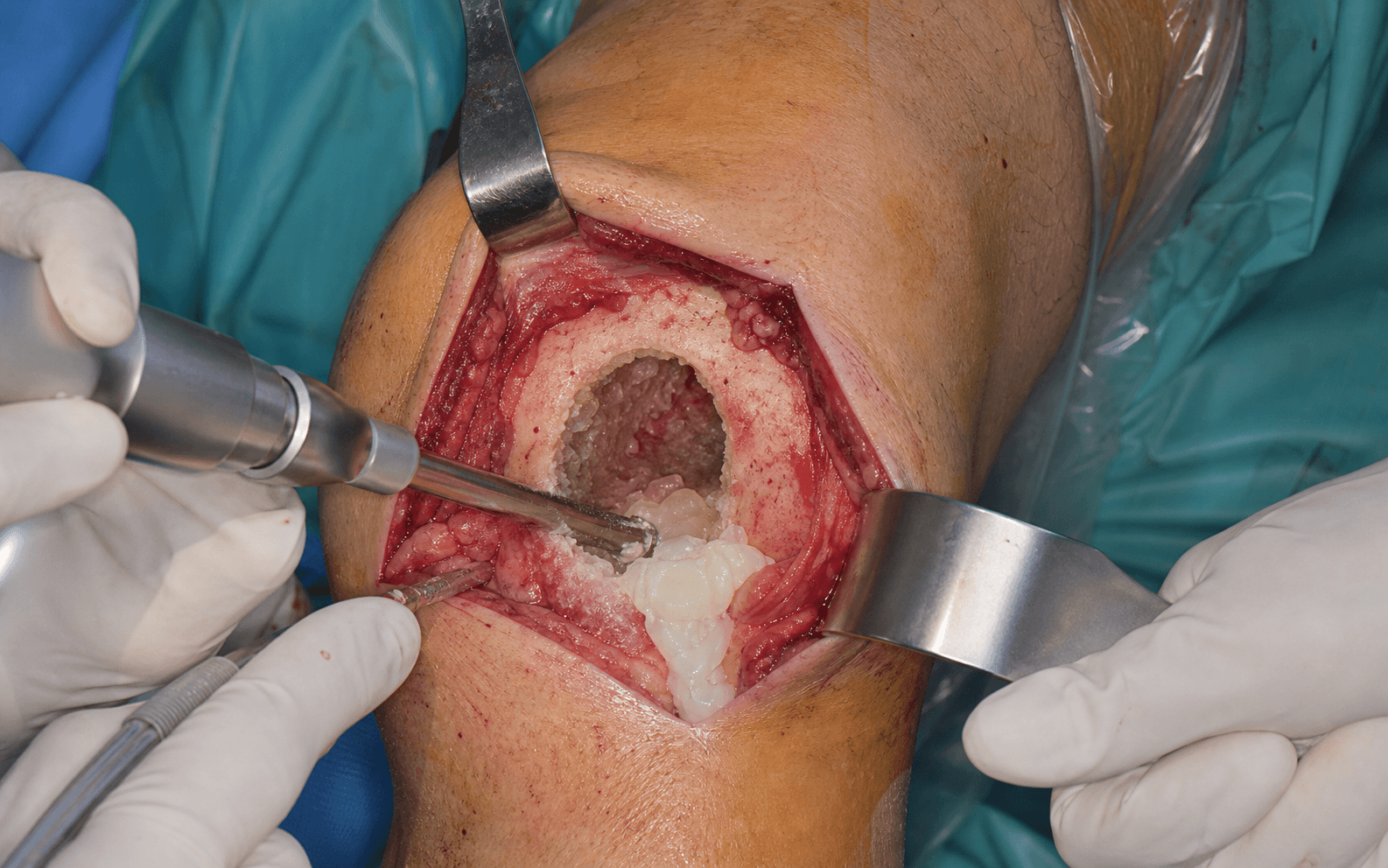
The goal: expose the lesion through a planned approach, make a cortical window large enough to see the whole cavity, mechanically clear every locule and septum, extend the margin with a high-speed burr, apply a local adjuvant to kill residual cells, and reconstruct the defect — all while protecting the adjacent joint. The exposure and window are laid out as the first steps below: they are the heart of the operation and the single biggest determinant of recurrence.


Operative sequence
- Use a direct approach over the lesion that respects future resection planes — include the biopsy tract so it can be excised, and do not contaminate extra compartments unnecessarily.
- Expose the cortex overlying the lesion through an extensile incision.
- Identify and protect adjacent neurovascular structures, and pack off the soft tissues with swabs to create a sealed field. This protects the soft tissues from tumour spill now and from the chemical or thermal adjuvant later.
- Outline the window with an osteotome or oscillating saw and remove the cortical lid.
- The window must expose the entire cavity from one end to the other — not just the lytic centre. It has to be large enough for curettes and a burr to reach every recess under direct vision.
- Plan the window to protect the articular cartilage and the growth plate where relevant.
- Using a full range of sharp curettes (straight, curved, ring, angled), systematically remove all tumour, locules and bony septa, working circumferentially and into every recess.
- Send curettings for histology to confirm the diagnosis and grade.
- Continue until only macroscopically normal bleeding bone remains; directly visualise and clear any subchondral recess, working carefully near the joint surface to avoid penetrating the subchondral plate.
- After mechanical curettage, use a high-speed burr to take the cavity walls down a further few millimetres into macroscopically normal bone.
- The burr removes the microscopic tumour layer the curette leaves behind and is itself a mechanical adjuvant.
- Burr the whole circumference, including under overhanging edges. Treat curettage and burring as two distinct steps: the curette clears the bulk, the burr extends the margin.
- Irrigate copiously with pulsed lavage to flush tumour debris from the cavity and bone interstices before applying the chemical or thermal adjuvant.
- Lavage also dislodges loose cells the burr has liberated.
- A local adjuvant treats the residual tumour cells in the bony interstices that surgery cannot physically remove. Adjuvants are chemical, thermal, or mechanical.
- Chemical: phenol (5 percent) applied on cotton-tipped applicators in a sealed, soft-tissue-protected field and neutralised with absolute alcohol — never where the cavity communicates with the joint; hydrogen peroxide is a gentler oxidising agent that also aids haemostasis.
- Thermal: cryotherapy with liquid nitrogen gives the largest necrotic margin (about 1-2 cm) but weakens bone; the PMMA polymerisation exotherm (about 60-90 degrees Celsius) is itself a thermal adjuvant.
- Mechanical / surface: argon beam or electrocautery coagulation of the cavity wall; the high-speed burr already performed is the mechanical adjuvant.
- The dominant variable in lowering recurrence is meticulous removal with the high-speed burr — a systematic review and meta-analysis (Algawahmed) did not show improved local control from a chemical or thermal adjuvant once meticulous burring was performed, while registry or series evidence favours PMMA cement specifically. Adjuvant choice is therefore institution- and site-dependent.
- PMMA cement gives immediate structural stability, an adjuvant exotherm, and a sharp cement-bone interface that makes radiographic surveillance easy — but it does not biologically restore bone and, against the subchondral plate, transmits heat and stiffness to cartilage.
- Bone graft or substitute (autograft, allograft, calcium phosphate/sulphate, bioactive ceramics) incorporates and remodels and is joint-friendly, but it obscures radiographic surveillance (incorporating graft mimics recurrence), has no adjuvant effect, and risks resorption or donor-site morbidity.
- When the cavity abuts the subchondral plate, lay a thin layer of subchondral cancellous bone graft (or a deliberate gap) between the cement and the articular surface to buffer the cartilage from the exotherm and the stiffness mismatch.
- Add a plate and/or screws (a composite construct, with cement augmentation around screws) when the window and curettage have left the bone structurally compromised, the lesion is peri-articular with a thin subchondral plate, or there is an actual or impending pathological fracture.
- Layered closure; protect weight-bearing where the construct is weakened.
- Plan surveillance: most recurrences appear within 2 years and are detected at the cement-bone interface or as new lysis on follow-up films, so watch that interface at every review.
- Mechanism
- Protein coagulation (chemical necrosis)
- Strengths
- Cheap, effective, widely available
- Hazards / cautions
- Caustic burn to skin/nerve/joint; never if joint communication
- Mechanism
- Oxidative cell lysis + haemostasis
- Strengths
- Lower caustic risk, aids haemostasis
- Hazards / cautions
- Gas embolism risk if forced into vascular bone (rare)
- Mechanism
- Freeze-thaw necrosis, 1-2 cm margin
- Strengths
- Largest necrotic margin of the adjuvants
- Hazards / cautions
- Bone weakening/fracture, skin necrosis, nerve injury
- Mechanism
- Thermal necrosis (60-90 C set)
- Strengths
- Adjuvant + reconstruction + surveillance in one
- Hazards / cautions
- Thermal joint/cartilage injury if no subchondral buffer
- Mechanism
- Thermal coagulation of cavity wall
- Strengths
- Controlled, surface-directed
- Hazards / cautions
- Limited depth; smoke/plume; equipment-dependent
- Mechanism
- RANKL blockade, anti-osteoclast
- Strengths
- Reossifies/controls axial and unresectable GCT
- Hazards / cautions
- Obscures plane, rebound on stopping, ONJ; not for routine appendicular curettage
- PMMA cement
- Immediate, high
- Bone graft / substitute
- Lower until incorporation
- PMMA cement
- Yes (exotherm 60-90 C)
- Bone graft / substitute
- None
- PMMA cement
- Easy (sharp cement-bone interface)
- Bone graft / substitute
- Difficult (graft mimics recurrence)
- PMMA cement
- None (permanent foreign body)
- Bone graft / substitute
- Yes (incorporates/remodels)
- PMMA cement
- Thermal + stiffness (buffer with subchondral graft)
- Bone graft / substitute
- Low
- PMMA cement
- GCT, adults, peri-articular needing surveillance
- Bone graft / substitute
- Children, growing skeleton, small benign cysts
A window that is too small leaves blind locules and septa uncurettaged and is the single commonest cause of local recurrence. Make the window as long as the cavity so that every part of it is seen directly, and anticipate fixation if the window leaves the bone structurally weakened. Plan the window to avoid breaching an uninvolved joint or growth plate. Phenol must never be used where the cavity communicates with the joint; cryotherapy mandates skin protection and construct augmentation because it weakens bone.
For a peri-articular GCT around the knee, lay a thin layer of subchondral cancellous graft (or leave a small gap) between the cement and the articular surface before cementing. It buffers the cartilage from the polymerisation exotherm and from the stiffness of a direct cement-on-subchondral-bone construct, reducing the risk of post-operative joint degeneration. If the construct is weak, add a buttress plate and accept that you are building a composite.
The curette clears the bulk; the burr extends the margin and reaches what the curette flattens against the wall. Burring the entire cavity wall into macroscopically normal bone is one of the simplest things that reliably lowers recurrence — curettage alone leaves microscopic tumour in the bony interstices and gives recurrence rates of 25-50 percent in GCT, falling to roughly 10-20 percent once made "extended".
Aftercare & Complications
Follow-up and surveillance. Local recurrence is the central concern after intralesional surgery. For GCT after extended curettage (window + curette + burr + adjuvant + reconstruction) recurrence is approximately 10-20 percent; curettage alone gives 25-50 percent. Most recurrences present within 2 years and are detected at the cement-bone interface or as new lysis on surveillance imaging. Recurrence is treated by repeat extended curettage or, if extensive or multiply recurrent, by en bloc resection. Stage and follow the chest in GCT.
- Approximate rate
- 10-20% extended curettage; 25-50% curettage alone
- Recognition
- New lysis or lucent rim at cement-bone interface; recurrent pain or swelling, usually within 2 years
- Prevention and management
- Prevention: large window, full curettage, high-speed burr, local adjuvant, surveillance. Management: repeat extended curettage; en bloc resection if extensive or multiply recurrent
- Approximate rate
- 5-10% (higher after cryotherapy)
- Recognition
- New pain, deformity or fracture on loading post-op, especially after a large window or cryo-weakened bone
- Prevention and management
- Prevention: augment construct (cement and/or fixation); protect weight-bearing. Management: fixation plus or minus revision reconstruction
- Approximate rate
- uncommon with technique
- Recognition
- Skin/soft-tissue necrosis, nerve dysfunction, chondrolysis if phenol reached the joint
- Prevention and management
- Prevention: sealed field, soft-tissue protection, alcohol neutralisation, never with joint communication. Management: debride necrosis; treat nerve injury; supportive joint care
- Approximate rate
- uncommon with closed systems
- Recognition
- Skin necrosis over the freeze zone; neurapraxia of adjacent nerve
- Prevention and management
- Prevention: warm saline irrigation of skin, gauze gutter, closed-probe systems, protect adjacent nerve. Management: wound care; most neurapraxias recover
- Approximate rate
- variable, long-term
- Recognition
- Progressive peri-articular pain and radiographic joint-space narrowing over years
- Prevention and management
- Prevention: subchondral cancellous graft buffer layer under cement; avoid direct cement on cartilage. Management: analgesia; arthroplasty for end-stage degeneration
- Approximate rate
- GCT on denosumab
- Recognition
- Rapid osteolysis after stopping the drug; viable tumour beneath a reossified rind found at surgery
- Prevention and management
- Prevention: reserve for axial/unresectable disease; do not rely on it for appendicular cure. Management: timely surgery; treat hypocalcaemia; monitor for ONJ and atypical fracture
- Approximate rate
- 3-5%
- Recognition
- Lung nodules on staging or surveillance CT; histologically identical to primary, often indolent
- Prevention and management
- Prevention: not preventable. Management: observe many; resect symptomatic or growing nodules; systemic denosumab for progressive disease
- Approximate rate
- 1-3% (higher post-radiotherapy)
- Recognition
- Aggressive growth, new soft-tissue mass, high-grade histology on re-biopsy
- Prevention and management
- Prevention: avoid radiotherapy where surgery is feasible. Management: re-stage and treat as a high-grade sarcoma (wide resection plus or minus chemotherapy)
- Approximate rate
- 1-3%
- Recognition
- Wound erythema, discharge, fever, raised inflammatory markers
- Prevention and management
- Prevention: meticulous technique, prophylactic antibiotics. Management: washout, antibiotics; retain or revise construct depending on stability
Viva & Exam Focus
CURETTECURETTE — the extended curettage sequence
GIANTGIANT — giant cell tumour of bone essentials
Critical principles and exam traps
The trap: making a window the size of the radiographic lytic area, or a keyhole through intact cortex. Residual tumour hides in locules and behind septa you cannot see. The fix: create a cortical window that exposes the WHOLE cavity from end to end. The single most important technical factor in reducing recurrence is direct visualisation of every part of the cavity.
The trap: mechanical curettage alone leaves microscopic tumour in the bony interstices and gives recurrence rates of 25-50 percent in giant cell tumour. The fix: always follow curettage with a high-speed burr to extend the margin, then a local adjuvant. Extended curettage lowers GCT recurrence to roughly 10-20 percent.
The risk: phenol is a caustic protein coagulant. Spillage onto skin, the neurovascular bundle, or the joint causes chemical burns, nerve injury, and chondrolysis. The fix: protect soft tissues with swabs and a sealed field, apply phenol with a cotton-tipped applicator, then neutralise with absolute alcohol. Never use it where the cavity communicates with the joint.
The risk: liquid nitrogen freezes a 1-2 cm margin of bone but weakens it — postoperative fracture risk is real. Spillage causes skin necrosis; overflow near a nerve causes neurapraxia. The fix: protect skin (warm saline irrigation, gauze gutter), augment the construct (cement and/or fixation), and protect weight-bearing. Modern closed-probe systems reduce spillage versus pour techniques.
The trade-off: PMMA gives immediate stability, an adjuvant exotherm, and a sharp radiographic interface that makes early recurrence easy to detect. Bone graft is biological but OBSCURES the cavity radiographically and has no adjuvant effect. The implication: for benign-aggressive lesions where recurrence surveillance matters (GCT), cement is often preferred. Graft is favoured in children, smaller benign cysts, and when biological restoration is the priority.
The trap: treating a pain-causing, endosteally-scalloped cartilage lesion as a benign enchondroma. Intralesional surgery of a true (intermediate or high-grade) chondrosarcoma seeds the field and compromises cure. The fix: confirm low grade before curettage. Only low-grade central (atypical cartilaginous, grade 1) lesions are curetted in selected centres; deep endosteal scalloping, pain, and growth point to formal resection.
Clinical Decision Scenarios
Practise clinical reasoning and management decisions out loud
“A 32-year-old presents with knee pain. Radiographs show an eccentric, lytic, expansile lesion in the distal femur extending to the subchondral bone, with a thinned but intact cortex (Campanacci grade II). Biopsy confirms giant cell tumour of bone. How do you manage this?”
“During curettage of a giant cell tumour you are planning to use a chemical adjuvant. Talk me through the principles of local adjuvants — what options exist, and what are the specific hazards of each?”
“You are reconstructing the cavity after extended curettage of a peri-articular giant cell tumour. You have a choice between PMMA cement and bone graft. How do you decide, and what are the trade-offs?”
Core principle
- Intralesional EXTENDED curettage preserves the joint and accepts a low recurrence (GCT 10-20%) in exchange for function
- En bloc resection cures but sacrifices the joint — reserved for extensive destruction, multiply recurrent disease, or malignancy
- Extended = LARGE window + full curettage + high-speed burr + local adjuvant + reconstruction
- The size of the cortical WINDOW is the single biggest determinant of recurrence
- Always confirm a benign or benign-aggressive diagnosis histologically before curetting
Lesions amenable
- Giant cell tumour of bone — the exemplar benign-aggressive lesion
- Aneurysmal bone cyst, chondroblastoma, simple bone cyst, symptomatic enchondroma
- Low-grade central chondrosarcoma (atypical cartilaginous tumour, grade 1) in selected centres only
- NEVER intralesional surgery for confirmed grade 2-3 chondrosarcoma or osteosarcoma — wide resection
Technique — key steps
- 1. Approach including the biopsy tract; pack off and protect soft tissues (sealed field)
- 2. LARGE cortical window exposing the ENTIRE cavity end to end
- 3. Thorough mechanical curettage of all locules and septa; send curettings
- 4. High-speed burr to extend the margin into normal bone (mechanical adjuvant)
- 5. Pulsed lavage to flush debris before adjuvant
- 6. Local adjuvant (chemical or thermal) in a protected field
- 7. Reconstruct (cement plus or minus subchondral graft buffer, or graft) plus or minus internal fixation
Local adjuvants
- Chemical: phenol (5%) with absolute alcohol neutralisation; hydrogen peroxide
- Thermal: cryotherapy (liquid N2, 1-2 cm margin, weakens bone); PMMA exotherm 60-90 C
- Mechanical or other: high-speed burr; argon beam coagulation
- Phenol caustic — sealed field, never if joint communication, protect nerve and skin
- Cryotherapy: largest margin but fracture, skin and nerve risk — protect and augment construct
Reconstruction — cement vs graft
- PMMA: immediate stability, exotherm adjuvant, easy surveillance at cement-bone interface; no biology, heat and stiffness to joint
- Subchondral cancellous graft layer UNDER cement protects the articular cartilage
- Bone graft or substitute: biological, joint-friendly, preferred in children and small cysts
- Graft downside: obscures surveillance (mimics recurrence), no adjuvant effect
- Add plate or screws (composite) if the window leaves the bone structurally compromised
Giant cell tumour specifics
- Eccentric lytic epiphyseal/metaphyseal lesion in a mature skeleton; around the knee and distal radius
- Campanacci grade (I-III) reflects cortical integrity, not malignancy
- Neoplastic stromal cell expresses RANKL; reactive osteoclast-like giant cells; H3F3A G34W mutation
- Denosumab (RANKL inhibitor): neoadjuvant for axial or unresectable disease; obscures plane, rebounds, NOT a curettage substitute
- Benign pulmonary metastases 3-5% (stage the chest); malignant transformation 1-3% (higher post-radiotherapy)
Complications and recurrence
- Local recurrence: 10-20% extended; 25-50% curettage alone; usually within 2 years
- Iatrogenic or pathological fracture: 5-10%, higher after cryotherapy — augment and protect loading
- Adjuvant injury: phenol chemical burn; cryo skin necrosis or nerve injury — protect the field
- Joint degeneration from cement on subchondral bone — buffer with a graft layer
- Recurrence management: repeat extended curettage; en bloc resection if extensive or multiply recurrent
Background & Evidence
The lesion. Giant cell tumour of bone is benign but locally aggressive — an eccentric, lytic, epiphyseal or metaphyseal lesion in a skeletally mature patient, typically around the knee and at the distal radius. The neoplastic stromal cell expresses RANKL, which recruits the reactive, osteoclast-like giant cells that drive the destructive osteolysis; the H3F3A G34W mutation is characteristic. GCT can produce benign pulmonary "metastases" (3-5 percent) that are histologically identical and often indolent, and true malignant transformation is rare (1-3 percent, higher after radiotherapy) — which is why the chest is staged and followed.
- Cortical integrity
- Intact, thickened cortex; well-defined margin
- Typical share
- ~4%
- Cortical integrity
- Cortex thinned and expanded but intact
- Typical share
- ~74%
- Cortical integrity
- Cortical breach with soft-tissue extension
- Typical share
- ~22%
References
Giant-cell tumor of bone — defining series and radiographic grading
- 327 patients from the Rizzoli Institute; radiographic grade was I in 4%, II in 74%, III in 22%, and did NOT correlate with recurrence risk
- Local recurrence 27% after intralesional procedures, 8% after marginal excision, 0% after wide or radical resection
- 90% of local recurrences appeared within the first 3 years; growth-plate adjacency in only 2%, articular invasion and trans-articular spread documented
Giant cell tumor of bone: risk factors for recurrence
- 118 patients with benign GCT; intralesional surgery recurred in 25% versus 5% after wide resection
- PMMA cement reconstruction lowered recurrence after intralesional surgery compared with bone grafting; phenol alone had NO independent effect on recurrence
- Pulmonary metastases occurred in 4% and were controllable, so were not by themselves an indication for wide resection of the primary
High-speed burring with and without surgical adjuvants in intralesional management of GCT — systematic review and meta-analysis
- Six paired-cohort studies, 387 patients, all treated with curettage plus high-speed burring with or without a chemical or thermal adjuvant
- Pooled data did NOT show improved local control from adding a surgical adjuvant once meticulous burring was performed
- Conclusion: the burr is the dominant variable; a chemical or thermal adjuvant may add little when tumour removal is meticulous
Denosumab may increase the risk of local recurrence in GCT treated with curettage
- Local recurrence 60% (15 of 25) after curettage plus denosumab versus 16% (36 of 222) after curettage alone
- Joint preservation lower with denosumab (80% versus 94%); denosumab was the only independent factor associated with poor recurrence-free survival
- Viable tumour was present in all 30 denosumab-treated specimens despite radiographic reossification
Denosumab in patients with giant-cell tumour of bone — multicentre, open-label, phase 2 study
- 532 patients across 12 countries; in the surgically salvageable cohort 92% had no surgery within the first 6 months of denosumab
- Disease control achieved in unresectable disease; grade 3 or worse events included hypophosphataemia (5%) and osteonecrosis of the jaw (3%), with sarcomatous transformation in 1%
- Confirms reossification and pain control but documents the toxicity profile and need for caution
Cryosurgery in the treatment of giant cell tumors of bone — 52 consecutive cases
- Liquid-nitrogen cryotherapy after curettage achieved durable local control in a 52-case series with joint motion usually preserved
- Documented the characteristic hazards: postoperative pathological fracture, delayed healing, skin necrosis and infection
- Foundational description of freeze-thaw necrosis as a local adjuvant in benign-aggressive bone tumour surgery