Open reduction of the dysplastic hip when closed reduction fails or the child presents late — remove the obstacles, achieve a concentric reduction, hold in the human position, avoid AVN | advanced
- APPROACH SELECTION BY AGE: the medial (Ludloff/Ferguson) approach suits the young child (roughly 6-18 months) with no acetabular procedure needed; the anterior (Smith-Petersen/bikini) approach is favoured in the older child (greater than 18-24 months) because it allows capsulorrhaphy and a concurrent pelvic osteotomy through the same exposure.
- OBSTACLES TO REDUCTION (extra-articular then intra-articular): a tight iliopsoas tendon and capsular constriction (hourglass) are the EXTRA-articular obstacles; the inverted limbus/labrum, hypertrophied ligamentum teres, pulvinar (fibrofatty tissue), and a hypertrophied transverse acetabular ligament (TAL) are the INTRA-articular obstacles. All must be addressed to seat the head.
- AVASCULAR NECROSIS of the femoral head is the most feared and most discussed complication — it is iatrogenic, related to forced or abducted positioning, excessive pressure on the reduced head, and damage to the medial femoral circumflex artery (MFCA). Hold in the safe HUMAN position (flexion with moderate abduction), NOT the extreme frog-leg/Lorenz position.
- The MEDIAL APPROACH places the MEDIAL FEMORAL CIRCUMFLEX ARTERY at direct risk as it passes between pectineus and iliopsoas — the anatomical reason some surgeons reserve the medial approach for the youngest children. NOTE the evidence nuance: a meta-analysis (Novais et al., 2016) found NO significant difference in osteonecrosis rates between the medial and anterior approaches (roughly 19% each), so the MFCA argument is anatomical/theoretical rather than proven in pooled outcome data.
When & Why
Indication. Open reduction is required when a concentric, stable reduction of the dislocated hip cannot be achieved or maintained by non-operative (closed) means, or when the child presents too late for those methods to succeed. Principal indications - Failed Pavlik harness in the infant — persistent dislocation after about 3 weeks, or the "Pavlik harness disease" of a posteriorly subluxed femoral head eroding the posterior acetabulum (abandon the harness)
- Failed closed reduction under anaesthesia — an irreducible hip, or one reducible only in an unsafe (extreme or forced) position outside the safe zone of Ramsey
- Unstable or non-concentric closed reduction — arthrogram shows an excessive medial dye pool (greater than the contralateral side), indicating interposed soft tissue
- Late-presenting DDH — the walking child, or any child presenting beyond the age at which closed methods reliably succeed (commonly cited beyond 18-24 months)
- Teratologic / syndromic dislocation — fixed antenatal dislocations (e.g. arthrogryposis, myelomeningocele) that are irreducible closed Age-based strategy — the classic exam framework. Management of DDH is stratified by age, and approach selection follows from it:
Pavlik harness (closed, dynamic). Open reduction is rarely needed at this age.
Closed reduction with arthrogram and spica under anaesthesia; if it fails or is non-concentric, open reduction. The medial (Ludloff) approach is an option in this window.
Open reduction, generally via the anterior (Smith-Petersen/bikini) approach, which allows capsulorrhaphy and a concurrent pelvic osteotomy.
Anterior open reduction plus a femoral shortening/derotation osteotomy and frequently a pelvic (Salter) osteotomy for the dysplastic, shallow acetabulum.
Approach selection. The medial approach suits the young child who needs no acetabular procedure; the anterior approach is favoured in the older child because it allows capsulorrhaphy and a concurrent pelvic osteotomy through the same exposure:
- Medial (Ludloff/Ferguson)
- Younger child (roughly 6-18 months)
- Anterior (Smith-Petersen/bikini)
- Older child (greater than 18-24 months)
- Medial (Ludloff/Ferguson)
- Medial; between adductors/pectineus and iliopsoas; direct route to inferomedial obstacles
- Anterior (Smith-Petersen/bikini)
- Anterior; between sartorius/TFL with iliac apophysis split; broad acetabular exposure
- Medial (Ludloff/Ferguson)
- Excellent — direct release at the lesser trochanter
- Anterior (Smith-Petersen/bikini)
- Good — tenotomy through the wound
- Medial (Ludloff/Ferguson)
- No — cannot effectively plicate the superolateral capsule
- Anterior (Smith-Petersen/bikini)
- Yes — T-capsulotomy and plication is a key advantage
- Medial (Ludloff/Ferguson)
- No — needs a separate exposure
- Anterior (Smith-Petersen/bikini)
- Yes — same incision allows Salter or acetabuloplasty
- Medial (Ludloff/Ferguson)
- MFCA lies in the interval (anatomical concern); pooled AVN about 19%
- Anterior (Smith-Petersen/bikini)
- Less direct MFCA exposure; pooled AVN about 20% — similar; positioning still drives AVN
- Medial (Ludloff/Ferguson)
- Hidden medial scar
- Anterior (Smith-Petersen/bikini)
- Bikini variant gives a cosmetic transverse scar
- Medial (Ludloff/Ferguson)
- Young child, no acetabular procedure needed, simple reduction
- Anterior (Smith-Petersen/bikini)
- Older child, capsulorrhaphy and/or pelvic osteotomy required
Relative contraindications and cautions. A hip that achieves a safe, concentric, stable CLOSED reduction should not be opened. A very high, long-standing bilateral dislocation in an older child may be left if it is painless and function is acceptable — an individualised decision, as some advocate leaving a painless bilateral dislocation. Active local infection or unfitness for anaesthesia also stay the surgeon's hand. Consent specifically for avascular necrosis (the principal long-term risk), redislocation or loss of reduction, residual dysplasia needing later surgery, lateral femoral cutaneous nerve injury (anterior approach), MFCA injury (medial approach), infection, and the prolonged spica cast with its hygiene implications. Setup. Supine. For the medial approach the hip is flexed, abducted and externally rotated for access (an access position, not the cast position). For the anterior approach a sandbag or bump under the ipsilateral buttock tilts the pelvis; the hemipelvis and whole leg are prepped free with the image intensifier available. Examination under anaesthesia with an arthrogram is performed first to confirm whether the hip reduces closed and concentrically.
The Operation
The goal is to remove every obstacle, seat the femoral head concentrically in the true acetabulum, and hold it in the safe human position — while protecting the blood supply to the femoral head. The exposure IS the heart of the operation: the timeline below runs through the shared start (EUA and arthrogram, setup), then the MEDIAL approach for the young child and the ANTERIOR approach for the older child as two parallel branches, finishing with the shared spica and imaging steps.


Operative sequence
- Examination under anaesthesia with an arthrogram FIRST. If the hip reduces concentrically within the safe zone of Ramsey, hold it closed in a spica — do not open it.
- An excessive medial dye pool ("rose-thorn"), greater than the contralateral side, means interposed soft tissue — proceed to open reduction.
- Medial: supine, hip flexed, abducted and externally rotated for access (this is the access position, not the casting position).
- Anterior: supine with a bump under the ipsilateral buttock; whole leg prepped free; image intensifier available.
- Transverse incision over the adductor mass, just distal to the groin crease. Identify adductor longus.
- Ludloff (anteromedial): develop the interval anterior to adductor longus, between pectineus (with the neurovascular bundle, anteriorly) and adductor brevis/iliopsoas.
- Ferguson (true medial): the interval between adductor brevis (anterior) and adductor magnus (posterior), behind adductor longus.
- Both give a direct route to the inferomedial capsule, the iliopsoas tendon, the ligamentum teres and the transverse acetabular ligament — exactly the obstacles blocking reduction in the young child.
- Perform an adductor longus tenotomy to improve access and reduce the deforming force.
- Follow the plane down to the lesser trochanter; identify and release or recess the iliopsoas tendon to relieve the hourglass capsular constriction.
- Protect the medial femoral circumflex artery in the pectineus-iliopsoas interval (see the safety alert below).
- Open the inferomedial capsule.
- Excise the ligamentum teres (follow it down to the true acetabular floor), clear the pulvinar, and divide the hypertrophied transverse acetabular ligament.
- Evert any inverted limbus (do NOT excise the labrum — it aids acetabular development).
- Reduce the head into the cleared true acetabulum and confirm a concentric, stable reduction with a generous safe arc of motion.
- The medial approach does NOT permit capsulorrhaphy — stability relies on the cleared acetabulum and the cast.
- Bikini (transverse) or oblique incision below the iliac crest.
- Superficial internervous interval: between sartorius (femoral nerve, medial) and tensor fasciae latae (superior gluteal nerve, lateral).
- Protect the lateral femoral cutaneous nerve near the ASIS (meralgia paraesthetica if injured).
- Deep internervous interval: between rectus femoris (femoral nerve) and gluteus medius (superior gluteal nerve).
- Split the iliac apophysis; reflect the abductors subperiosteally off the lateral ilium (for the pelvic osteotomy) and reflect the iliacus off the inner table to expose the anterior capsule.
- Identify and divide the iliopsoas tendon. Dividing it proximally at the pelvic brim (rather than distally at the lesser trochanter) may protect the MFCA — see the evidence alert below.
- Perform a T-shaped capsulotomy (along the femoral neck axis, then along the acetabular rim) to fully expose the joint.
- Excise the ligamentum teres and pulvinar, divide the TAL, and evert or radially incise the inverted limbus.
- Reduce the head into the true acetabulum and confirm concentricity and stability.
- If the head sits proximal or the reduction is tense, perform a subtrochanteric femoral shortening osteotomy, removing a segment and correcting excessive anteversion (derotation) and coxa valga (varus) as needed; fix with a paediatric plate.
- This decompresses the reduction and lowers AVN risk — the modern alternative to pre-operative traction.
- Add a Salter innominate osteotomy (or a Pemberton/Dega acetabuloplasty) to provide anterolateral coverage of the now-reduced head.
- Excise the redundant superolateral capsule and plicate (capsulorrhaphy) to reinforce the reduction — the defining advantage of the anterior approach.
- Apply a hip spica in the SAFE human position: about 95-100 degrees flexion, about 40-50 degrees abduction (MODERATE, not extreme), neutral-to-slight internal rotation.
- AVOID the extreme abducted frog-leg (Lorenz) position — it compresses the head and kinks the MFCA, the classic precipitant of AVN.
- Obtain a post-reduction CT or limited MRI through the cast to confirm a concentric reduction (and exclude posterior redislocation, which plain films miss).
- Typical immobilisation is about 12 weeks (often 6 weeks, then EUA/cast change and re-imaging, then a further 6 weeks).

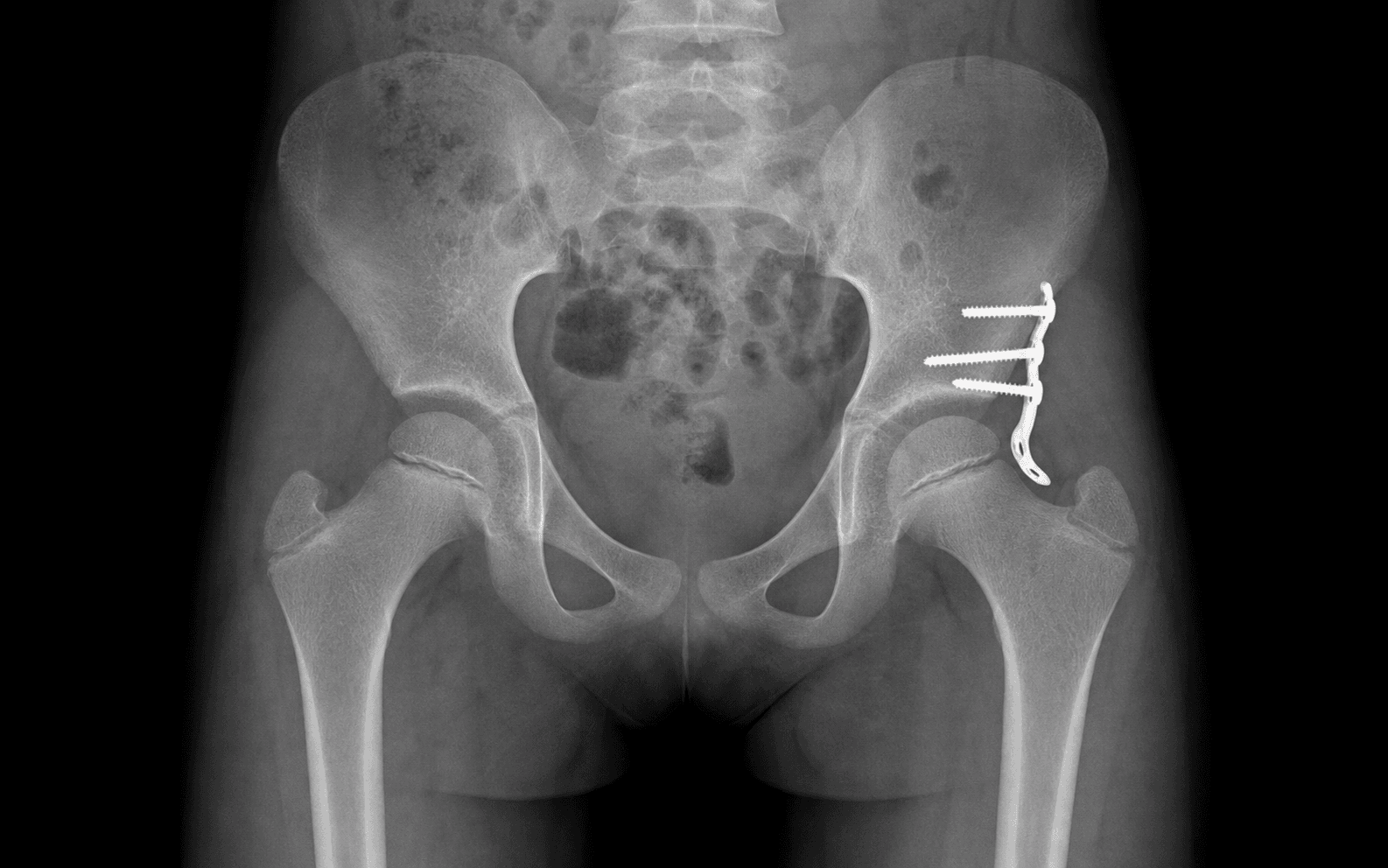
In the medial (Ludloff) approach the medial femoral circumflex artery runs between pectineus and iliopsoas, close to the lesser trochanter and the inferomedial capsule — directly in the operative interval. Stay on the iliopsoas tendon, divide it under vision, and avoid blind deep medial retraction. The anterior branch of the obturator nerve runs on or near adductor brevis and is also at risk; do not stray posteriorly toward the profunda. Note the evidence nuance: a meta-analysis (Novais et al., 2016, PMID 26472583) found NO significant difference in osteonecrosis between the medial and anterior approaches (roughly 19% each), so the MFCA argument is anatomical rather than proven in pooled outcomes. An RCT (Doski, 2025, PMID 39853427) found that dividing the iliopsoas PROXIMALLY at the pelvic brim (rather than distally at the lesser trochanter) caused fewer MFCA injuries and less AVN — directly relevant when choosing the level of release.
Capsulorrhaphy (T-capsulotomy then plication of the redundant superolateral capsule) is a KEY step of the anterior approach and a major reason to choose it in the older child. The medial approach does NOT allow effective capsulorrhaphy — a frequent viva point.
The extreme abducted frog-leg (Lorenz) position compresses the reduced head and kinks the medial femoral circumflex artery — the classic precipitant of AVN. Hold the reduction in the HUMAN position: roughly 100 degrees flexion, 40-50 degrees abduction, neutral rotation. If the reduction is only stable in extreme abduction, add a femoral shortening osteotomy rather than forcing it.
Concentric reduction is confirmed intra-operatively (a stable arc of motion, the head seated against the medial wall, no telescoping) AND post-operatively with CT or limited MRI through the spica. A persistently widened medial joint space (greater than the contralateral side) suggests retained pulvinar or interposed soft tissue.
Aftercare & Complications
Rehabilitation | Phase | Timing | Immobilisation | Focus | |-------|--------|----------------|-------| | Immediate | 0-6 weeks | Spica in the human position; post-op CT or MRI confirms reduction | Meticulous cast and perineal hygiene; parental education; watch for pressure areas and soiling; simple analgesia | | Cast change | around 6 weeks | EUA to confirm stability; re-image; reapply spica or transition to an abduction orthosis | Confirm a maintained concentric reduction | | After cast removal | 6-12 weeks | Removable abduction brace (e.g. nights and naps) | Gentle mobilisation; hips are stiff after casting — gradual return of motion, physiotherapy if slow | | Return to function | from 3 months | None | Age-appropriate weight-bearing; full activity returns over weeks to months | Surveillance imaging is critical and long-term. Serial radiographs (3-6 monthly initially, then yearly) monitor AVN (ossific nucleus appearance and growth, fragmentation; Kalamchi-MacEwen grading), acetabular development (acetabular index trending down toward normal), and concentricity and head coverage. Follow to skeletal maturity — residual dysplasia may declare late and require pelvic osteotomy years after a "successful" reduction. Complications
- Recognition
- Pooled roughly 18-20% after open reduction (variable, single series 0-60%); failure of the ossific nucleus to appear or grow, fragmentation, later coxa magna/breva/vara (Kalamchi-MacEwen)
- Prevention
- Tension-free reduction (add femoral shortening), avoid extreme abduction (human position), protect the MFCA
- Management
- Cannot be reversed; protected weight-bearing, observation, containment surgery if collapse or deformity develops
- Recognition
- Up to about 5-10%; loss of concentric reduction on cast-change imaging; clinical shortening or asymmetry after cast removal
- Prevention
- Address ALL obstacles, capsulorrhaphy (anterior), femoral shortening to reduce tension, sound human-position spica
- Management
- EUA and re-reduction, revision open reduction, consider an added osteotomy
- Recognition
- Common, especially if reduced late; persistent shallow or oblique acetabulum, rising acetabular index, lateralised head on serial films
- Prevention
- Concentric reduction early gives the best remodelling; pelvic osteotomy when the acetabulum will not remodel
- Management
- Later Salter, Pemberton or Dega, or a periacetabular osteotomy in the older child or adolescent
- Recognition
- Widened medial joint space (greater than the contralateral side) on post-op CT or MRI; persistent telescoping
- Prevention
- Clear the pulvinar, excise the ligamentum teres, divide the TAL, evert the inverted limbus before declaring reduced
- Management
- Return to theatre to remove retained interposed tissue
- Recognition
- Postoperative AVN; intra-operative bleeding from the pectineus-iliopsoas interval
- Prevention
- Stay on the iliopsoas tendon, avoid blind deep medial retraction; consider a proximal (brim) release
- Management
- As for AVN once established
- Recognition
- Common, often transient; numbness or dysaesthesia over the anterolateral thigh (meralgia paraesthetica)
- Prevention
- Identify and protect or retract the LFCN medial to the ASIS; develop the interval slightly lateral
- Management
- Usually resolves; reassurance; rarely persistent
- Recognition
- Variable; limb-length discrepancy, coxa vara or valga, trochanteric overgrowth on serial films
- Prevention
- Accurate osteotomy; protect the physis and the triradiate cartilage
- Management
- Growth monitoring, guided growth, later corrective osteotomy or epiphysiodesis
- Recognition
- Reduced arc after cast removal; affects roughly a third of hips (Desai 2024)
- Prevention
- Avoid a tense reduction — add femoral shortening whenever a pelvic osteotomy is used in the older or high-dislocated child
- Management
- Physiotherapy; rarely manipulation
- Recognition
- Common, usually minor; pressure sores, skin breakdown, cast soiling, transient stiffness
- Prevention
- Well-moulded human-position spica, generous padding, parental cast-care education
- Management
- Cast windows or changes, skin care, physiotherapy after removal
Viva & Exam Focus
TIP-TOPTIP-TOP — the obstacles to reduction
SAFESAFE — holding the reduction without causing AVN
- The trap / location
- Most feared; largely iatrogenic from forced abduction, excessive pressure on the head, and MFCA compromise
- How to avoid it
- Tension-free reduction (femoral shortening), human position, protect the MFCA; grade with Kalamchi-MacEwen and follow to maturity
- The trap / location
- Runs between pectineus and iliopsoas in the medial approach — directly in the operative interval
- How to avoid it
- Stay on the iliopsoas tendon; divide under vision; prefer a proximal (brim) release; no blind medial retraction
- The trap / location
- An infolded labrum physically blocks reduction; forcing the head against it damages the head
- How to avoid it
- Evert or radially incise; do NOT excise the labrum (it aids acetabular development)
- The trap / location
- Crosses anterior to the capsule and indents it (hourglass or figure-of-eight), trapping the head superiorly
- How to avoid it
- Recess or release at the lesser trochanter (medial) or the brim (anterior) to relieve the constriction
- The trap / location
- Lies near the ASIS in the Smith-Petersen interval; injury causes meralgia paraesthetica
- How to avoid it
- Develop the interval slightly lateral; protect or retract the nerve; warn parents pre-operatively
- The trap / location
- Fibrofatty pulvinar and a hypertrophied TAL prevent medial seating, leaving a widened medial joint space
- How to avoid it
- Clear the pulvinar from the true acetabulum and divide the hypertrophied TAL
Clinical Decision Scenarios
Practise clinical reasoning and management decisions out loud
“An 11-month-old girl has a left hip that failed Pavlik harness treatment. Under anaesthesia you perform an arthrogram and the hip does not reduce concentrically — there is an excessive medial dye pool. Talk me through how you would proceed and which approach you would choose.”
“A 3-year-old boy presents with a high, long-standing left hip dislocation that was never treated. He walks with a Trendelenburg gait and apparent shortening. How does your surgical plan differ from that of a 1-year-old, and why?”
“Six months after an open reduction and spica for DDH, the post-cast radiograph shows a fragmented, poorly ossified femoral head. The parents ask what has happened. How do you explain and manage avascular necrosis in this setting?”
Indications for open reduction
- Failed Pavlik harness (persistent dislocation, or Pavlik harness disease)
- Failed or non-concentric closed reduction — excessive medial dye pool on arthrogram
- Reducible only in an unsafe (extreme) position outside the safe zone of Ramsey
- Late-presenting DDH (typically beyond 18-24 months or the walking child)
- Teratologic or syndromic fixed dislocation (arthrogryposis, myelomeningocele)
Approach selection by age
- 0-6 months: Pavlik harness (closed) — open reduction rarely needed
- 6-18 months: closed reduction and arthrogram; if it fails, OPEN — the medial (Ludloff/Ferguson) approach is an option
- 18 months to 2 or 3 years: anterior (Smith-Petersen/bikini) — allows capsulorrhaphy and a pelvic osteotomy
- Older than 2-3 years: anterior plus femoral shortening/derotation and a Salter pelvic osteotomy
Obstacles to reduction
- EXTRA-articular: tight iliopsoas tendon (hourglass capsular constriction); contracted adductors
- INTRA-articular: inverted limbus or labrum (evert, do not excise)
- INTRA-articular: hypertrophied ligamentum teres (excise to find the true floor)
- INTRA-articular: pulvinar (fibrofatty — clear it)
- INTRA-articular: hypertrophied transverse acetabular ligament (divide to open the inferomedial entrance)
- Capsular constriction or redundancy — T-capsulotomy then capsulorrhaphy (anterior approach)
Medial approach (Ludloff / Ferguson)
- Young child (roughly 6-18 months); a direct route to the inferomedial obstacles
- Ludloff: anterior to adductor longus; Ferguson: between adductor brevis and magnus
- Adductor longus tenotomy plus iliopsoas release at the lesser trochanter
- The MFCA lies in the pectineus-iliopsoas interval — the recognised AVN concern
- CANNOT do capsulorrhaphy or a pelvic osteotomy through this approach
Anterior approach (Smith-Petersen / bikini)
- Superficial interval: sartorius (femoral n.) and TFL (superior gluteal n.)
- Deep interval: rectus femoris (femoral n.) and gluteus medius (superior gluteal n.)
- Split the iliac apophysis; protect the lateral femoral cutaneous nerve (meralgia paraesthetica)
- Allows iliopsoas tenotomy, T-capsulotomy, CAPSULORRHAPHY and a Salter osteotomy through one incision
- The workhorse for the older child needing reduction plus coverage
Adjunctive osteotomies
- Femoral shortening osteotomy: decompresses a tense or proximal reduction; lowers AVN — it replaces routine traction
- Derotation (for excessive anteversion) and varus (for coxa valga) are often combined with shortening
- Salter innominate osteotomy: redirects the acetabulum for anterolateral coverage
- Pemberton or Dega acetabuloplasty: reshapes the acetabular roof (an incomplete osteotomy hinging on the triradiate cartilage)
Holding the reduction — human position
- Hip spica: roughly 95-100 degrees flexion, 40-50 degrees abduction (MODERATE), neutral rotation
- AVOID the extreme frog-leg or Lorenz position — it kinks the MFCA and causes AVN
- Confirm reduction with post-op CT or limited MRI (plain films miss posterior redislocation)
- Typical immobilisation about 12 weeks (often 6 weeks, EUA and cast change, then 6 more)
Complications
- AVN (most feared, iatrogenic): tension-free reduction, human position, protect the MFCA; grade with Kalamchi-MacEwen
- Redislocation or loss of reduction: address all obstacles, capsulorrhaphy, femoral shortening, sound spica
- Residual acetabular dysplasia: pelvic osteotomy when it will not remodel; follow to maturity
- Non-concentric reduction: a widened medial space on CT or MRI means retained interposed tissue — re-explore
- LFCN injury (anterior approach) — meralgia paraesthetica; MFCA injury (medial approach) — AVN
Background & Evidence
Epidemiology. Developmental dysplasia of the hip covers a spectrum from a clinically unstable neonatal hip (Barlow or Ortolani positive) through to a fixed dislocation in the walking child. It is more common in girls, in the left hip, and in first-born children; breech presentation, oligohydramnios and a positive family history are recognised risk factors. The instances that reach open reduction are typically those that have failed harness or closed management, or that presented late. Pathoanatomy of the dysplastic hip. A chronically dislocated hip remodels in characteristic ways the surgeon must understand to achieve and hold a reduction: - The femoral head is displaced supero-laterally (and posteriorly), is smaller and often slightly flattened, with excessive femoral anteversion and coxa valga.
- The true acetabulum is shallow, anteverted and antero-laterally deficient; it is filled with pulvinar and obscured by the hypertrophied ligamentum teres and transverse acetabular ligament.
- The transverse acetabular ligament (TAL) is the inferior continuation of the labrum across the acetabular (cotyloid) notch; hypertrophied and tight, it tightens with the ligamentum teres and must be divided to deepen the true acetabulum — failure to divide it is a cause of failed or incomplete reduction.
- The labrum (limbus) is infolded (the "inverted limbus"), blocking the entrance.
- The capsule is stretched and redundant supero-laterally but constricted in the middle by the overlying iliopsoas tendon, producing the classic hourglass (figure-of-eight) deformity. Why the head will not reduce — the obstacles. Classically divided into extra-articular (a tight iliopsoas producing the hourglass capsular constriction; a contracted adductor mass) and intra-articular (inverted limbus or labrum, hypertrophied ligamentum teres, pulvinar, hypertrophied transverse acetabular ligament). A concentric reduction is impossible until each relevant obstacle is dealt with — hence the TIP-TOP mnemonic. Femoral shortening rather than traction. Historically, pre-operative skin or skeletal traction was used to bring the head down to the level of the acetabulum and was believed to reduce AVN; modern evidence does NOT support routine traction as protective. Primary femoral shortening osteotomy has largely replaced it in the older child: it decompresses the reduction, allows correction of anteversion and coxa valga, and is associated with LOWER AVN rates than forcing a tense reduction. Pelvic osteotomy as an adjunct. In the older child the acetabulum is shallow and antero-laterally deficient. A redirectional Salter innominate osteotomy (or a Pemberton or Dega acetabuloplasty, which hinges on the triradiate cartilage to reshape the roof) is added to provide anterolateral coverage once a concentric reduction is achieved. Special situations. In bilateral DDH the procedures may be staged or combined with careful anaesthetic and positioning planning; bilateral disease may present later because a symmetrical gait masks the diagnosis. The older child (greater than 3 years) or a very high dislocation should be planned for femoral shortening with derotation and a pelvic osteotomy routinely, with counselling that AVN and residual dysplasia risks rise with age at reduction. Teratologic dislocations (arthrogryposis, myelomeningocele) carry higher redislocation and stiffness rates and are individualised. Key evidence. The pooled data (Novais 2016) show no difference in osteonecrosis between the medial and anterior approaches (roughly 19% each) or by age — so approach choice can rest on surgeon preference and the need for capsulorrhaphy or a pelvic osteotomy rather than on AVN risk, and deliberately delaying reduction past one year to protect the head is not supported. The under-24-month comparison by Ergin (2021) confirms comparable mid- to long-term outcomes between approaches. Doski's RCT (2025) favours a proximal (brim) iliopsoas release for fewer MFCA injuries and less AVN. Desai (2024) identifies older age, a high dislocation, and a pelvic osteotomy without femoral shortening as independent stiffness risk factors. Balioglu (2015) demonstrates durable acetabular index correction with a Pemberton acetabuloplasty.
References
- Salter RB (1961). Innominate osteotomy in the treatment of congenital dislocation and subluxation of the hip. J Bone Joint Surg Br. — Foundational description of the innominate (Salter) osteotomy and the principles of concentric reduction in DDH. 2. Ludloff K (1908/1913). The open reduction of the congenital hip dislocation by an anterior incision (medial approach). — The original description of the medial approach to open reduction. 3. Ferguson AB Jr (1973). Primary open reduction of congenital dislocation of the hip using a median adductor approach. J Bone Joint Surg Am 55(4):671-689. — The original description of the medial (adductor) approach and its outcomes. 4. Kalamchi A, MacEwen GD (1980). Avascular necrosis following treatment of congenital dislocation of the hip. J Bone Joint Surg Am 62(6):876-888. — The widely used radiographic classification of AVN and growth disturbance after DDH treatment. 5. Tachdjian MO. Pediatric Orthopaedics — the standard textbook reference for the obstacles to reduction and the surgical approaches to DDH. 6. Novais EN, Hill MK, Carry PM, Heyn PC (2016). Is age or surgical approach associated with osteonecrosis in patients with developmental dysplasia of the hip? A meta-analysis. Clin Orthop Relat Res 474(5):1166-1177. PMID 26472583. 7. Ergin ON, Demirel M, Meric E, Sensoy V, Bilgili F (2021). A comparative study of clinical and radiological outcomes of open reduction using the anterior and medial approaches for DDH. Indian J Orthop 55(1):130-141. PMID 33569107. 8. Doski J (2025). Proximal versus distal tenotomy of the iliopsoas tendon in the surgical treatment of DDH: a randomized clinical trial. Int Orthop 49(3):581-588. PMID 39853427. 9. Desai VM, Hall CE, Cardin S, et al. (2024). Prevalence and risk factors for stiffness following open reduction for DDH. J Pediatr Orthop 44(10):e908-e914. PMID 39021118. 10. Balioglu MB, Oner A, Aykut US, Kaygusuz MA (2015). Mid-term results of Pemberton pericapsular osteotomy. Indian J Orthop 49(4):418-424. PMID 26229162.
Is Age or Surgical Approach Associated With Osteonecrosis in DDH? A Meta-analysis
- Pooled 24 studies; 584 hips by open reduction (364 medial, 220 anterior) with at least 2 years follow-up
- NO difference in osteonecrosis (Grade II+) between medial and anterior approaches: 18.7% medial vs 19.6% anterior (OR 1.1, 95% CI 0.5-2.2, p=0.9)
- Osteonecrosis after open reduction did NOT differ by age over 12 months vs at or before 12 months (OR 1.1, 95% CI 0.7-1.9)
- Delaying reduction past 1 year as a strategy to avoid osteonecrosis was NOT supported
Clinical and Radiological Outcomes of Open Reduction Using the Anterior versus Medial Approaches for DDH
- 61 children (70 hips) under 24 months: 31 anterior (AOR) vs 39 medial (MOR); mean follow-up roughly 10-11 years
- Similar McKay clinical and Severin radiographic outcomes between approaches (p=0.76 and p=0.28)
- AVN 32% AOR vs 20% MOR (p=0.26) and further corrective surgery 22% vs 12% (p=0.46) — differences NOT statistically significant
- No significant difference in centre-edge angle between groups
Proximal versus Distal Tenotomy of the Iliopsoas Tendon in the Surgical Treatment of DDH
- RCT, 38 patients (54 hips), anterior open reduction; iliopsoas divided at the pelvic brim (proximal) vs at the lesser trochanter (distal)
- Distal (lesser trochanter) release had MORE complications: 48% vs the proximal group, including 5 medial circumflex femoral vessel bleeds and 8 AVN cases
- Proximal (brim) release: only 4 AVN cases and earlier recovery of hip-flexion strength (grade 5 vs grade 4 at 24 months, p=0.007)
- Distal tenotomy near the lesser trochanter sits closer to the MFCA — the proposed mechanism for the extra vascular injuries
Prevalence and Risk Factors for Stiffness Following Open Reduction for DDH
- 170 hips (mean age 21.6 months), 92% anterior approach; arthrofibrosis in 36% (22% mild, 13.5% significant)
- Older age, higher dislocation, and a concomitant pelvic osteotomy WITHOUT femoral shortening were independent risk factors for stiffness
- Children over 18 months had 4.7x and high dislocations (over 16% of pelvic width) 2.7x the risk of stiffness
- Highlights stiffness as an under-recognised morbidity distinct from AVN and redislocation
Mid-term Results of Pemberton Pericapsular Osteotomy for DDH
- 14 hips (age 16-83 months), single-stage Pemberton osteotomy, often combined with open reduction and femoral shortening
- Mean acetabular index improved from 41.9 degrees to 19.5 degrees (p less than 0.001) at mean 83-month follow-up
- McKay clinical result very good or good in 100% and Severin radiographic grade I in 86%
- Kalamchi-MacEwen AVN limited to type I-II (no severe head involvement) in this series