Portal Anatomy | OCD Capitellum | Contracture Release | 6 Nerve Danger Zones
- Radial nerve / PIN is the most frequently injured nerve — the posterior interosseous nerve lies only 7 mm from the anterolateral portal in a neutral forearm; distend the joint before any portal.
- Working in a well-distended joint reduces nerve risk — distend with 20–30 mL of saline through the soft-spot before any portal insertion; it tenses the capsule away from neurovascular structures.
- Proximal anteromedial portal first — it is the safest anterior portal (furthest from all three anterior nerves) and establishes viewing before any lateral work.
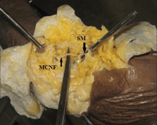
- Mark the ulnar nerve before ANY medial portal — palpate it in the cubital tunnel at 90° flexion; flexing the elbow moves it posteriorly away from medial portal entry.
- Posterior compartment is safer — nerves run anteriorly; the posterior central and posterolateral portals carry lower neurovascular risk.
- OCD capitellum Grade 3–4 — unstable or free-fragment lesions need arthroscopic fixation (headless compression screw or bioabsorbable pin) or osteochondral autograft transfer (OAT) for larger defects.
When & Why
Elbow arthroscopy gives diagnostic and therapeutic access to all three compartments of the elbow through a handful of portals, and its indications have expanded considerably since Andrews and Carson first described the technique in 1985. Its strength is low morbidity, multi-compartment visualisation and the ability to combine procedures — its singular danger is the dense neurovascular envelope around the joint, which makes portal technique the whole game.
- Role of arthroscopy
- First-line for unstable lesions in skeletally immature athletes — fixation, microfracture or OAT
- Key evidence / outcome
- Bexkens 2017: 62% same-level return to sport; Lu 2018 meta-analysis: 91.4% RTS arthroscopic vs 86.4% open
- Role of arthroscopy
- Most common historical indication; superior to open for multiple loose bodies; both compartments inspected
- Key evidence / outcome
- Synovial osteochondromatosis, post-traumatic bodies
- Role of arthroscopy
- Arthroscopic capsular release for extrinsic contracture without bridging HO
- Key evidence / outcome
- Ball 2002: arc under 100° improved 69° to 119°, no neurovascular injury; Cohen & Hastings 1998: 74° to 129° open LCL-sparing
- Role of arthroscopy
- Comminuted Mason III not amenable to fixation in selected patients; less soft-tissue morbidity than open
- Key evidence / outcome
- Johnson 2005: excision alters kinematics and stability — case selection is critical
- Role of arthroscopy
- ECRB debridement after 6 months of failed conservative care; assess intra-articular pathology at the same sitting
- Key evidence / outcome
- Allows simultaneous joint assessment
- Role of arthroscopy
- Rheumatoid synovectomy, PVNS, posterolateral plica resection
- Key evidence / outcome
- Delays inflammatory progression; definitive for plica syndrome
- Role of arthroscopy
- Unexplained elbow pain or suspected chondral injury when imaging is inconclusive
- Key evidence / outcome
- Staging of articular pathology before definitive treatment
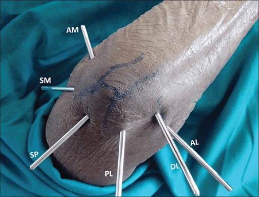
Consent specifically for transient nerve palsy (the PIN and ulnar nerve most commonly), the small risk of permanent nerve injury, fluid extravasation and compartment syndrome, infection, and — after contracture release — recurrence of stiffness and heterotopic ossification. Counsel the rheumatoid or contracted-elbow patient that nerve-palsy risk is higher (Kelly 2001). Setup. General anaesthesia with a high upper-arm tourniquet; regional supplementation optional. The standard position is lateral decubitus with the arm over a bolster and the elbow flexed to 90°, which gives free anterior and posterior access without a traction device. Before draping, mark the medial and lateral epicondyles, the radial head (palpated with forearm rotation), the olecranon tip — and palpate and mark the ulnar nerve in the cubital tunnel at 90° flexion.
The Operation
The goal is to enter the joint safely, inspect every compartment systematically, and complete the therapeutic task — loose-body removal, OCD fixation, capsular release or synovectomy — without injuring a nerve. The elbow's tiny working volume of only 3–5 mL is the reason every portal is preceded by distension: 20–30 mL of saline confirms intra-articular placement, tenses the capsule away from the neurovascular structures, and creates working space. Getting the portals right — the exposure — is the dominant skill and the dominant examination topic.
- Location
- 2 cm proximal, 1 cm anterior to medial epicondyle
- Primary use
- Viewing — anterior compartment (FIRST portal)
- Key nerve relation
- Median nerve 7 mm medial; safest anterior portal
- Location
- 3 cm distal, 1 cm anterior to lateral epicondyle
- Primary use
- Working — anterior compartment (SECOND, under vision)
- Key nerve relation
- PIN 7 mm neutral, 13–14 mm distended and supinated
- Location
- 3 cm proximal to olecranon tip, midline
- Primary use
- Viewing — posterior compartment
- Key nerve relation
- Ulnar nerve 25 mm medial; safest posterior portal
- Location
- 3 cm proximal to olecranon, lateral to triceps
- Primary use
- Working — posterior compartment
- Key nerve relation
- No significant nerve risk laterally
- Location
- Centre of triangle: lateral epicondyle, radial head, olecranon
- Primary use
- Outflow / lateral compartment / distension
- Key nerve relation
- Lateral antebrachial cutaneous nerve superficially
- Nearest portal
- Anterolateral
- Undistended
- 7 mm
- Distended / protected
- 13–14 mm
- Nearest portal
- Anterolateral
- Undistended
- 10 mm
- Distended / protected
- 16 mm
- Nearest portal
- Proximal anteromedial
- Undistended
- 7 mm
- Distended / protected
- 12 mm
- Nearest portal
- Proximal anteromedial
- Undistended
- 10 mm
- Distended / protected
- 15 mm
- Nearest portal
- Proximal anteromedial
- Undistended
- 25 mm
- Distended / protected
- 30 mm
- Nearest portal
- Proximal anteromedial
- Undistended
- 3 mm (superficial)
- Distended / protected
- —
The posterior interosseous nerve lies only 7 mm from the anterolateral portal in a neutral forearm. Three protections, in order: distend the joint with 20–30 mL of saline first (moves the nerve to 13–14 mm), supinate the forearm when creating the anterolateral portal (moves the PIN anteriorly), and create it under direct vision through the already-established proximal anteromedial portal, localising with a spinal needle before the trocar. Never make the anterolateral portal blind, and never before distension.
Patient lateral, arm over a bolster, elbow at 90°. Easy anterior and posterior access with no traction device — the default position for most surgeons.
Patient prone, arm hanging over the table edge. Gravity assists joint distraction and posterior access is excellent, but anaesthetic access is limited and the orientation is unfamiliar.
A traction device suspends the forearm. Familiar orientation and easy conversion to open, but adds complexity and changes portal angles.
Operative sequence
- Lateral decubitus, arm over a bolster, elbow at 90°, tourniquet high on the arm.
- Mark the medial and lateral epicondyles, the radial head (palpate with forearm rotation), the olecranon tip, and palpate and mark the ulnar nerve in the cubital tunnel at 90° flexion before draping.
- Insert an 18-gauge needle through the soft-spot (direct lateral portal site) and inject 20–30 mL of saline until firm resistance is felt.
- This confirms intra-articular placement and pushes the capsule and neurovascular structures away from the portals you are about to make.
- 2 cm proximal to the medial epicondyle, just anterior to the medial intermuscular septum.
- Stab the skin only; pass a blunt trocar toward the centre of the joint, staying close to the anterior humerus; loss of resistance signals intra-articular entry.
- Insert the 4 mm, 30° arthroscope and confirm position by visualising the capitellum and trochlea. This is the safest anterior portal and the primary viewing portal.
- 3 cm distal and 1 cm anterior to the lateral epicondyle.
- Supinate the forearm (moves the PIN from 7 mm to about 14 mm away) and keep the joint distended.
- Insert an 18-gauge spinal needle first and watch it enter the joint on the monitor from the proximal anteromedial view; only then make a skin-only stab and advance a blunt trocar under arthroscopic guidance.
- Capitellum and radial head — OCD, articular wear, loose bodies.
- Radiocapitellar joint — motion, plica, soft tissue.
- Coronoid tip and coronoid fossa — erosion, osteophytes, loose bodies.
- Trochlea and medial gutter — articular surface.
- Anterior capsule — thickness and contracture.
- Lateral collateral ligament complex from inside.
- Switch the arthroscope to the posterior central portal (3 cm proximal to the olecranon tip, midline); the posterolateral portal becomes the working portal.
- Inspect the olecranon tip and fossa, the posterior trochlea, and the medial and lateral gutters.
- A posteromedial portal is added only if needed for the medial gutter — mark the ulnar nerve again immediately beforehand.
- Grade 1–2: debridement and microfracture of a stable lesion; restrict overhead activities for 3–6 months.
- Grade 3: curettage of the base, drill channels for vascular ingrowth, and fixation with a bioabsorbable pin or headless compression screw under arthroscopic and fluoroscopic guidance.
- Grade 4: remove the free fragment and assess the defect — microfracture for small defects, OAT from the ipsilateral knee for defects greater than 1 cm².
- Grasp through the working portal; search systematically — coronoid fossa, radiocapitellar recess, posterior gutter, olecranon fossa.
- Multiple loose bodies require inspection of both anterior and posterior compartments.
- Anterior release: with a radiofrequency ablator or arthroscopic blade, work lateral to medial keeping close to the anterior humerus; stay anterior to the anterior band of the MCL and do not violate the lateral ulnar collateral ligament — it destabilises the elbow.
- Clear the coronoid fossa of fibrous tissue and osteophytes.
- Posterior release: resect the posterior capsule transversely and debride olecranon-tip osteophytes and the olecranon fossa.
- Do not release the MCL — it destabilises the elbow to valgus load.
- Close portal sites with skin sutures or steri-strips.
- A small drain for 24 hours is optional after a bloody synovectomy.
- Bulky compression dressing and a sling for 24 hours of comfort.
Twenty to thirty millilitres of saline through the soft-spot is the single most protective step in elbow arthroscopy — it confirms you are intra-articular and moves the radial, median and ulnar nerves and the brachial artery an extra 5–8 mm away from every subsequent portal.
Supinating the forearm swings the PIN anteriorly and increases its distance from the anterolateral portal from 7 mm (neutral) to about 14 mm. Combined with distension and direct-vision needle localisation, this is how the most dangerous portal is made safe.
Establish the proximal anteromedial portal before any lateral portal. It is the furthest of the anterior portals from all three major nerves, it gives you a viewing portal to make the anterolateral portal safely under direct vision, and it sets the tone for the whole procedure.
Aftercare & Complications
Rehabilitation is procedure-specific. The general principles are early range of motion (the first 4–6 weeks determine long-term outcome), active-assisted rather than forced passive stretching (forced stretching fuels heterotopic ossification), and strengthening in a pain-free arc as motion returns.
- Immobilisation
- Sling 24 h
- First ROM
- Immediate active ROM
- Return to sport
- 2–4 weeks
- Immobilisation
- Sling 24 h
- First ROM
- Immediate
- Return to sport
- 1–2 weeks
- Immobilisation
- Sling 48–72 h
- First ROM
- Day 3–5 active ROM
- Return to sport
- 3–6 months
- Immobilisation
- Posterior slab 2 weeks, then hinged brace
- First ROM
- Week 2
- Return to sport
- 3–6 months (restricted overhead)
- Immobilisation
- Posterior slab 4 weeks
- First ROM
- Week 4–6
- Return to sport
- 9–12 months for overhead athletes
- Immobilisation
- Immediate active ROM — critical
- First ROM
- Day 1 physiotherapy
- Return to sport
- 3–6 months depending on baseline
- Immobilisation
- Sling 1–2 weeks
- First ROM
- Day 3–5 active ROM
- Return to sport
- 6–8 weeks
- Immobilisation
- Sling 48 h
- First ROM
- Day 3 active ROM
- Return to sport
- 4–6 weeks
Contracture release — special protocol. Postoperative motion is the single most important determinant of outcome, so physiotherapy starts within 24–48 hours: therapist-guided active and passive ROM three times daily, dynamic extension splinting at night (turnbuckle or Dynasplint), serial static splinting for persistent deficits at 6 weeks, and a target of full terminal extension and 130° flexion by week 6. Return to throwing (OCD). Grades 1–2 begin an interval throwing programme at 3 months; Grades 3–4 after fixation begin at 6 months with full overhead competition at 9–12 months. Better outcomes are predicted by an open capitellar physis, a shorter symptom duration, and loose-body removal or advanced lesions (Bexkens 2017: 62% same-level return to sport; Lu 2018 meta-analysis: 91.4% return to sport in arthroscopically treated patients). Complications. Elbow arthroscopy carries a higher complication rate than shoulder or knee arthroscopy because of the neurovascular density — reported overall rates range from 0.8 to 14% across series.
- Incidence
- 0.5–2% (most common nerve injury)
- Prevention
- Distend 20–30 mL first; supinate forearm; proximal anteromedial portal first; create anterolateral under direct vision
- Management
- Document at surgery; EMG at 6–8 weeks; most neurapraxias resolve by 3–6 months; rarely nerve exploration
- Incidence
- 0.5–1%
- Prevention
- Mark ulnar nerve before medial portals; flex elbow 90°; blunt dissection medially; never make an extended medial portal
- Management
- Monitor grip and ring/small-finger sensation; EMG; observe; consider cubital tunnel decompression if persistent
- Incidence
- Less than 0.5% (rare but catastrophic)
- Prevention
- Proximal anteromedial portal only (not direct anteromedial); blunt trocar; distend joint; stay close to anterior humerus
- Management
- Immediate vascular surgery consult for arterial injury; median nerve injury may need exploration and repair
- Incidence
- 1–3%
- Prevention
- Limit pump pressure (less than 40 mmHg); use gravity inflow when possible; monitor arm girth; time limit on procedure
- Management
- Immediate recognition — firm arm with tense compartments; measure compartment pressures; fasciotomy if indicated
- Incidence
- Less than 0.5%
- Prevention
- Inspect instruments before use; replace worn or bent instruments; avoid levering
- Management
- Retrieve fragments arthroscopically or via mini-open; document in the operative note
- Incidence
- 0.2–0.8%
- Prevention
- Perioperative antibiotics; strict sterile technique; limit portal dilations
- Management
- IV antibiotics, arthroscopic washout; culture-directed therapy, often 6 weeks
- Incidence
- 2–5% (higher after contracture release)
- Prevention
- Indomethacin 75 mg/day for 6 weeks; low-dose radiation (700 cGy) in high-risk patients
- Management
- Surgical excision after maturation (greater than 12 months, cold bone scan); repeat arthroscopic release possible
- Incidence
- 10–15% after contracture release
- Prevention
- Aggressive early ROM; dynamic splinting; physiotherapy within 24–48 hours
- Management
- Revision arthroscopic release if it fails; assess for heterotopic ossification
During or after the procedure, watch for a firm, tense forearm, pain out of proportion on passive stretch of the fingers, and paraesthesiae in the median or ulnar distribution — fluid extravasation through capsular defects or portal sites is the usual cause. The threshold is an absolute compartment pressure greater than 30 mmHg or a delta pressure (diastolic minus compartment) less than 30 mmHg. If confirmed, perform an immediate four-compartment forearm fasciotomy (volar and dorsal). Prevent it by limiting pump pressure to 40 mmHg, preferring gravity inflow, watching arm girth, and keeping operative time under 90 minutes.
Viva & Exam Focus
PALMPALM — order of portal establishment
Hook:PALM reminds you to work from the safest portal inward — proximal anteromedial first, never anterolateral blind.
OCDLOCDL — OCD capitellum grading and management
Hook:OCDL — from cartilage softening all the way to a loose body; the grade determines whether you fix or replace.
Elbow arthroscopy — exam viva scenarios
Practise clinical reasoning and management decisions out loud
“A 15-year-old elite baseball pitcher has 4 months of lateral elbow pain and locking. MRI shows a 12 mm Grade 3 OCD lesion of the capitellum with a partially detached articular fragment in situ. Describe your management.”
“A 32-year-old manual worker had a terrible-triad injury 18 months ago treated non-operatively. His elbow moves from 45° to 95° of flexion — he cannot straighten the last 45° or flex beyond 95° — with no ligamentous instability. Describe your operative management.”
“You are 45 minutes into an elbow arthroscopy for loose-body removal when the scrub nurse notes the limb looks swollen and firm and the anaesthetist reports sudden difficulty maintaining the pulse-oximeter reading on the ipsilateral hand. Describe your immediate management.”
Portal anatomy and nerve distances
- Proximal anteromedial: 2 cm proximal and anterior to the medial epicondyle — FIRST portal — median nerve 7 mm, ulnar nerve 25 mm
- Anterolateral: 3 cm distal and 1 cm anterior to the lateral epicondyle — PIN 7 mm undistended, 14 mm with distension and supination
- Posterior central: 3 cm proximal to the olecranon tip, midline — ulnar nerve 25 mm medial — safest posterior portal
- Posterolateral: lateral to the triceps, 3 cm proximal to the olecranon — working portal for the posterior compartment
- Direct lateral (soft-spot): centre of the triangle of lateral epicondyle, radial head and olecranon — outflow and lateral compartment
OCD capitellum grading
- Grade 1: intact articular cartilage, softening only — conservative, restrict throwing 3–6 months
- Grade 2: cartilage fissuring, fragment stable on probing — conservative vs debridement and microfracture
- Grade 3: partially detached, unstable on probing, in situ — arthroscopic fixation (Herbert screw or bioabsorbable pin)
- Grade 4: free fragment within the joint — remove the loose body and OAT for defects greater than 1 cm²
- Bexkens 2017 (AJSM): 62% same-level return to sport; open physis predicts better outcome. Lu 2018 (Int Orthop) meta-analysis: 91.4% RTS arthroscopic vs 86.4% open
Arthroscopic steps (in order)
- Step 1: position (lateral decubitus most common) and mark landmarks including the ulnar nerve
- Step 2: distend the joint with 20–30 mL saline before any portal
- Step 3: proximal anteromedial portal FIRST — blunt trocar close to the anterior humerus
- Step 4: anterolateral portal UNDER DIRECT VISION — supinate the forearm, needle localisation first
- Step 5: systematic anterior compartment inspection (capitellum, trochlea, coronoid, capsule)
- Step 6: posterior compartment via posterior central and posterolateral portals
- Step 7: therapeutic procedure (loose-body removal / OCD fixation / contracture release)
- Step 8: close portals — drain optional — sling 24 h
Indications summary
- Loose-body removal — the most common historical indication
- OCD capitellum Grade 3–4 — arthroscopic fixation or OAT
- Post-traumatic contracture / arthrofibrosis — arthroscopic capsular release
- Lateral epicondylitis — arthroscopic ECRB debridement after 6 months of conservative failure
- Radial head excision — comminuted Mason III in selected patients
- Synovitis / plica — rheumatoid, PVNS, posterolateral plica syndrome
- Diagnostic — unexplained elbow pain with inconclusive imaging
Danger zones — 6 key structures
- PIN: 7 mm from the anterolateral portal undistended — MOST COMMONLY INJURED
- Radial nerve main trunk: 10 mm from the anterolateral portal
- Median nerve: 7 mm from the proximal anteromedial portal
- Brachial artery: 10 mm from the proximal anteromedial portal
- Ulnar nerve: must be marked before ANY medial portal — flex the elbow to 90°
- Medial and lateral antebrachial cutaneous nerves: superficial — protect with blunt dissection at the skin
OCD management by grade
- Grade 1–2 (stable): conservative — restrict throwing, physiotherapy, serial imaging; consider debridement and drilling if no improvement at 6 months
- Grade 3 (unstable in situ): arthroscopic curettage of the base plus fixation with a headless screw or bioabsorbable pin and subchondral drilling
- Grade 4 (free fragment): remove the fragment and assess the defect — microfracture for small defects (less than 1 cm²) and OAT for larger defects
- Return to throwing: 3–6 months for Grades 1–2, 9–12 months for Grades 3–4 after fixation
Contracture release key points
- CT scan first — exclude bridging heterotopic ossification (a contraindication to arthroscopic release)
- Mark the ulnar nerve before all medial portals
- Anterior capsulectomy: lateral to medial, close to the anterior humerus — do NOT violate the LUCL
- Posterior capsulectomy and olecranon-fossa debridement for an extension deficit
- Do NOT release the MCL (destabilises the elbow to valgus load)
- Post-op: physiotherapy within 24–48 hours, dynamic splinting, indomethacin HO prophylaxis 6 weeks
- Ball 2002 (JSES): arthroscopic release improved arc from 69° to 119° (arc under 100° group), no neurovascular complications; Cohen & Hastings 1998 (JBJS Br): open LCL-sparing release 74° to 129°
Post-op and complications
- Nerve injury (most common: PIN 0.5–2%) — EMG at 6–8 weeks — most neurapraxias resolve
- Compartment syndrome — stop the pump, assess pressures, fasciotomy if delta pressure less than 30 mmHg
- Pump pressure: maximum 40 mmHg for the elbow (lower than shoulder 60 mmHg)
- Heterotopic ossification: prophylaxis with indomethacin 75 mg/day for 6 weeks after contracture release
- Return to sport: loose bodies 2–4 weeks; OCD Grade 3–4 fixation 9–12 months; contracture release 3–6 months
Background & Evidence
Surgical anatomy. The elbow shares a single joint cavity across three articulations — the ulnohumeral hinge (flexion-extension), the radiocapitellar joint (rotation and valgus load) and the proximal radioulnar joint. For arthroscopy it is divided into anterior and posterior compartments with no true medial/lateral division as in the knee. The anterior compartment holds the capitellum (the site of OCD in young athletes), the trochlea, the coronoid process, the radial head and the anterior capsule (the target in contracture release); the posterior compartment holds the olecranon tip and fossa (where loose bodies collect), the posterior trochlea, the gutters and a thicker, more fibrous posterior capsule. Why the elbow is dangerous. The joint's tiny working volume of only 3–5 mL sits within the densest neurovascular envelope of any major joint — the radial and median nerves, the PIN, the ulnar nerve, the brachial artery and the medial and lateral antebrachial cutaneous nerves all pass within millimetres of the working portals. This is why distension, portal order and elbow position — not the procedure itself — determine nerve safety (Lynch 1986), and why overall complication rates of 0.8–14% are reported across series, higher than shoulder or knee arthroscopy. Epidemiology of the indications. Capitellar OCD presents in adolescent overhead or throwing athletes (open physis) with lateral elbow pain and locking; post-traumatic contracture follows elbow trauma in young adults, for whom a functional 30°–130° arc is the benchmark; loose bodies arise from synovial osteochondromatosis, OCD or trauma; and refractory lateral epicondylitis brings the middle-aged patient after at least 6 months of failed conservative care.
- Radiographic / arthroscopic features
- Articular cartilage intact, softening only
- Management
- Conservative — restrict throwing 3–6 months, physiotherapy, serial imaging
- Radiographic / arthroscopic features
- Cartilage fissuring; fragment stable on probing
- Management
- Conservative, or arthroscopic debridement and microfracture if no improvement by 6 months
- Radiographic / arthroscopic features
- Partially detached, unstable fragment in situ
- Management
- Arthroscopic curettage of the base and fixation (headless compression screw or bioabsorbable pin) plus subchondral drilling
- Radiographic / arthroscopic features
- Free fragment within the joint
- Management
- Remove the loose body; microfracture for small defects (less than 1 cm²); OAT for larger defects
- Indication
- Portal technique
- Key finding
- First systematic portal description; loose-body removal gave the best results
- Level
- IV
- Indication
- Nerve anatomy
- Key finding
- Cadaveric mapping of neurovascular structures to portals — injury is driven by portal placement, direction and elbow position
- Level
- IV
- Indication
- Portal safety
- Key finding
- Proximal medial and lateral portals safer than anteromedial/anterolateral — recommended as standard anterior portals
- Level
- IV
- Indication
- Contracture release
- Key finding
- Arc under 100° improved from 69° to 119°; no neurovascular complications
- Level
- IV
- Indication
- OCD capitellum
- Key finding
- 75 elbows; 62% return to sport; open physis and shorter symptoms predicted better outcome
- Level
- IV
- Indication
- Complications
- Key finding
- 473 cases: 0.8% serious (infection), 11% minor; transient nerve palsy linked to rheumatoid arthritis and contracture
- Level
- IV
The consistent evidence message is that elbow arthroscopy is effective and mostly safe when portal discipline is meticulous: loose-body removal remains its most reliable indication (Andrews 1985), arthroscopic contracture release matches open release with lower morbidity (Ball 2002; Cohen & Hastings 1998), and arthroscopic OCD management gives good function with a majority return to sport (Bexkens 2017; Lu 2018) — but nerve-palsy risk rises sharply in rheumatoid and contracted elbows (Kelly 2001), who warrant extra caution and counselling.
References
- Andrews JR, Carson WG. Arthroscopy of the elbow. Arthroscopy. 1985;1(2):97–107. PMID: 4091924. DOI: 10.1016/s0749-8063(85)80038-4. — First systematic description of elbow arthroscopy portals and technique; loose-body removal gave the best results. 2. Lynch GJ, Meyers JF, Whipple TL, Caspari RB. Neurovascular anatomy and elbow arthroscopy: inherent risks. Arthroscopy. 1986;2(3):190–197. PMID: 3768116. DOI: 10.1016/s0749-8063(86)80067-6. — Landmark cadaveric study mapping neurovascular structures to portals; injury driven by portal placement, entry direction and elbow position. 3. Poehling GG, Whipple TL, Sisco L, Goldman B. Elbow arthroscopy: a new technique. Arthroscopy. 1989;5(3):222–224. PMID: 2775398. DOI: 10.1016/0749-8063(89)90176-x. — Prone position with a proximal medial portal improving scope mobility and visualisation. 4. Stothers K, Day B, Regan WR. Arthroscopy of the elbow: anatomy, portal sites, and a description of the proximal lateral portal. Arthroscopy. 1995;11(4):449–457. PMID: 7575879. DOI: 10.1016/0749-8063(95)90200-7. — Proximal medial and proximal lateral portals safer than anteromedial/anterolateral; recommended as standard anterior portals. 5. Cohen MS, Hastings H 2nd. Post-traumatic contracture of the elbow. Operative release using a lateral collateral ligament sparing approach. J Bone Joint Surg Br. 1998;80(5):805–812. PMID: 9768890. DOI: 10.1302/0301-620x.80b5.8528. — Open LCL-sparing release improved total ulnohumeral movement from 74° to 129°. 6. Ball CM, Meunier M, Galatz LM, Calfee R, Yamaguchi K. Arthroscopic treatment of post-traumatic elbow contracture. J Shoulder Elbow Surg. 2002;11(6):624–629. PMID: 12469091. DOI: 10.1067/mse.2002.126770. — Arthroscopic release: arc under 100° improved from 69° to 119°; no neurovascular complications. 7. Lindenhovius ALC, Linzel DS, Doornberg JN, Ring DC, Jupiter JB. Comparison of elbow contracture release in elbows with and without heterotopic ossification restricting motion. J Shoulder Elbow Surg. 2007;16(5):621–625. PMID: 17644008. DOI: 10.1016/j.jse.2007.01.005. — Motion gains greater when motion-blocking HO is removed than with capsular contracture alone. 8. Bexkens R, van den Ende KIM, Ogink PT, van Bergen CJA, van den Bekerom MPJ, Eygendaal D. Clinical outcome after arthroscopic debridement and microfracture for osteochondritis dissecans of the capitellum. Am J Sports Med. 2017;45(10):2312–2318. PMID: 28520461. DOI: 10.1177/0363546517704842. — 75 elbows; 62% return to sport; open physis predicts better outcome. 9. Lu Y, Li YJ, Guo SY, Zhang HL. Is there any difference between open and arthroscopic treatment for osteochondritis dissecans (OCD) of the humeral capitellum: a systematic review and meta-analysis. Int Orthop. 2018;42(3):601–607. PMID: 29349503. DOI: 10.1007/s00264-018-3768-3. — 91.4% return to sport arthroscopic vs 86.4% open; no complications in arthroscopic group. 10. Kelly EW, Morrey BF, O'Driscoll SW. Complications of elbow arthroscopy. J Bone Joint Surg Am. 2001;83(1):25–34. PMID: 11205854. DOI: 10.2106/00004623-200101000-00004. — 473 cases: 0.8% serious (infection), 11% minor; transient nerve palsy linked to rheumatoid arthritis and contracture. 11. Safran MR. Ulnar collateral ligament injury in the overhead athlete: diagnosis and treatment. Clin Sports Med. 2004;23(4):643–663. PMID: 15474227. DOI: 10.1016/j.csm.2004.05.002. — Overhead-athlete elbow pathology relevant to the throwing population presenting with OCD and loose bodies. 12. Johnson JA, Beingessner DM, Gordon KD, Dunning CE, Stacpoole RA, King GJW. Kinematics and stability of the fractured and implant-reconstructed radial head. J Shoulder Elbow Surg. 2005;14(1 Suppl S):195S–201S. PMID: 15726082. DOI: 10.1016/j.jse.2004.09.034. — Radial head excision markedly alters elbow kinematics and stability — relevant when considering arthroscopic radial head excision.
Arthroscopy of the elbow
- First systematic description of elbow arthroscopy portals and the normal intra-articular anatomy seen from anterolateral, anteromedial and posterolateral portals
- In 12 patients, removal of loose bodies produced the best objective and subjective results; chondroplasty results were less satisfactory
- The only complication was a transient median nerve palsy from extracapsular extravasation of local anaesthetic — establishing the neurovascular caution central to the procedure
Neurovascular anatomy and elbow arthroscopy: inherent risks
- Cadaveric dissection of 5 elbows defined the relationship of superficial cutaneous and deep neurovascular structures to standard portals
- Neurovascular injury is driven by inappropriate portal placement, wrong direction of entry, or elbow position — not the procedure itself
- Defined a safe, reproducible portal technique that remains the basis of modern practice
Arthroscopy of the elbow: anatomy, portal sites, and a description of the proximal lateral portal
- Cadaveric assessment (12 specimens) plus a clinical series confirming the proximal medial and proximal lateral portals are safer than the anteromedial and anterolateral portals
- All areas of the anterior compartment can be visualised using the two proximal portals — recommended as the standard anterior portals
- All posterior approaches were found to be safe
Complications of elbow arthroscopy
- 473 consecutive elbow arthroscopies: serious complication (joint-space infection) in 0.8%; minor complications in 11%
- 12 transient nerve palsies in 10 patients (ulnar most common, then superficial radial, then PIN); no permanent neurovascular injuries and no compartment syndromes
- Rheumatoid arthritis and a pre-existing contracture were the strongest risk factors for transient nerve palsy
Arthroscopic treatment of post-traumatic elbow contracture
- 14 patients: flexion improved from a mean 117.5° to 133° and extension from 35.4° to 9.3° after arthroscopic capsular release
- In the subgroup with a preoperative arc under 100° (10 patients), the mean arc improved from 69° to 119°
- No neurovascular complications; mean satisfaction 8.4 out of 10 — outcomes compared favourably with open release
Clinical outcome after arthroscopic debridement and microfracture for osteochondritis dissecans of the capitellum
- 75 elbows (mean age 16) treated by arthroscopic debridement and microfracture for advanced capitellar OCD; mean postoperative Oxford Elbow Score 40.8
- An open capitellar physis, loose-body removal/advanced lesions, and shorter symptom duration independently predicted better outcome
- 62% returned to their primary sport (55% same level, 7% lower); no complications recorded