Modular segmental replacement after resection for bone sarcoma or metastatic disease · advanced
- An endoprosthesis gives immediate stability, early weight-bearing and reproducible function; biological options (allograft, allograft-prosthetic composite, vascularised fibula) are more durable long-term but carry nonunion, fracture and infection risk and slower recovery.
- Soft-tissue reattachment is the determinant of function: extensor mechanism reconstruction with a gastrocnemius flap at the proximal tibia, and abductor reattachment at the proximal femur, dictate the functional result more than the implant itself.
- The Henderson failure classification has 5 modes — (1) soft-tissue failure, (2) aseptic loosening, (3) structural or implant failure, (4) infection, (5) tumour progression — and is the universal language for reporting megaprosthesis failure.
- Infection is the dominant catastrophic complication (approximately 10%, higher at the proximal tibia and after chemo or radiotherapy) and is the leading cause of secondary amputation; silver-coated implants and gastrocnemius coverage reduce this risk.
- “Always plan resection length and limb-length restoration on a whole-bone MRI — the proximal and distal tumour margin defines the prosthesis size, and a missed skip lesion is a planning failure.
- “The proximal tibia and post-radiation cases carry the highest infection rate — a medial gastrocnemius flap provides both extensor mechanism reconstruction and durable soft-tissue coverage.
- “Extracortical bone bridging and hydroxyapatite collars at the bone-implant junction reduce aseptic loosening; compress-type fixation uses controlled compression to promote bone hypertrophy at the transcortical interface.
- “In skeletally immature children a growing (expandable) prosthesis maintains limb-length equality, but reoperation and mechanical failure rates are high; rotationplasty or amputation must be discussed honestly as alternatives.
When & Why
Indication. Segmental reconstruction of a long bone after wide resection for a primary bone sarcoma, for aggressive benign or metastatic disease with massive bone loss, or for a failed prior reconstruction. The reconstruction is always secondary to an oncologically adequate wide (Enneking) margin — the operation is planned from the tumour outwards. Primary bone sarcoma — the classic indications in children and young adults: osteosarcoma and Ewing sarcoma (reconstruction follows wide resection after neoadjuvant chemotherapy), chondrosarcoma (surgical resection is the mainstay as it is chemo- and radio-resistant), and high-grade spindle-cell sarcoma of bone or dedifferentiated chondrosarcoma. Aggressive benign or metastatic disease — metastatic bone disease with extensive bone loss or an impending or pathological fracture not amenable to internal fixation (especially a solitary renal or thyroid metastasis with a good prognosis), aggressive giant cell tumour with joint destruction unsuitable for curettage, and failed prior reconstruction with massive bone loss (revision megaprosthesis). Common anatomical sites — distal femur (the commonest site, rotating-hinge knee), proximal femur (bipolar or total-hip articulation; abductor reattachment critical), proximal tibia (highest infection risk; extensor mechanism and gastrocnemius flap mandatory), proximal humerus (commonest upper-limb site; shoulder stability and abduction limited), and total femur for massive disease or a failed prior segmental reconstruction. The one decision that matters — what do you rebuild with? The choice balances immediate function (favours an endoprosthesis) against long-term biological durability (favours allograft or biological options), modified by patient age, oncological prognosis, defect location and surgeon expertise.
- Modular Endoprosthesis
- Immediate
- Osteoarticular Allograft
- Delayed (union needed)
- Allograft-Prosthetic Composite
- Intermediate
- Vascularised Fibula
- Delayed (union or hypertrophy)
- Modular Endoprosthesis
- Early
- Osteoarticular Allograft
- Protected, slow
- Allograft-Prosthetic Composite
- Intermediate
- Vascularised Fibula
- Prolonged protection
- Modular Endoprosthesis
- Mechanical wear or loosening
- Osteoarticular Allograft
- Most durable if it unites
- Allograft-Prosthetic Composite
- Durable
- Vascularised Fibula
- Excellent if it hypertrophies
- Modular Endoprosthesis
- Loosening, infection, structural
- Osteoarticular Allograft
- Nonunion, fracture, infection
- Allograft-Prosthetic Composite
- Nonunion at host junction, infection
- Vascularised Fibula
- Stress fracture, donor morbidity
- Modular Endoprosthesis
- High (off-the-shelf modular)
- Osteoarticular Allograft
- Variable (size and availability)
- Allograft-Prosthetic Composite
- Variable
- Vascularised Fibula
- Technically demanding (microsurgery)
- Modular Endoprosthesis
- Older or metastatic, rapid function
- Osteoarticular Allograft
- Younger, joint-sparing intercalary
- Allograft-Prosthetic Composite
- Young adult, biological plus stable
- Vascularised Fibula
- Intercalary or diaphyseal defects, children
Pre-operative planning is the operation on paper. Staging and imaging: a whole-bone MRI defines intramedullary tumour extent, the soft-tissue mass, neurovascular involvement and skip lesions, and the resection length is planned from this; CT chest (pulmonary metastases) and bone scan or PET-CT complete systemic staging; plain radiographs of the whole bone are used for templating and contralateral length comparison; the biopsy is image-guided (core or open) with the tract in line with the definitive incision and planned for en-bloc excision. Templating and implant selection: measure the resection length from the planned osteotomy (a wide margin from the tumour edge on MRI), restore limb length and joint offset templated against the contralateral limb, order a modular system so segment length and articulation can be adjusted intra-operatively, and plan fixation (cemented versus uncemented stem, compress-type fixation, extracortical bridging). Consent and setup. Positioning and approach follow the resection: an extensile utility approach incorporating the biopsy tract, allowing wide en-bloc resection and protection of the major neurovascular bundle. The limb-salvage decision is made at the multidisciplinary tumour board, and the honest discussion of limb salvage versus amputation — or, in a child with a distal femoral tumour, rotationplasty — is mandatory.
My priority is an oncologically adequate wide margin planned on the whole-bone MRI, with the biopsy tract excised en bloc. The reconstruction is secondary to the resection. I template the resection length and restore limb length and offset against the contralateral side, and I choose a modular system so I can fine-tune intra-operatively. I discuss limb salvage versus amputation or, in a child with a distal femoral tumour, rotationplasty, at the multidisciplinary tumour board.
The Operation
The goal is an oncologically adequate wide resection with en-bloc excision of the biopsy tract, protection of the major neurovascular bundle, and a modular reconstruction that restores limb length and offset with stable fixation — then a soft-tissue reconstruction that dictates the functional result. The exposure and resection are laid out in the timeline below; the site-specific detail and the dangers at each site follow it.
Operative sequence
- Position per the site and approach; mark an extensile utility incision that incorporates the biopsy tract so it is excised en bloc with the tumour.
- Plan the incision to allow wide extracompartmental resection and proximal/distal control of the major neurovascular bundle.
- Develop the extracompartmental planes around the tumour, preserving the major neurovascular bundle: the popliteal bundle at the knee, the sciatic nerve at the hip, the common peroneal nerve at the proximal tibia, and the axillary nerve and brachial plexus at the shoulder.
- Mobilise and isolate the tumour en bloc, dividing only what must be divided to achieve a wide margin.
- Perform the planned osteotomy at the templated level taken from the whole-bone MRI, with intra-operative margin assessment (and frozen section where indicated).
- Confirm an adequate wide (Enneking) margin before reconstruction begins — the reconstruction never compromises the margin.
- Prepare the medullary canal for a cemented stem (immediate fixation, early weight-bearing) or an uncemented or press-fit stem (biological fixation).
- Apply extracortical bone bridging or a hydroxyapatite collar at the bone-implant junction to promote ongrowth and reduce aseptic loosening.
- Assemble the modular segments to the measured resection length and restore limb length and joint offset against the contralateral side.
- Choose the articulation appropriate to the site and the soft-tissue loss (rotating-hinge knee, bipolar or dual-mobility hip, hemiarthroplasty or reverse shoulder).
- Trial the articulation, reduce it, and check stability, range of motion and soft-tissue tension.
- Adjust constraint (for example a constrained or dual-mobility liner where abductors or capsule are lost) before final assembly.
- Reconstruct the soft tissues that determine function — abductor reattachment at the proximal femur, extensor mechanism reconstruction plus a medial gastrocnemius flap at the proximal tibia, and a suspension reconstruction at the proximal humerus.
- This step dictates the functional result more than the implant itself, and it is also central to infection control.
- Provide muscle coverage over the implant, meticulous dead-space management and haemostasis, and a layered closure over a drain.
- Confirm limb perfusion and neurological status before leaving theatre.
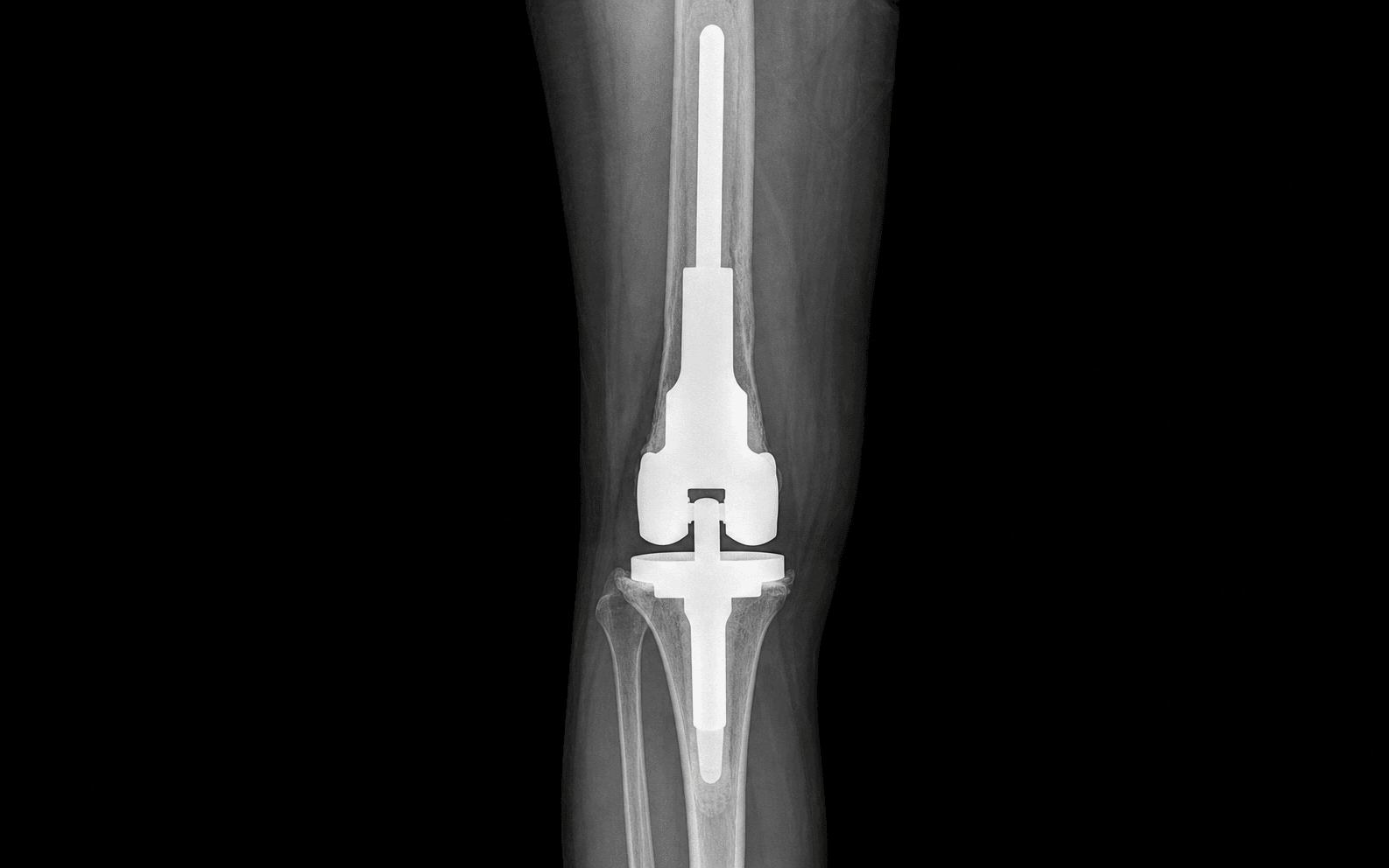
The general sequence is common to every site, but the articulation, the key soft-tissue problem and the danger structures differ at each. The four common sites are laid out below. Distal femur — the commonest site. A rotating-hinge knee tolerates the loss of the collateral ligaments after resection. The extensor mechanism is usually preservable, so protect the quadriceps and patellar tendon. Cemented or uncemented femoral and tibial stems are used, with extracortical bone bridging or an HA collar at the bone-implant junction to reduce aseptic loosening; reattach the gastrocnemius and soft tissues and close over a drain.

- The popliteal neurovascular bundle lies posteriorly during resection — develop the plane carefully and protect the popliteal artery and the tibial and peroneal nerves.
- An inadequate margin at the proximal osteotomy — confirm the level against the MRI-planned osteotomy.
- Aseptic loosening at the cement-bone or implant-bone junction over time — optimise fixation and use extracortical bridging.
Proximal femur. A bipolar or total hip articulation is used; a dual-mobility or constrained liner reduces dislocation given the loss of the abductors and capsule. Reattach the abductors to the prosthesis via a trochanteric attachment device or synthetic mesh, and consider capsular reconstruction. Restore offset and limb length to tension the soft tissues and stabilise the hip. A cemented or uncemented stem runs to the femoral diaphysis.

- Dislocation from abductor and capsular loss — use a dual-mobility or constrained articulation and meticulous soft-tissue reattachment.
- The sciatic nerve during posterior resection or retraction.
- An abductor lurch and Trendelenburg gait even with reattachment — counsel the patient pre-operatively.
Proximal tibia — the highest-risk site. It has the highest infection rate of all sites because of the thin anteromedial soft-tissue envelope and frequent prior radiotherapy. Extensor mechanism reconstruction is mandatory: reattach the patellar tendon to the prosthesis or a tendon anchor, then rotate a medial gastrocnemius flap to reinforce the repair and provide durable soft-tissue coverage. A rotating-hinge knee is used; protect the common peroneal nerve laterally.

At the proximal tibia I reconstruct the extensor mechanism by securing the patellar tendon to the prosthesis, then I rotate a medial gastrocnemius flap to both cover the implant and reinforce the extensor reconstruction. This single manoeuvre tackles the two things that wreck proximal tibial replacements — extensor lag and infection from a thin soft-tissue envelope.
- The highest infection risk — gastrocnemius flap coverage and a silver-coated implant mitigate it.
- Extensor lag from inadequate extensor mechanism reconstruction — protect the repair in extension post-operatively.
- The common peroneal nerve laterally during resection.
Proximal humerus — the commonest upper-limb site. Resection sacrifices the rotator cuff and deltoid attachments and the capsule. Articulation options are hemiarthroplasty, a reverse total shoulder (if the deltoid and axillary nerve are intact), or a constrained spacer, with a suspension reconstruction using mesh or tendon. Active abduction is usually limited, but hand, wrist and elbow function are preserved, which is the main functional goal of upper-limb salvage. Protect the axillary and radial nerves and the brachial plexus and axillary vessels.
- The axillary nerve and brachial plexus during resection — preserve them for any reverse-shoulder option.
- Instability or subluxation from cuff and capsular loss — use a suspension reconstruction and soft-tissue tensioning.
- Limited shoulder abduction is expected — set realistic functional expectations.
Fixation and growing prostheses. The fixation strategy is chosen for the patient and the site: cemented stems give immediate fixation and early weight-bearing and suit older or metastatic patients and the post-chemotherapy setting; uncemented stems give biological fixation, with extracortical bone bridging and hydroxyapatite collars at the transcortical junction promoting bone ongrowth and reducing aseptic loosening; compress-type fixation applies controlled compression across the bone-implant interface to stimulate cortical hypertrophy and osseointegration and is useful in young patients. In skeletally immature children a growing (expandable) prosthesis maintains limb-length equality (lengthened non-invasively by electromagnetics or with minor procedures) at the cost of higher mechanical-failure and reoperation rates. Silver-coated megaprostheses provide an antimicrobial surface that reduces periprosthetic infection, especially at high-risk sites.
- Typical articulation
- Rotating-hinge knee
- Key soft-tissue issue
- Quadriceps and extensor preservation
- Headline functional outcome (MSTS)
- Generally good (often 75-85% of normal)
- Typical articulation
- Bipolar or total hip (dual-mobility or constrained)
- Key soft-tissue issue
- Abductor reattachment; dislocation risk
- Headline functional outcome (MSTS)
- Good ambulation; frequent abductor lurch
- Typical articulation
- Rotating-hinge knee
- Key soft-tissue issue
- Extensor mechanism plus gastrocnemius flap
- Headline functional outcome (MSTS)
- Variable; extensor lag limits the result
- Typical articulation
- Hemiarthroplasty, reverse or spacer
- Key soft-tissue issue
- Deltoid, cuff and capsule loss
- Headline functional outcome (MSTS)
- Limited active abduction; preserved hand and elbow function
Aftercare & Complications
Rehabilitation. In the lower limb (distal femur, proximal femur, proximal tibia), mobilise early when fixation allows — immediately with cemented stems, and protected weight-bearing with biological fixation. At the proximal tibia, protect the extensor reconstruction in an extension brace and delay active knee flexion until the patellar tendon and gastrocnemius repair is secure. At the proximal femur, observe hip precautions and abductor protection and expect a period of abductor lurch. Co-ordinate rehabilitation with the chemotherapy schedule, because wound healing and infection risk are higher during myelosuppression. In the upper limb (proximal humerus), use sling immobilisation initially to protect the suspension reconstruction, focus rehabilitation on the preserved hand, wrist and elbow function, accept limited shoulder abduction, and begin pendulum and assisted-range exercises as the soft-tissue reconstruction allows. Complications — the Henderson failure modes. The Henderson classification is the universal language for reporting megaprosthesis failure and frames the differential when a reconstruction fails.
- Description
- Dislocation, tendon or extensor mechanism failure, wound dehiscence
- Recognition
- Instability or dislocation; extensor lag (proximal tibia); abductor lurch (proximal femur); wound breakdown over the implant
- Prevention and management
- Prevention: meticulous reattachment of abductors and patellar tendon, gastrocnemius flap, dual-mobility or constrained articulation, muscle coverage. Management: bracing or extension splinting, revision of the soft-tissue reconstruction, articulation change
- Description
- Failure of fixation at the bone-implant interface
- Recognition
- Progressive pain, radiolucent lines, implant migration or subsidence on serial radiographs
- Prevention and management
- Prevention: optimal cement technique or press-fit, extracortical bone bridging, HA collars, compress fixation. Management: revision of the loose component with re-fixation
- Description
- Fracture of the implant, modular junction or periprosthetic bone
- Recognition
- Sudden mechanical failure, deformity, an audible event; a broken component or periprosthetic fracture on radiograph
- Prevention and management
- Prevention: robust modular junctions, avoid undersized stems, manage bone stock. Management: revision or exchange of the fractured component; fixation of a periprosthetic fracture
- Description
- Periprosthetic infection — the dominant catastrophic mode (approximately 10%)
- Recognition
- Pain, swelling, sinus, raised CRP or ESR; positive aspiration or cultures; worst at the proximal tibia and after radiotherapy
- Prevention and management
- Prevention: silver-coated implants, muscle-flap coverage, dead-space management, prophylactic antibiotics, meticulous haemostasis. Management: DAIR if early; two-stage revision; suppression; secondary amputation if uncontrolled — the leading cause of amputation
- Description
- Local recurrence or soft-tissue or extracompartmental progression
- Recognition
- A new mass, pain, recurrent disease on MRI or biopsy; usually within the first few years
- Prevention and management
- Prevention: an adequate wide margin at the index resection, en-bloc biopsy excision, adjuvant therapy. Management: re-resection and revision reconstruction, or amputation if margins cannot be achieved
Modes 1 to 3 are mechanical; modes 4 and 5 are non-mechanical. Infection (Type 4) and tumour progression (Type 5) are the modes most likely to end in amputation. The proximal tibia has the highest combined failure rate, driven by infection and extensor mechanism (Type 1) failure.
Viva & Exam Focus
These are the critical decisions, danger structures and exam traps that recur in the limb-salvage viva.
The trap: sizing the prosthesis from plain radiographs or a localised MRI. Intramedullary tumour and skip metastases extend beyond what radiographs show. The fix: plan the resection on a whole-bone MRI with the staging biopsy tract included in the resection. A wide Enneking margin is the oncological priority and the reconstruction is secondary. Measure the planned osteotomy and order the implant length before theatre.
The principle: the biopsy is placed by, or in discussion with, the resecting surgeon, in line with the definitive incision, so the whole tract is excised en bloc with the tumour. The risk: a poorly placed or transverse biopsy contaminates compartments and neurovascular planes, converting a limb-salvage candidate into an amputation. Never delegate the biopsy to a non-tumour unit.
The problem: resection of the proximal tibia detaches the patellar tendon and tibial tubercle, destroying active knee extension. The fix: reconstruct the extensor mechanism (patellar tendon to prosthesis or tendon anchor) and rotate a medial gastrocnemius flap to reinforce it and provide soft-tissue coverage. Failure produces an extensor lag and a non-functional limb.
The principle: resection sacrifices the abductor insertion on the greater trochanter and the hip capsule, leaving a high dislocation risk. The fix: reattach the abductors to the prosthesis (trochanteric attachment device or mesh), use a dual-mobility, constrained or bipolar articulation, and consider capsular reconstruction. Restore offset and limb length to tension the soft tissues.
Why it dominates: large metal surface area, big dead space, immunosuppression from chemotherapy, irradiated tissue and long operative times all raise infection risk to roughly 10% overall. The implication: infection is the commonest reason for secondary amputation. Mitigate it with silver-coated implants, muscle-flap coverage, meticulous haemostasis and dead-space management.
The decision: limb salvage is appropriate only when a wide margin can be achieved with preservation of a functional, sensate, vascularised limb. The alternatives: amputation, or rotationplasty in children with distal femoral tumours, give durable, low-maintenance function and may outperform a salvaged limb facing repeated reoperations. The honest discussion is an examiner favourite.
SALVAGESALVAGE — limb-salvage planning checklist
1–5Megaprosthesis failure — the 5 Henderson modes
Clinical Decision Scenarios
Practise clinical reasoning and management decisions out loud
“A 16-year-old presents with a high-grade osteosarcoma of the distal femur. After neoadjuvant chemotherapy, how do you decide between an endoprosthetic reconstruction and the alternatives, and how do you plan the operation?”
“Why does the proximal tibia have the worst outcomes of all endoprosthetic sites, and how do you specifically address this in your reconstruction?”
“Take me through the Henderson classification of endoprosthetic failure and explain why it is useful in clinical practice and the exam.”
Indications and sites
- Primary bone sarcoma (osteosarcoma, Ewing, chondrosarcoma) after wide resection; metastatic disease with massive bone loss; aggressive GCT; failed prior reconstruction
- Commonest sites: distal femur (commonest overall), then proximal femur, then proximal tibia, then proximal humerus; total femur for massive disease
- Reconstruction is always secondary to an oncologically adequate wide (Enneking) margin
- An honest discussion of amputation and, in children with a distal femoral tumour, rotationplasty is mandatory
Endoprosthesis vs biological reconstruction
- Endoprosthesis: immediate stability, early weight-bearing, reproducible — but a finite lifespan and lifelong revision burden
- Allograft, APC or vascularised fibula: more durable biologically but slower, with nonunion, fracture and infection
- Older or metastatic patients favour an endoprosthesis; younger patients with intercalary defects may favour biological options
- Oncological survival is equivalent between salvage and amputation when margins are adequate
Pre-operative planning
- A whole-bone MRI defines intramedullary extent and skip lesions — the basis for resection length
- Systemic staging: CT chest, bone scan or PET; biopsy tract in line with the incision for en-bloc excision
- Template the resection length and restore limb length and offset against the contralateral side
- Choose a modular system; plan fixation (cemented versus uncemented) and infection prevention (silver coating)
Site-specific technique
- Distal femur: rotating-hinge knee; protect the popliteal neurovascular bundle; extracortical bridging or HA collar
- Proximal femur: dual-mobility or constrained articulation; reattach the abductors (trochanteric device or mesh); restore offset to prevent dislocation
- Proximal tibia: reconstruct the extensor mechanism plus a medial gastrocnemius flap; highest infection risk
- Proximal humerus: hemi, reverse or spacer; suspension reconstruction; expect limited abduction but preserved hand and elbow function
Fixation strategies
- Cemented stems: immediate fixation, early weight-bearing, good in older or metastatic patients
- Uncemented stems: biological fixation; extracortical bone bridging and HA collars reduce aseptic loosening
- Compress-type fixation: controlled compression stimulates cortical hypertrophy and osseointegration (young patients)
- Growing (expandable) prostheses for skeletally immature children maintain limb-length equality, with a high reoperation rate
- Silver-coated megaprostheses reduce periprosthetic infection at high-risk sites
Henderson failure classification
- Type 1 soft-tissue failure — dislocation, extensor or tendon failure, wound dehiscence (early, function-limiting)
- Type 2 aseptic loosening — bone-implant interface; reduced by extracortical bridging, HA collars and compress fixation
- Type 3 structural failure — implant, modular junction or periprosthetic bone fracture
- Type 4 infection — dominant catastrophic mode (around 10%), worst at the proximal tibia or after radiation; leading cause of amputation
- Type 5 tumour progression — local recurrence; Types 1 to 3 mechanical, Types 4 and 5 non-mechanical
Outcomes and rehab
- The MSTS score is the standard functional outcome measure (lower- and upper-limb versions)
- Distal and proximal femur tend to score best; proximal tibia is limited by extensor lag; proximal humerus preserves hand and elbow
- Co-ordinate rehabilitation with the chemotherapy schedule (wound healing and infection risk during myelosuppression)
- Proximal tibia: protect the extensor repair in an extension brace; proximal femur: hip precautions and abductor protection
High-yield danger points
- The biopsy tract must be planned and excised en bloc — a poor biopsy can convert salvage to amputation
- Proximal tibia infection risk is highest — gastrocnemius flap coverage plus a silver implant
- Proximal femur dislocation from abductor and capsular loss — dual-mobility or constrained plus abductor reattachment
- Infection (Type 4) is the leading cause of secondary amputation — prevention is paramount
Background & Evidence
Setting the reconstruction in context. Endoprosthetic reconstruction is one limb of the reconstructive spectrum after segmental bone resection. The choice between it and the biological options (osteoarticular allograft, allograft-prosthetic composite, vascularised fibula) turns on the balance between immediate, reproducible function and long-term biological durability, weighed against patient age, oncological prognosis, the defect location and surgeon expertise. Functional outcomes (MSTS). The Musculoskeletal Tumour Society (MSTS) score is the standard functional outcome measure; its domains include pain, function, emotional acceptance, supports, walking and gait (lower limb), and hand positioning, dexterity and lifting (upper limb). Limb-salvage survival at 5 to 10 years is acceptable but punctuated by reoperations, and many young patients undergo multiple revisions over a lifetime. By site, the distal and proximal femur tend to achieve the best functional scores, the proximal tibia is limited by extensor lag, and the proximal humerus preserves hand and elbow function but has limited shoulder abduction. Endoprosthesis versus amputation and rotationplasty. Limb salvage generally gives equal or better function than amputation in selected patients, but at the cost of more reoperations and complications. Rotationplasty (in young children with distal femoral tumours) converts the ankle into a knee, giving a durable, high-function, low-maintenance reconstruction that tolerates high activity and growth, and is chosen where a growing prosthesis would face years of reoperations. Amputation remains appropriate when a wide margin cannot preserve a functional, sensate limb, after failed salvage, or for uncontrolled infection, and gives reliable, durable function. Critically, oncological survival is equivalent between limb salvage and amputation when margins are adequate — the choice is functional, not oncological.
I counsel patients that endoprosthetic reconstruction gives immediate stability and early function, but it is a lifelong commitment with a real reoperation burden — infection and aseptic loosening in particular. In a young child with a distal femoral osteosarcoma I would discuss rotationplasty honestly, because it can outperform a growing prosthesis over a lifetime of activity. Oncological survival is equivalent to amputation when margins are adequate, so the decision is driven by function and patient priorities.
Key evidence. The Henderson classification (2011) defined the five universal failure modes and was extended (2014) to biological and expandable reconstructions; it is the standard reporting framework. Jeys (2005) quantified periprosthetic infection at 11% in 1240 tumour prostheses with amputation in 37% of infected cases; Schmolders (2016) reported a lower infection rate with silver-coated lower-limb megaprostheses. Zhang (2018) showed the distal femur outperforms the proximal tibia for implant survival (86.1% versus 66.9% at five years) and validated patellar-tendon reattachment with a medial gastrocnemius flap, with a mean MSTS of 22.9 of 30. Enneking (1993) described the MSTS scoring system.
References
Failure mode classification for tumor endoprostheses: retrospective review of five institutions and a literature review
- Multicentre review of 2174 skeletally mature patients receiving a large tumour endoprosthesis, identifying 534 failures
- Defined the five universal failure modes: Type 1 soft-tissue, Type 2 aseptic loosening, Type 3 structural, Type 4 infection, Type 5 tumour progression
- Mode of failure was significantly dependent on anatomic location and on time to failure; infection was the commonest mode in this series (aseptic loosening commonest in the pooled literature)
Classification of failure of limb salvage after reconstructive surgery for bone tumours: a modified system including biological and expandable reconstructions
- ISOLS committee consensus revision adding subclassification to all five primary endoprosthetic failure types
- Added a complementary classification for biological reconstruction failure and a separate paediatric (expandable or growth) failure system
- Extends the 2011 system beyond endoprostheses to allow comparable reporting across the whole reconstructive spectrum
Periprosthetic infection in patients treated for an orthopaedic oncological condition
- Single-centre series of 1240 tumour prostheses: infection in 136 patients (11.0%), most commonly coagulase-negative Staphylococcus
- Two-stage revision eradicated infection in 72%; isolated debridement succeeded in only 6%; amputation was required in 37% of infected cases
- Tibial and pelvic sites, radiotherapy and paediatric expandable prostheses were significant risk factors for infection
Lower limb reconstruction in tumor patients using modular silver-coated megaprostheses with regard to perimegaprosthetic joint infection
- Case series of 100 patients (31% primary bone tumour, 69% metastasis) with silver-coated lower-limb megaprostheses
- Periprosthetic joint infection occurred in 10 patients (10%) at a median 24-month follow-up
- Reported infection among silver-coated implants was lower than historical non-silver controls in the same setting
Survival, complications and functional outcomes of cemented megaprostheses for high-grade osteosarcoma around the knee
- 108 cemented knee megaprostheses for osteosarcoma; mean MSTS 22.9 of 30 (good or excellent function)
- Extensor mechanism reconstructed by suturing the patellar tendon or hamstrings to the tibial component and reinforcing with a medial gastrocnemius flap
- Five-year implant survival favoured the distal femur over the proximal tibia (86.1% versus 66.9%), with worse complication rates at the proximal tibia
A system for the functional evaluation of reconstructive procedures after surgical treatment of tumors of the musculoskeletal system (MSTS score)
- MSTS and ISOLS consensus instrument scoring six domains 0 to 5 each (pain, function, emotional acceptance; plus supports, walking and gait for the lower limb and hand positioning, dexterity and lifting for the upper limb)
- Field-tested in 220 patients with low interobserver variability
- Adopted jointly by the MSTS and ISOLS as the standard functional outcome measure for limb-salvage reconstruction