Permanent growth-plate arrest of the longer limb to equalise leg length · intermediate
- Epiphysiodesis SHORTENS the long limb by arresting its growth — it is a destructive procedure with a narrow timing window, ideal for predicted discrepancies of roughly 2 to 5 cm where enough growth remains in the long leg to make up the difference.
- Get the timing wrong and you cannot undo it: too early causes overcorrection (the short leg overtakes), too late causes undercorrection (not enough growth left to equalise) — accurate growth prediction and bone age are everything.
- Distal femoral physis contributes roughly 9 to 10 mm of growth per year and the proximal tibial physis roughly 6 mm per year (rule of thumb: distal femur three-eighths inch, proximal tibia one-quarter inch per year) — these rates drive timing calculations.
- Asymmetric (incomplete) arrest produces angular deformity — a partial medial or lateral arrest tilts the joint — so a planned epiphysiodesis must achieve COMPLETE, symmetric arrest across the whole physis.
- “Skeletal (bone) age — Greulich-Pyle atlas or Sauvegrain elbow method — not chronological age drives every prediction chart; a child can be one to two years off their birthday.
- “Three classic prediction methods: Green-Anderson growth-remaining charts (Menschik data), the Moseley straight-line graph, and the Paley multiplier method — be ready to name and compare them.
- “Epiphysiodesis avoids the morbidity, hardware and prolonged frame time of lengthening but costs the patient final height — counsel the family that the child will be shorter than their genetic potential.
- “Distinguish PERMANENT epiphysiodesis (for length) from temporary hemiepiphysiodesis with a tension-band plate or staples (for ANGULAR correction, e.g. genu valgum) — same physis, different goal.
When & Why
Indication. A symptomatic or progressive leg-length discrepancy predicted at skeletal maturity of roughly 2 to 5 cm, in a skeletally immature child with enough growth remaining in the long limb to make up the difference, where the family accepts the loss of final height in exchange for avoiding a lengthening. The short limb is left alone — epiphysiodesis SHORTENS the long limb and lets the short one catch up. The ideal candidate has a predicted discrepancy in the 2 to 5 cm window, a bone age before the end of growth (broadly girls up to about 14 years and boys up to about 16 years bone age, but always calculated individually), and serial measurements that project reliably to maturity. Relative indications. A discrepancy of less than 2 cm rarely needs surgery — a shoe raise or observation is usually sufficient. A 2 to 2.5 cm discrepancy may be managed non-operatively; epiphysiodesis is offered if it is symptomatic or clearly progressive. A mild discrepancy in a child with an underlying syndrome, where lengthening morbidity is undesirable, is another relative indication. Contraindications. Absolute: skeletal maturity already reached (the physis is closed, so there is no growth left to arrest); a predicted discrepancy greater than 5 cm where shortening the long limb would leave the patient unacceptably short (favour lengthening); and active infection at the operative site. Relative: very large discrepancies better served by lengthening (or combined lengthening plus contralateral epiphysiodesis); significant short stature where any height loss is poorly tolerated; and uncertain or unreliable growth prediction from insufficient serial data or an ambiguous bone age. Why timing is everything. The whole operation succeeds or fails on doing it at the moment when the growth the long limb has left to give exactly equals the discrepancy to correct. Arrest is permanent and immediate — the physis stops growing from the day of surgery. Too early and the long limb stops while too much growth remains, so the short limb overtakes it (overcorrection); too late and too little growth remains to make up the difference (undercorrection). A one-year error in bone age alters the final correction by roughly 1 cm at the distal femur.
Skeletal (bone) age — read from a Greulich-Pyle hand and wrist radiograph, or the Sauvegrain elbow method around the pubertal growth spurt — drives every prediction chart, not the child's chronological age. A child can be one to two years off their birthday, which is enough to shift the final answer by about a centimetre of correction.
- Epiphysiodesis (shorten long limb)
- Roughly 2 to 5 cm
- Lengthening (lengthen short limb)
- Greater than 5 cm, or refusal to lose height
- Epiphysiodesis (shorten long limb)
- Reduces final height
- Lengthening (lengthen short limb)
- Preserves or increases height
- Epiphysiodesis (shorten long limb)
- Narrow — must have growth remaining
- Lengthening (lengthen short limb)
- Can be done at or near maturity
- Epiphysiodesis (shorten long limb)
- Low — day-case, no frame
- Lengthening (lengthen short limb)
- High — frame or nail, prolonged, pin sites
- Epiphysiodesis (shorten long limb)
- Low (mistiming, angular deformity)
- Lengthening (lengthen short limb)
- Higher (non-union, contracture, infection, neurovascular)
- Epiphysiodesis (shorten long limb)
- None — permanent arrest
- Lengthening (lengthen short limb)
- Not applicable — additive procedure
Consent specifically for the irreversible nature of the arrest, the risk of over- or under-correction from mistiming, angular deformity from incomplete or asymmetric arrest, the loss of final height (the child will be shorter than their genetic potential and the operated knee will sit at a slightly lower level), infection, and — for a proximal tibial arrest — common peroneal nerve injury. Setup. Supine on a radiolucent table with the affected (long) limb prepared free and the image intensifier brought in from the opposite side (a small bump under the ipsilateral hip helps for lateral imaging). General anaesthesia; a thigh tourniquet may be applied but is not always required for percutaneous work. Image intensifier in BOTH AP and lateral is mandatory, because the physis is not flat.
The Operation
The goal is to ablate the targeted physis completely and symmetrically so that bony bridging occurs and the long limb stops growing, while protecting the joint surface, the neurovascular structures, and (at the proximal tibia) the common peroneal nerve. The dominant modern technique is the percutaneous drill-and-curette epiphysiodesis (Canale and Bowen) — minimally invasive, low morbidity, day-case — laid out step by step below; the open Phemister and percutaneous transphyseal screws (PETS) are the alternatives.
Percutaneous drill / curette epiphysiodesis (Canale / Bowen)
- Supine on a radiolucent table, long limb prepared free, image intensifier from the opposite side, bump under the ipsilateral hip for lateral imaging.
- Using AP and lateral fluoroscopy, mark the medial and lateral physeal positions on the skin — for the distal femur just above the femoral condyles, for the proximal tibia just below the joint line (and the proximal fibula if combined).
- The distal femoral physis is undulating: confirm its anterior and posterior extent on the lateral view, because both must be reached.
- Make small medial and lateral stab incisions over the physis.
- Advance a drill or cannula down to the physis under fluoroscopic control.
- Confirm the instrument sits in the CENTRE of the physis on both AP and lateral views before any ablation.
- Through each portal, drill across the physis and then curette thoroughly to remove physeal cartilage centrally, anteriorly and posteriorly, creating a cavity that bridges epiphysis to metaphysis.
- Curette in a fan shape from each portal so the medial and lateral cavities meet in the centre; the aim is complete destruction of the growth plate so that bony bridging occurs.
- The single governing principle is COMPLETE ablation — any residual peripheral physis will keep growing and tilt the joint.
- Confirm symmetric, complete ablation across the whole physis (including the periphery) on AP and lateral fluoroscopy.
- Irrigate and close the stab incisions with absorbable sutures; apply a simple dressing.
- No internal fixation is required for the drill-and-curette technique.
- Mislocalising the physis — entering metaphysis or epiphysis instead of the growth plate; always confirm position in two planes before ablation.
- Incomplete ablation — residual peripheral physis causes progressive angular deformity (a medial residue gives valgus, a lateral residue gives varus).
- For a proximal tibial arrest, the common peroneal nerve at the fibular neck — keep instrumentation anterior and avoid posterolateral passes.
- Plunging through the physis into the joint or epiphysis, or thermal injury from uncontrolled drilling — stay within the physeal plane confirmed on imaging.
Mark the physis on the skin in both planes before any incision. The distal femoral physis is undulating, so confirm its anterior and posterior position on the lateral view — you must reach both. Keep the proximal tibial entry anterior and avoid the posterolateral corner to stay clear of the common peroneal nerve. Curette in a fan shape from each portal so the medial and lateral cavities meet centrally, and check the cavity in both planes to confirm you have reached the anterior and posterior extents.

- Invasiveness
- Minimal — stab incisions
- Hardware
- None
- Key advantage
- Day-case, low morbidity, most common
- Key drawback
- Relies on complete curettage; no visual block
- Invasiveness
- Open incisions
- Hardware
- None (bone block)
- Key advantage
- Direct visual confirmation of arrest
- Key drawback
- Larger wounds, longer recovery
- Invasiveness
- Percutaneous
- Hardware
- Two screws
- Key advantage
- Controllable, partially reversible if removed early
- Key drawback
- Hardware issues; possible asymmetric arrest
Open Phemister (classic). The original described technique — reliable but more invasive than percutaneous methods. Through medial and lateral longitudinal incisions over the physis, expose the plate and cut a rectangular block of bone spanning from epiphysis to metaphysis with an osteotome, curette the remaining physeal cartilage from the bed, then rotate the bone block 90 to 180 degrees and replace it so cortical bone bridges the physis and promotes bony arrest, and close in layers. Phemister gives direct visual confirmation of physeal removal but at the cost of larger incisions and longer recovery than the percutaneous drill-and-curette method. Percutaneous transphyseal screws (PETS). Cannulated screws are placed obliquely across the physis — typically two screws, medial and lateral — to bridge and arrest the growth plate. The screws are inserted percutaneously under fluoroscopy from the metaphysis, across the physis, into the epiphysis; the attraction is a technique that is theoretically more controllable and described as relatively reversible if the screws are removed early before the bridge consolidates, though true reversibility is debated, and the hardware can later be removed. The key outcome caveat is that arrest with PETS is GRADUAL rather than immediate and is frequently incomplete: in the Ilharreborde maturity series the final loss of growth was only about 66 percent of that predicted pre-operatively, so the planned correction should be OVER-estimated to compensate. Crucially, tibial PETS produced a valgus deformity in 20 percent of patients and a high revision rate, leading those authors to ABANDON PETS in the proximal tibia while continuing to use it in the femur — in contrast to the immediate, complete arrest of a well-performed drill-and-curette epiphysiodesis.

With PETS I place two screws to span the physis medially and laterally and confirm in both planes that each crosses the growth plate. The attraction is that, if removed early, growth may resume — so it is the option I discuss when I want a margin of reversibility. But I counsel the family that arrest may still be permanent once the screws have been in place.
Aftercare & Complications
Rehabilitation | Phase | Timing | Weight-bearing and wounds | Monitoring | |-------|--------|---------------------------|------------| | Early recovery | 0 to 2 weeks | Weight-bearing as comfort allows (percutaneous); protected weight-bearing if open Phemister. Simple dressings over stab incisions; absorbable sutures; simple analgesia. Return to school in 1 to 2 weeks for the percutaneous technique | Wound check | | Surveillance | Every 3 to 6 months until maturity | Normal activity | Standing long-leg radiograph or scanogram — confirm lengths are converging, watch for angular deformity, recalculate actual versus predicted | Expected outcomes. When timing and growth prediction are accurate, epiphysiodesis reliably equalises discrepancies in the 2 to 5 cm range with low morbidity and an excellent day-case recovery profile. The percutaneous drill-and-curette technique gives results comparable to the open Phemister technique with smaller scars and faster recovery, and is the most commonly used method today. The principal determinant of success is the accuracy of the timing decision, not the technique used — accurate timing with complete arrest gives reliable equalisation, whereas an arrest performed too early overcorrects (the opposite discrepancy), too late leaves a residual discrepancy, and an incomplete or asymmetric arrest produces angular deformity.
- Setting
- Mistiming — arrest too early
- Recognition
- At maturity the previously short limb is now longer; a new discrepancy in the opposite direction
- Prevention and management
- Prevention: accurate bone age and growth prediction; do not arrest while excessive growth remains. Management: contralateral (compensatory) epiphysiodesis of the now-longer limb if growth remains; otherwise lengthening or a shoe raise at maturity
- Setting
- Mistiming — arrest too late
- Recognition
- A residual discrepancy persists at maturity because insufficient growth was left to equalise
- Prevention and management
- Prevention: act before too little growth remains; recalculate timing at each visit. Management: shoe raise for a small residual, lengthening, or shortening osteotomy of the long limb for a larger one
- Setting
- Incomplete or asymmetric arrest
- Recognition
- Progressive valgus (residual medial physis) or varus (residual lateral physis); mechanical-axis deviation on long-leg films
- Prevention and management
- Prevention: complete, symmetric ablation across the entire physis including the periphery. Management: complete the arrest, or corrective osteotomy or guided growth for established deformity
- Setting
- Inadequate physeal destruction
- Recognition
- Continued growth of the limb that was meant to be arrested; the discrepancy fails to correct as predicted
- Prevention and management
- Prevention: thorough curettage reaching the anterior and posterior extents; confirm on imaging. Management: revision epiphysiodesis to complete the arrest
- Setting
- Proximal tibial or fibular epiphysiodesis
- Recognition
- Foot drop and sensory loss over the dorsum of the foot post-operatively
- Prevention and management
- Prevention: keep proximal tibial instrumentation anterior; avoid the posterolateral fibular neck. Management: most neurapraxias recover; explore if transection is suspected
- Setting
- Around 1 to 2 percent
- Recognition
- Erythema, swelling, discharge, fever; raised inflammatory markers
- Prevention and management
- Prevention: sterile technique, prophylactic antibiotics, minimal soft-tissue trauma. Management: antibiotics; washout for deep infection; remove hardware if PETS and infected
- Setting
- Inherent to the procedure
- Recognition
- Final adult height below genetic potential; the operated knee sits at a slightly lower level
- Prevention and management
- Prevention: cannot be avoided — manage by counselling; consider lengthening if height loss is unacceptable. Management: informed consent and psychological support; lengthening only if height is critical
- Setting
- Screw technique
- Recognition
- Screw prominence, irritation, breakage, or asymmetric arrest from uneven screw placement
- Prevention and management
- Prevention: confirm both screws cross the physis symmetrically in two planes. Management: hardware removal; revision if asymmetric arrest is causing deformity
Viva & Exam Focus
ARRESTARREST — principles of permanent epiphysiodesis
ASSESSASSESS — work-up of leg-length discrepancy
Clinical Decision Scenarios
Practise clinical reasoning and management decisions out loud
“An 11-year-old girl is referred with a leg-length discrepancy following a previous distal femoral physeal injury. Clinically her right leg is 2 cm shorter. How would you assess her and decide whether epiphysiodesis is appropriate?”
“Talk me through the timing calculation for an epiphysiodesis. A boy has a predicted discrepancy of 3 cm at maturity and you plan to arrest the distal femur of the long limb. How do you decide the moment to operate?”
“You performed a percutaneous distal femoral epiphysiodesis 18 months ago. At follow-up the child has developed a progressive valgus deformity of that knee. What has happened and how do you manage it?”
Core principles
- Epiphysiodesis arrests the physis of the LONG limb to let the short limb catch up — it SHORTENS the long leg
- Ideal for a predicted discrepancy at maturity of roughly 2 to 5 cm with growth remaining
- Permanent and irreversible — timing is the single most important variable
- Too early means overcorrection; too late means undercorrection
- Costs final height — counsel against lengthening for larger discrepancies (greater than 5 cm)
Growth prediction
- Use BONE age (Greulich-Pyle hand and wrist; Sauvegrain elbow near puberty), NOT chronological age
- Green-Anderson growth-remaining charts (Menschik data) — read off remaining growth at a bone age
- Moseley straight-line graph — linearises growth; integrates serial measurements and simulates timing
- Paley multiplier method — single time-point estimate; best for congenital proportionate discrepancies
- The predicted discrepancy at maturity drives the decision, not the current discrepancy
Growth rates
- Distal femur: roughly 9 to 10 mm per year (three-eighths inch) — fastest physis in the body, about 70 percent of femoral length
- Proximal tibia: roughly 6 mm per year (one-quarter inch)
- Combined distal femur plus proximal tibia of the long limb: about 1.5 to 1.6 cm per year
- Match the arrest level to where growth needs to be limited
- Include the proximal fibula when arresting the proximal tibia to avoid relative fibular overgrowth
Techniques
- Percutaneous drill and curette (Canale and Bowen) — minimally invasive, day-case, most common; no hardware
- Open Phemister — bone block rotated across the physis; direct visual confirmation; larger wounds
- Percutaneous transphyseal screws (PETS) — two screws span the physis; partially reversible if removed early
- All techniques require COMPLETE, symmetric ablation across the whole undulating physis
- Image intensifier in AP AND lateral is mandatory — the physis is not flat
Complications
- Overcorrection (arrest too early) — contralateral epiphysiodesis if growth remains
- Undercorrection (arrest too late) — shoe raise, lengthening, or shortening osteotomy
- Angular deformity from incomplete or asymmetric arrest — medial residue gives valgus, lateral residue gives varus
- Incomplete arrest with rebound growth — revision to complete the arrest
- Common peroneal nerve injury (proximal tibia or fibula) — keep instrumentation anterior
- Loss of final height — inherent; manage by counselling
Permanent vs temporary
- Permanent epiphysiodesis destroys the whole physis to STOP LENGTH (drill and curette, Phemister, PETS)
- Hemiepiphysiodesis tethers ONE side of the physis to correct ANGULAR deformity (tension-band plate or staples)
- Hemiepiphysiodesis (guided growth) is typically reversible by removing the implant
- Same physis, different goal — do not confuse the two in the viva
Follow-up
- Serial standing long-leg radiographs or scanogram (every 3 to 6 months) until skeletal maturity
- Confirm limb lengths are converging as predicted
- Watch for developing angular deformity (signals incomplete arrest) — address early
- Recalculate actual versus predicted; plan a contralateral procedure or lengthening if undercorrecting
- Percutaneous technique: return to school or activity within one to two weeks
Background & Evidence
Aetiology of leg-length discrepancy. Common causes to identify during the work-up include congenital limb hypoplasia or hemihypertrophy, post-traumatic physeal arrest, previous infection involving a growth plate, neuromuscular conditions, skeletal tumours or their treatment, and constitutional or vascular anomalies. The aetiology determines whether the discrepancy is progressive and proportionate, which in turn governs which prediction method applies. Bone age assessment. Skeletal maturity — not the calendar — determines remaining growth. The Greulich-Pyle atlas compares a left hand and wrist radiograph to standard plates and is the standard method across childhood. The Sauvegrain (elbow) method uses ossification of the elbow during the pubertal growth spurt and is more discriminating around puberty, when timing decisions are most sensitive. A discrepancy of one to two years between bone age and chronological age is common and clinically important. Normal physeal growth rates. To remove length you arrest the physis (or physes) contributing the relevant growth. For a femoral-side discrepancy, arrest the distal femur; for a combined correction, arrest the distal femur AND proximal tibia on the long side. | Physis | Growth per year | Rule of thumb | Share of limb growth | |--------|-----------------|---------------|----------------------| | Distal femur | 9 to 10 mm | three-eighths inch | About 70 percent of femoral length; the fastest-growing physis in the body | | Proximal tibia | 6 mm | one-quarter inch | About 55 to 60 percent of tibial length | | Proximal femur | About 3 mm | — | Minor contribution | | Distal tibia | About 3 to 5 mm | — | Minor contribution |

Growth-prediction methods. Three validated methods are used; all require bone age, not chronological age, and a delayed or advanced skeletal age of one to two years substantially changes the remaining-growth estimate. - Green-Anderson growth-remaining charts are built on longitudinal data (Menschik) of normal limb growth. They plot the remaining growth of the distal femur and proximal tibia against skeletal age, so the surgeon reads off how much growth is left at a given bone age and chooses when to arrest to remove the desired length. Their strength is that they directly give growth-remaining figures; their weakness is that they assume the child follows a normal percentile.
- The Moseley straight-line graph transforms the non-linear growth data into a straight line so that serial measurements of the short and long limbs plot as lines, with skeletal-age reference lines drawn and the effect of epiphysiodesis shown as a change in slope of the long-limb line. It visually integrates multiple serial measurements and lets you simulate the timing of surgery, but needs at least two or three data points for accuracy.
- The Paley multiplier method uses a multiplier (derived from databases of normal growth, specific for age and sex) applied to the CURRENT discrepancy or current bone length to predict the value at maturity: the predicted discrepancy at maturity equals the current discrepancy multiplied by the multiplier (for congenital, proportionate discrepancies). It is a single time-point calculation with no need for serial data — quick and widely used — but assumes the discrepancy grows proportionately, so it is best for congenital causes.
- Basis
- Menschik growth-remaining data
- Data needed
- Bone age and percentile
- Best used for
- Reading off remaining growth at a given bone age
- Basis
- Linearised growth data
- Data needed
- Serial measurements (2 to 3)
- Best used for
- Integrating serial data and simulating timing
- Basis
- Age and sex specific multipliers
- Data needed
- Single time-point measurement
- Best used for
- Congenital proportionate discrepancies; quick estimate
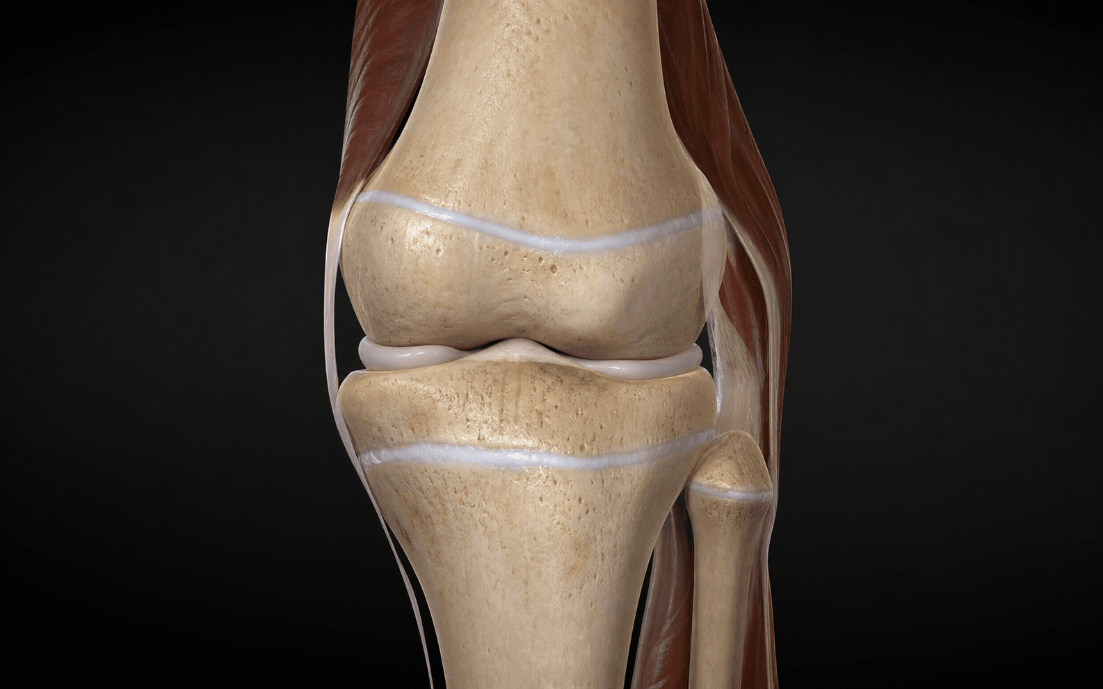
Relevant surgical anatomy. The distal femoral physis is undulating, with anterior and posterior portions — the most complex physis, which is why open Phemister or percutaneous drilling must address its full three-dimensional extent; it is approached medially and laterally just above the joint line, anterior to the collateral ligament origins, and the peripheral perichondrial ring (ring of LaCroix and zone of Ranvier) must be ablated for complete arrest, since residual peripheral physis causes deformity. The proximal tibial physis lies just distal to the joint line and the tibial tubercle apophysis anteriorly is part of this growth complex; the common peroneal nerve wraps around the fibular neck laterally and is at risk with lateral proximal tibial dissection, and the proximal tibiofibular relationship matters because combined tibial epiphysiodesis often includes the proximal fibular physis to avoid a relatively long fibula. On fluoroscopy the physis is seen as a lucent transverse line in both AP and lateral views (it is not flat), and the curette and drill must reach the central, anterior and posterior portions, not just the easily accessed peripheral margins.
References
Growth and predictions of growth in the lower extremities (Green-Anderson data)
- Foundational longitudinal study of normal lower-limb growth in children from Boston, generating the growth-remaining charts for the distal femur and proximal tibia plotted against skeletal age.
- Established that remaining growth must be read against skeletal (bone) age rather than chronological age.
- Provides the data underpinning the classic growth rates used in timing calculations (distal femur the largest contributor to lower-limb length).
A straight-line graph for leg-length discrepancies
- Introduced the straight-line graph, which linearises non-linear growth so serial measurements of the short and long limbs plot as straight lines and the effect of epiphysiodesis appears as a change in slope.
- Automatically incorporates the child's growth percentile and the degree of growth inhibition in the short limb.
- In the original series the straight-line graph was significantly more accurate than the growth-remaining method, particularly when there was growth inhibition.
Multiplier method for predicting limb-length discrepancy
- Derived an age- and sex-specific multiplier from growth databases so that, for a congenital discrepancy, LLD at maturity equals the current discrepancy multiplied by the multiplier — from as few as one measurement.
- Multiplier values were independent of percentile, height, generation, ethnicity and race, validated across radiographic, clinical and anthropological datasets.
- Predictions correlated well with the Moseley method and with actual outcomes in both lengthening and epiphysiodesis cohorts; the method also gives the timing of epiphysiodesis.
Percutaneous epiphysiodesis: experimental study and preliminary clinical results
- Animal study (immature rabbits) showed percutaneous drilling and pneumatic burring under image intensification produced complete arrest with bony fusion of the distal femoral physis in eight of nine limbs.
- Thirteen children then underwent percutaneous epiphysiodesis with radiographic fusion of all treated physes.
- Established the percutaneous drill-and-curette technique as a feasible, low-morbidity alternative to open arrest (companion technique to Bowen, PMID 6488627).
Efficacy and late complications of percutaneous epiphysiodesis with transphyseal screws (PETS)
- In 45 consecutive patients followed to maturity, growth arrest was DELAYED and incomplete: final loss of growth was only about 66 percent of that predicted pre-operatively for both femur and tibia.
- Tibial PETS caused valgus deformity in 9 patients (20 percent), leading the authors to ABANDON the technique in the tibia; femoral PETS was safe.
- Overall revision rate was 18 percent (8 patients), of which 7 (87.5 percent) involved the tibia.
Additional references: - Métaizeau JP, Wong-Chung J, Bertrand H, Pasquier P (1998). Percutaneous epiphysiodesis using transphyseal screws (PETS). J Pediatr Orthop 18:363-9. PMID 9600565. — Original description of PETS; mean bone-length inequality fell from 2.47 cm to 0.51 cm at maturity across 32 cases.
- Bowen JR, Johnson WJ (1984). Percutaneous epiphysiodesis. Clin Orthop Relat Res 190:170-3. PMID 6488627. — Original clinical description of percutaneous drill-and-curette epiphysiodesis under image intensification.
- Phemister DB (1933). Operative arrestment of longitudinal growth of bones in the treatment of deformities. J Bone Joint Surg Am 15:1-15. — Original description of the open bone-block (Phemister) epiphysiodesis technique (predates PubMed indexing).
- Aguilar JA, Paley D, Paley J, et al. (2005). Clinical validation of the multiplier method, part II. J Pediatr Orthop 25:192-6. PMID 15718900. DOI 10.1097/01.bpo.0000150808.90052.7c. — Validation cohort: multiplier method mean error for residual discrepancy about 0.9 to 1.0 cm, more accurate than Moseley for residual LLD after epiphysiodesis.