Basicervical and selected undisplaced femoral neck fractures · lateral (vastus lateralis) approach to the proximal femur
- The DHS is the fixation of choice for BASICERVICAL femoral neck fractures - at the base of the neck with no metaphyseal buttress. The lateral side-plate buttresses against varus collapse; the FAITH RCT (Lancet 2017) showed base-of-neck fractures and smokers were the subgroups that favoured the sliding hip screw over cancellous screws.
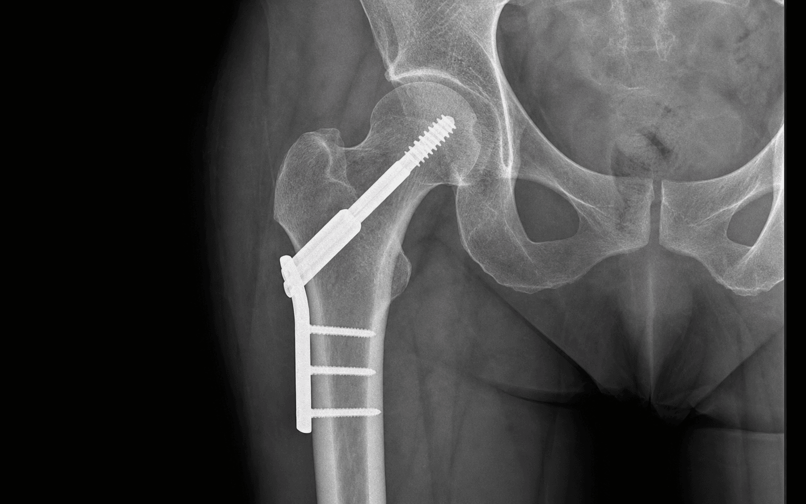
- Tip-apex distance (TAD) under 25mm is the single most important modifiable predictor. In Baumgaertner's JBJS 1995 study NONE of the 120 screws with a TAD of 25mm or less cut out; mean TAD was 38mm in cut-outs versus 24mm in healed fractures.
- Guide wire target: INFERIOR-CENTER or CENTER-CENTER on the AP view and CENTER-CENTER on the lateral (NEVER posterior). A superior position on AP increases cut-out risk 2-4 fold; a posterior position on lateral is the most common error.
- Garden alignment index assesses reduction quality: compressive trabeculae parallel to the medial cortex at 170-180 degrees on AP and 160-180 degrees on lateral. Posterior tilt is the most commonly missed malreduction - correct it by internally rotating the leg.
- Malreduction (outside 160-180 degrees) raises the failure rate to 30-50 percent versus 5-10 percent with anatomic reduction.
- Add an ANTI-ROTATION screw (6.5mm or 7.3mm cancellous screw, 1-1.5cm from and parallel to the lag screw) for basicervical patterns - it must not block the sliding mechanism.
When & Why
Primary indication. The dynamic hip screw (DHS) is most strongly indicated for a basicervical femoral neck fracture - a fracture at the base of the femoral neck, immediately proximal to the intertrochanteric line, with little or no medial metaphyseal buttress and a short, rotationally unstable proximal fragment. Biomechanically it behaves between a true femoral neck fracture and an unstable intertrochanteric fracture, with high varus and shear forces. Cannulated screws alone fail here through rotational instability and varus collapse; the DHS lateral side-plate directly buttresses the varus moment. In the FAITH randomised controlled trial (Lancet 2017) the sliding hip screw showed no overall reoperation advantage over cancellous screws across all hip fractures, but the pre-specified subgroups of base-of-neck (basicervical) fractures and smokers appeared to benefit from the sliding hip screw. The trade-off to disclose is that avascular necrosis was slightly more common with the sliding hip screw. Other indications.
Undisplaced or valgus-impacted fractures as an alternative to three parallel cannulated screws - surgeon preference. Consider a DHS if bone quality is poor or there is concern for displacement during screw insertion.
Cut-out or non-union after cannulated screw fixation, when adequate bone stock remains and the head is viable. Arthroplasty is more common if the head is not salvageable.
A combined pattern where the long DHS side-plate provides distal fixation; may require additional cerclage wiring or a longer plate.
What it is NOT for. A posterior (Moore) approach gives inadequate access for lateral plate placement and is not used for a DHS. A cephalomedullary nail is the main alternative many surgeons now prefer for basicervical and subtrochanteric-extension patterns, because the short unstable proximal fragment behaves like an unstable trochanteric fracture (Matre, Norwegian Hip Fracture Register). Patient factors and consent. Counsel for implant prominence and lateral thigh pain, and for the small but real risks of cut-out, non-union, avascular necrosis (related chiefly to the injury rather than the surgery), infection, and periprosthetic femoral shaft fracture, plus the prolonged protected weight-bearing. For displaced fractures in older, lower-demand patients, arthroplasty (hemiarthroplasty or total hip arthroplasty) is usually preferred over fixation - both NICE and the AAOS recommend this. Head-preserving fixation is favoured in younger patients (under about 60-65 years) regardless of displacement. Setup. Supine on a fracture table with a well-padded perineal post (check distal pulses and padding to protect the pudendal nerve). The uninjured leg is flexed 90 degrees at hip and knee, abducted and held in a leg holder to allow C-arm access from the opposite side. The injured leg sits in 10-15 degrees of abduction and neutral to slight (5-10 degrees) internal rotation to correct posterior tilt, with gentle longitudinal traction of 10-20kg. Confirm acceptable reduction on AP and lateral fluoroscopy BEFORE prepping.
The Operation
The goal is to anatomically reduce the fracture, place a lag screw centrally in the femoral head with a tip-apex distance under 25mm, seat a 135-degree barrel plate flush on the lateral cortex, apply interfragmentary compression, and - for basicervical patterns - add an anti-rotation screw. The exposure is a direct lateral approach to the proximal femur, elevating vastus lateralis off the lateral cortex to seat the plate; the Watson-Jones (anterolateral) and Hardinge (direct lateral, muscle-splitting) intervals are the relevant surgical planes (detailed in Background and Evidence).
Operative sequence
- Supine on a fracture table; well-padded perineal post - check distal pulses and padding to avoid pudendal nerve injury, and limit traction time.
- Uninjured leg flexed 90 degrees at hip and knee, abducted in a stirrup holder (pad the fibular head to protect the common peroneal nerve).
- Injured leg in 10-15 degrees abduction, neutral to 5-10 degrees internal rotation; 10-20kg longitudinal traction.
- C-arm from the opposite side, between the legs; obtain AP and lateral images BEFORE prepping to confirm reduction.
- Assess reduction on AP and lateral before prepping. AP: compressive trabeculae parallel to the medial cortex (170-180 degrees). Lateral: central axis to neck axis 160-180 degrees.
- Varus (less than 160 degrees) or valgus (greater than 180 degrees) on AP means malreduction. Posterior tilt (less than 160 degrees on lateral) is the MOST COMMON missed malreduction - correct it with internal rotation of the leg.
- Aim for a Garden alignment index of 160-180 degrees on both views, with a fracture gap less than 2-3mm.
- If closed reduction fails after 3-4 attempts, convert to open reduction. Anatomic reduction is essential - malreduction raises failure from 5-10 percent to 30-50 percent.
- Lateral thigh incision centred on the greater trochanter, 2-3cm distal to the GT tip, extending 8-10cm distally along the femoral shaft axis.
- Incise skin and subcutaneous tissue, then the fascia lata and iliotibial band in line with the skin incision.
- Identify vastus lateralis anteriorly (a thick belly with longitudinal fibres) and elevate its origin off the GT and proximal lateral femur with a Cobb or periosteal elevator, creating a subvastus pocket for the barrel plate (4-5cm distal to the GT tip).
- The guide-wire entry point is the lateral cortex 2-3cm distal to the vastus ridge (vastus tubercle).
- Dangers: an incision too anterior risks the lateral femoral cutaneous nerve (meralgia paresthetica); too proximal or posterior risks the superior gluteal nerve (exits 5cm above the GT tip); incomplete vastus elevation leaves the plate proud. Keep the dissection directly lateral.
- Use a 135-degree angled guide. Entry on the lateral cortex 2-3cm distal to the vastus ridge, directly lateral on AP, in the anterior third to middle of the shaft on lateral.
- Target: INFERIOR-CENTER or CENTER-CENTER on AP, CENTER-CENTER on lateral (NEVER posterior). Advance to within 5mm of subchondral bone without breaching the joint.
- Compute the tip-apex distance (TAD): the sum of the distances from the wire tip to the head apex on AP and lateral, corrected for magnification (divide by about 1.15-1.20). Target TAD under 25mm (Baumgaertner).
- If TAD is over 25mm or the position is superior or posterior, remove and reposition the wire BEFORE reaming - it is far easier to correct at the wire stage.
- Measure the wire length (accounting for magnification); typical lag screw 85-100mm. Select a screw ending 5mm short of subchondral bone (measured length minus 5-10mm).
- Triple ream over the wire: outer reamer for the lateral cortex, middle for the smooth barrel track, inner for the distal threads. Ream to 10mm short of the planned screw depth to protect subchondral bone.
- Advance steadily under fluoroscopy; withdraw carefully to avoid dislodging the wire.
- Remove the trocar, leaving the smooth wire. Insert the DHS lag screw over the wire with a T-handle or low-torque power driver (12-16mm lag screw; distal threads purchase head and neck bone, the proximal smooth shaft slides in the barrel).
- Advance to depth - ending 5mm from subchondral bone, fully seated on the lateral cortex. Confirm CENTER-CENTER on both views and TAD under 25mm before removing the wire.
- Avoid over-torquing (it strips the threads); a screw under 60mm into the head gives inadequate purchase.
- Select a 135-degree barrel plate (4-hole standard; 2-hole or longer variants available). Slide the barrel over the smooth shaft of the lag screw until fully engaged.
- The plate MUST sit completely flush on the lateral cortex - even a 1-2mm gap creates a varus moment, a stress riser, and hardware prominence. If proud, further elevate vastus lateralis or clear soft tissue from under the plate.
- Align parallel to the shaft; hold with bone or plate forceps; provisionally fix with a K-wire through the distal hole. Confirm on AP and lateral.
- Insert 4.5mm cortical screws through the distal holes: drill both cortices (3.2mm bit), measure, tap if needed, insert. A standard 4-hole plate needs a minimum of 2 bicortical screws (4 cortices); prefer 3-4 screws (6-8 cortices).
- Tighten sequentially, alternating holes; hold the plate firmly to prevent it lifting. Each screw should engage the far cortex fully without excessive medial protrusion (maximum 2-3mm).
- Dangers: unicortical screws are inadequate (varus collapse); screws that are too long threaten the profunda femoris vessels (8-10cm anteromedial to the GT) and perforators; medial protrusion over 5mm irritates soft tissue.
- Insert the compression (set) screw through the barrel into the back of the lag screw with a hex driver. It advances the lag screw into the head while the barrel (now fixed to the shaft) stays still, creating interfragmentary compression.
- Turn clockwise under fluoroscopy - typically 2-3 turns - watching the fracture gap close and the lag screw advance 2-3mm. Stop when the gap is closed.
- Do NOT over-compress (it can drive the screw through the head, or distract a comminuted fragment); ensure the plate is firmly fixed before compressing.
- For basicervical patterns add a 6.5mm or 7.3mm partially-threaded cancellous screw parallel to the lag screw, 1-1.5cm superior (preferred) or inferior.
- Place a parallel guide wire in the center-center position on lateral, measure to the same depth (5mm from subchondral bone), drill and insert.
- It must NOT block the lag screw sliding mechanism - maintain at least 1-1.5cm spacing. Avoid over-convergence (crowding the head and raising AVN risk) and posterior placement.
- Systematically verify on true AP and lateral: lag screw CENTER-CENTER on both views and within 5mm of subchondral bone; TAD confirmed under 25mm; plate flush and parallel to the shaft; all cortical screws bicortical; fracture gap closed (under 2mm); Garden index 160-180 degrees on both views; anti-rotation screw parallel if used.
- Release traction slowly, maintaining reduction; recheck fluoroscopy after release - reduction must be maintained. Save final images.
- Irrigate with at least 3 litres of saline (pulsatile lavage); achieve meticulous haemostasis.
- Close in layers: repair vastus lateralis over the plate where possible (reduces prominence); close the fascia lata and ITB securely with strong absorbable suture (e.g. No. 1 Vicryl) - the load-bearing layer; subcutaneous 2-0 or 3-0 absorbable to eliminate dead space; skin with staples or 3-0 nylon.
- A drain is not routine unless there is significant ongoing ooze; if used, place a 10-12Fr closed-suction drain deep to fascia, exiting posteriorly.
12-15cm posterior to the greater trochanter at the level of the lesser trochanter, deep to gluteus maximus. Stay anterior and lateral; place no posterior retractor more than 2-3cm behind the GT.
Exits above piriformis 5cm proximal to the GT tip, running between gluteus medius and minimus. Limit proximal dissection to less than 5cm above the GT; incise fascia directly lateral, not posterolateral.
8-10cm anteromedial to the GT in the femoral triangle beneath the inguinal ligament. Stay lateral on the shaft; avoid anterior dissection beyond the vastus ridge; take care with medial cortical screw length.
Subcutaneous; crosses the ASIS 2-3cm medial and runs 3-4cm anterior to the standard incision. Keep the incision directly lateral; avoid anterior extension (meralgia paresthetica if injured).
Exit with the superior gluteal nerve above piriformis, 4-5cm proximal to the GT. Avoid aggressive proximal dissection; if encountered, control immediately with bipolar at a low setting.
Tip-apex distance under 25mm is the single most modifiable predictor of success. In Baumgaertner's JBJS 1995 study NONE of the 120 screws with a TAD of 25mm or less cut out; cut-out rises to around 30 percent when TAD exceeds 25mm. Measure it on the guide wire before reaming, and reposition the wire if it would exceed 25mm.
The lateral view matters as much as the AP. Posterior tilt is the most commonly missed malreduction and a powerful predictor of failure - Palm (Acta Orthop 2009) found a tilt of 20 degrees or more was the only independent predictor of reoperation in undisplaced fractures. Correct it by internally rotating the leg 5-10 degrees on the fracture table before you prep.
Aftercare & Complications
Rehabilitation | Phase | Timing | Weight-bearing | Focus | |-------|--------|----------------|-------| | Immediate | 0-2 weeks | Touch weight-bearing (foot flat, no push-off), walking frame | Day-1 mobilisation; sit out, transfer; DVT prophylaxis (LMWH e.g. enoxaparin 40mg daily), extended to 28-35 days per NICE, AAOS and ACCP | | Early | 2-6 weeks | Touch WB, progress to partial (20-30kg) at 4-6 weeks if healing | Wound check at 2 weeks; hip ROM (avoid forced internal rotation and adduction); continue LMWH to 6 weeks | | Intermediate | 6-12 weeks | Weight-bearing as tolerated if callus visible and pain-free | Full ROM, abductor strengthening, gait re-education; basicervical patterns may progress earlier | | Late | 3-6 months | Full unrestricted WB by 10-12 weeks if union | Bridging callus on 3 cortices; graded return by bone quality | | Long-term | 6-24 months | Full | AVN surveillance (can present up to 3 years); osteoporosis management (DEXA, bisphosphonates e.g. alendronate 70mg weekly, or denosumab 60mg 6-monthly) | Avoid NSAIDs for the first 6 weeks, as they may impair fracture healing. Undisplaced fractures fixed with DHS return toward roughly 70-80 percent of pre-fracture function by one year; one-year mortality for fragility hip fractures is about 20-30 percent, driven chiefly by comorbidity rather than implant choice. Complications The commonest pathway to mechanical failure is a preventable technical error: malreduction accepted (varus under 160 degrees or posterior tilt under 160 degrees), poor guide-wire position (superior on AP, posterior on lateral, or TAD over 25mm), a plate not flush on the lateral cortex, or inadequate or over-compression.
- Recognition
- Groin pain at 2-6 months, loss of motion, screw prominence; radiographs show superolateral migration out of the head
- Prevention
- TAD under 25mm; inferior-center or center-center on AP; center-center on lateral (never posterior); anatomic reduction; good bone quality
- Management
- Conversion to arthroplasty (hemiarthroplasty if low demand, THA if active); remove the DHS; assess the acetabulum for penetration damage
- Recognition
- Persistent groin pain beyond 3-4 months, unable to weight-bear; no bridging callus at 6 months, persistent fracture line
- Prevention
- Anatomic reduction and compression; early mobilisation; smoking cessation; optimise nutrition
- Management
- Mostly conversion to arthroplasty (THA preferred); in the young, valgus osteotomy with revision fixation or bone graft (rare)
- Recognition
- Progressive groin pain at 12-24 months (up to 3 years); limited internal rotation; head sclerosis then collapse and crescent sign; MRI most sensitive early
- Prevention
- Minimise time to surgery (under 24 hours ideal); gentle, anatomic reduction; avoid multiple reduction attempts
- Management
- Pre-collapse: observe or core decompression (controversial). Collapsed: arthroplasty; remove the DHS at the time
- Recognition
- Progressive varus on serial films, falling neck-shaft angle, leg shortening, abductor limp, groin pain on weight-bearing
- Prevention
- Anatomic reduction; plate flush (a gap creates a varus moment); minimum 4 cortices, prefer 6-8; compression
- Management
- Accept if under 10 degrees and functional; revise to arthroplasty if symptomatic or over 10-15 degrees
- Recognition
- Superficial: erythema, drainage within 2-4 weeks. Deep: persistent pain, fever, raised CRP and ESR, sinus tract
- Prevention
- Pre-op IV cefazolin 2g within 60 min; minimise OR time; greater than 3L irrigation; meticulous haemostasis; glycaemic control
- Management
- Superficial: oral antibiotics, wound care. Deep early (under 3 weeks): washout, debride, retain hardware if stable, 6 weeks IV. Deep late: remove hardware, debride, consider arthroplasty
- Recognition
- Acute pain after a fall; fracture at or just distal to the last cortical screw
- Prevention
- Protected weight-bearing initially; fall-prevention counselling; use the shortest adequate plate
- Management
- ORIF with a plate extending beyond the DHS (3 screws above and below); cerclage if comminuted; bone graft if bone loss
- Recognition
- Lateral thigh pain over the GT, palpable plate, bursa, pain on abduction and external rotation
- Prevention
- Complete vastus elevation and repair over the plate; plate flush; warn thin patients
- Management
- Conservative first (NSAIDs, activity modification); removal after confirmed union (over 12 months) if persistent, with protected weight-bearing for 6 weeks afterwards
- Recognition
- Restriction of hip motion; usually incidental on radiograph
- Prevention
- Minimise soft-tissue stripping
- Management
- Observe if asymptomatic; excise if it limits motion (rare for DHS). Consider indomethacin 25mg TDS for 6 weeks in high-risk patients
Viva & Exam Focus
C-PLATEC-PLATE: DHS placement essentials
SCREW-ITSCREW-IT: preventing DHS failure
Clinical Decision Scenarios
Practise clinical reasoning and management decisions out loud
“Walk me through your decision-making for a 68-year-old with a basicervical femoral neck fracture. Why a DHS over cannulated screws?”
“Explain tip-apex distance. Why does it matter and how do you keep it under 25mm?”
“Describe the Garden alignment index. What does it assess and what is acceptable?”
Indications
- Basicervical femoral neck fracture - the base-of-neck subgroup that favoured the sliding hip screw in FAITH (Lancet 2017); the lateral buttress resists varus collapse (a cephalomedullary nail is the main alternative)
- Garden I or II undisplaced in patients over 65 - an alternative to cannulated screws (surgeon preference)
- Garden II valgus-impacted in the elderly - if choosing fixation over arthroplasty
- Failed screw fixation - if bone stock is adequate and the head is viable
- Neck fracture with subtrochanteric extension - the long DHS side-plate gives distal fixation
Key anatomy
- Blood supply: medial femoral circumflex artery (60-80 percent) via the posterosuperior retinacular vessels - disrupted by displacement
- AVN risk by Garden: I 0-5 percent, II 5-10 percent, III 20-30 percent, IV 25-40 percent - related to the injury, not the surgery
- Vastus ridge 2-3cm distal to the GT tip - the guide-wire entry point on the lateral cortex
- Compressive trabeculae run from medial cortex to superior head - the basis of the Garden alignment index
- Danger zones: sciatic 12-15cm posterior to GT, superior gluteal nerve 5cm proximal, LFCN 3-4cm anterior
Critical steps - say these phrases
- Assess reduction on the traction table BEFORE prepping using the Garden alignment index - 170-180 degrees AP, 160-180 degrees lateral; posterior tilt is the commonest error, correct with internal rotation
- The guide wire is the critical step - inferior-center or center-center on AP, center-center on lateral; NEVER posterior
- Confirm TAD will be under 25mm on the wire before reaming - Baumgaertner showed over 25mm triples cut-out risk
- Lag screw to 5mm from subchondral bone; plate FLUSH on the lateral cortex - any gap creates a varus moment
- Compression screw 2-3 turns watching the fracture gap close - converts shear to compression
- For basicervical fractures add an anti-rotation screw 1-1.5cm from the lag screw - prevents rotation while preserving sliding
- Final fluoroscopy: TAD under 25mm, center-center both views, plate flush, Garden index 160-180 degrees, compression achieved
Danger zones - location and protection
- Sciatic nerve: 12-15cm posterior to the GT at the lesser-trochanter level - stay anterior and lateral, no posterior retractor over 2-3cm
- Superior gluteal nerve: exits 5cm proximal to the GT between medius and minimus - limit proximal dissection to under 5cm
- Femoral neurovascular bundle: 8-10cm anteromedial in the femoral triangle - stay lateral, careful with medial screw length
- Lateral femoral cutaneous nerve: 3-4cm anterior, subcutaneous - keep the incision directly lateral
- Superior gluteal vessels: exit with the nerve 4-5cm proximal to the GT - avoid aggressive proximal dissection
Technique pearls
- TAD under 25mm is the single most important modifiable predictor - measure on the wire before reaming; in Baumgaertner none of 120 screws with TAD 25mm or less cut out
- Inferior-center on AP is ideal - a superior position increases cut-out 2-4 fold
- Posterior on lateral is the commonest error - eccentric loading, screw back-out
- The plate must be flush - a 1-2mm gap creates a varus moment and prominence
- Basicervical means add an anti-rotation screw 1-1.5cm away, preserving the sliding gap
- Garden index 160-180 degrees on both views before starting - malreduction raises failure to 30-50 percent
Complications - incidence and management
- Cut-out 5-10 percent (under 3 percent if TAD under 25mm, up to 30 percent if over) - convert to arthroplasty, remove the DHS
- Non-union 5-10 percent - persistent pain, no callus at 6 months; arthroplasty (valgus osteotomy rarely)
- AVN 10-15 percent Garden I or II, up to 40 percent IV - presents at 12-24 months, MRI early; arthroplasty
- Varus collapse 3-5 percent - accept if under 10 degrees and functional, else revise to arthroplasty
- Infection 1-3 percent - superficial oral antibiotics; deep washout, retain hardware if stable, 6 weeks IV
- Femoral shaft fracture 1-2 percent - ORIF with a long plate overlapping the DHS, cerclage if comminuted
Post-op protocol
- DVT prophylaxis: LMWH (e.g. enoxaparin 40mg daily) extended to 28-35 days per NICE, AAOS and ACCP
- Weight-bearing: touch WB (foot flat, no push-off) for 6-8 weeks, then WBAT if callus visible
- Follow-up: 2-week wound check, 6-week XR and progress WB, 3-month union, 6-12-month AVN surveillance
- Imaging: AP and lateral immediate post-op, then 2 and 6 weeks, 3 and 6 months; MRI at 12-24 months if symptoms
- Osteoporosis: DEXA, bisphosphonates (alendronate 70mg weekly) or denosumab (60mg 6-monthly)
What examiners want to hear
- On 'DHS versus screws': FAITH showed no overall difference, but basicervical or base-of-neck and smokers favoured the sliding hip screw - the lateral buttress resists varus collapse
- On technique: lead with TAD under 25mm and 'Baumgaertner showed none of 120 screws with TAD 25mm or less cut out'
- On reduction: 'Garden alignment index 160-180 degrees on both views; posterior tilt is the commonest missed malreduction, corrected by internal rotation'
- If shown a film with TAD over 25mm: 'I would revise - over 25mm triples cut-out risk'
- Always mention the anti-rotation screw for basicervical fractures
- On complications: lead with cut-out and TAD, then AVN (injury-related), then non-union
Background & Evidence
Epidemiology and outcomes. Femoral neck fractures are typical fragility fractures of the elderly. One-year mortality for fragility hip fractures is about 20-30 percent, driven predominantly by patient comorbidity rather than implant choice. Undisplaced fractures fixed with DHS return toward roughly 70-80 percent of pre-fracture function by one year, best when reduction is anatomic and mobilisation is early. Predictors of a good outcome are a younger physiological age, an undisplaced pattern, anatomic reduction (low posterior tilt), a TAD under 25mm, and early weight-bearing. Fixation versus arthroplasty (global guidance). Both NICE (UK) and the AAOS (US) recommend arthroplasty for most DISPLACED intracapsular fractures in older patients, and internal fixation for UNDISPLACED fractures with acceptable posterior tilt. In younger patients (under about 60-65 years) head-preserving anatomic reduction and fixation is favoured regardless of displacement, with arthroplasty reserved for salvage. The DHS is one fixation option in this paradigm; cannulated screws, cephalomedullary nails and fixed-angle devices are the alternatives. Femoral head blood supply. The medial femoral circumflex artery (MFCA) is the dominant supply (60-80 percent of the head); its deep branch passes posteriorly under piriformis and the ascending cervical (lateral epiphyseal) branches course along the posterosuperior neck - these are the vessels disrupted by a displaced fracture. The lateral femoral circumflex artery contributes 20-30 percent via ascending branches along the anterior capsule. The artery of the ligamentum teres is a minor contributor in adults (5-10 percent, more important in children). The intracapsular retinacular vessels ascend beneath the synovial reflection and enter the head at the subcapital junction; the posterosuperior retinacular vessels are the most vulnerable. AVN risk by Garden grade: I 0-5 percent, II 5-10 percent, III 20-30 percent, IV 25-40 percent. Proximal femur landmarks. The vastus ridge (tubercle), 2-3cm distal to the GT tip on the lateral cortex, is the guide-wire entry point. The calcar femorale is the dense vertical medial cortex providing medial support; Ward's triangle is the relatively weak area in the neck trabeculae. The compressive trabeculae run from the medial cortex to the superior head (the basis of the Garden alignment index); the tensile trabeculae run from the lateral cortex to the inferior head. Garden classification.
- Description
- Incomplete, valgus-impacted; trabeculae interrupted but not displaced
- AVN risk
- 0-5 percent
- Description
- Complete but undisplaced; trabeculae aligned on AP and lateral
- AVN risk
- 5-10 percent
- Description
- Complete with partial displacement; varus angulation and posterior tilt
- AVN risk
- 20-30 percent
- Description
- Complete with total displacement; no engagement, high AVN risk
- AVN risk
- 25-40 percent
Pauwels classification (biomechanical). Based on the angle of the fracture line from the horizontal: Pauwels I (less than 30 degrees) - stable, mainly compression; Pauwels II (30-50 degrees) - intermediate, mixed compression and shear; Pauwels III (greater than 50 degrees) - unstable, mainly shear, high failure risk. A higher Pauwels angle increases shear across the fracture and raises non-union and cut-out risk with any fixation method. Surgical approach planes. The DHS is placed through a direct lateral approach, elevating vastus lateralis off the lateral femur. The relevant named intervals are the Watson-Jones (anterolateral) approach, working between tensor fascia lata and gluteus medius (both supplied by the superior gluteal nerve, L4-S1 - not a true internervous plane, but more anatomic with less muscle splitting); the direct lateral (Hardinge) approach, splitting the anterior third of gluteus medius and minimus for direct access (simpler, but with abductor damage and a Trendelenburg risk if split excessively); and the posterior (Moore) approach, which is NOT used for a DHS because access is inadequate for a lateral plate.
References
The value of the tip-apex distance in predicting failure of fixation of peritrochanteric fractures of the hip
- 198 peritrochanteric fractures fixed with a sliding hip screw; 19 failures, 16 of which were lag-screw cut-out
- Mean TAD was 24mm in successfully healed fractures versus 38mm in those that cut out (p=0.0001)
- NONE of the 120 screws with a TAD of 25mm or less cut out; risk rose steeply with increasing TAD regardless of other variables
A new measurement for posterior tilt predicts reoperation in undisplaced femoral neck fractures: 113 consecutive patients treated by internal fixation and followed for 1 year
- 113 consecutive undisplaced (Garden I-II) femoral neck fractures fixed with two parallel implants; 26 (23 percent) reoperated within 1 year
- Posterior tilt of 20 degrees or more on the preoperative lateral radiograph: 14 of 25 reoperated versus 12 of 88 with less tilt (p less than 0.001)
- On multivariable analysis posterior tilt of 20 degrees or more was the ONLY significant independent predictor of reoperation
Fracture fixation in the operative management of hip fractures (FAITH): an international, multicentre, randomised controlled trial
- 1108 patients aged 50 or older randomised to sliding hip screw versus multiple cancellous screws for fixation
- No overall difference in 24-month reoperation: 20 percent (SHS) versus 22 percent (cancellous screws), HR 0.83 (95 percent CI 0.63-1.09)
- Avascular necrosis was MORE common with the sliding hip screw (9 percent versus 5 percent, HR 1.91); pre-specified subgroups of smokers and displaced or base-of-neck fractures appeared to favour the sliding hip screw
Operative treatment of femoral neck fractures in patients between the ages of fifteen and fifty years
- 73 femoral neck fractures in young patients (15-50 years) followed to union, conversion, or minimum 2 years
- Osteonecrosis in 23 percent and nonunion in 8 percent; 10-year survival of the native head free of conversion to THA was 85 percent
- Outcome was driven by initial displacement and quality of reduction - poor reduction was strongly associated with osteonecrosis and nonunion
Sliding hip screw versus IM nail in reverse oblique trochanteric and subtrochanteric fractures: a study of 2716 patients in the Norwegian Hip Fracture Register
- 2716 transverse or reverse-oblique trochanteric and subtrochanteric fractures from a national registry
- 1-year reoperation 6.4 percent (SHS) versus 3.8 percent (IM nail), p=0.011, with slightly more pain and lower satisfaction in the SHS group
- Registry signal favours intramedullary nailing for biomechanically unstable, lateral-wall-deficient patterns