Proximal femoral osteotomy correcting coxa valga and excessive anteversion to improve hip containment | advanced
- VDRO corrects TWO deformities simultaneously: varus (reducing an elevated neck-shaft angle of coxa valga down to roughly 110-120 degrees) and derotation (reducing excessive femoral anteversion to a physiological 10-15 degrees) — both improve femoral head containment within the acetabulum.
- The osteotomy is performed at the intertrochanteric or subtrochanteric level. Fixation is with a fixed-angle device — a 90-degree blade plate, a proximal femoral locking plate, or a paediatric hip screw — that converts the planned correction into a stable construct.
- In neuromuscular (cerebral palsy) hip subluxation and in older DDH, VDRO alone is frequently insufficient: it is combined with a pelvic osteotomy (Salter, Dega, or Pemberton) and often an open reduction and soft-tissue release for the migrated or dislocated hip.
- Shortening is commonly added at the osteotomy to decompress the joint, reduce joint reaction force, and ease concentric reduction — but excessive varus plus shortening weakens the abductors and produces a Trendelenburg gait and limb-length discrepancy.
When & Why
Indications. VDRO is a containment and realignment operation used across three paediatric settings. Developmental dysplasia of the hip (DDH).
- Residual dysplasia with femoral deformity — excessive anteversion and coxa valga preventing concentric, stable reduction.
- To aid concentric reduction — derotation and varus redirect the head deep into the acetabulum after open or closed reduction.
- Combined with a pelvic osteotomy in the older child (typically over 18-24 months and especially over 4 years) where the acetabulum will not remodel. Perthes disease (containment).
- Lateral pillar B and B/C border hips in children over 8 years — the poorer-prognosis groups that benefit most from containment.
- Femoral head at risk (Catterall or lateral pillar head-at-risk signs) in the fragmentation stage with a still-spherical, containable head.
- VDRO redirects the femoral head under the lateral acetabular margin during biological healing. Neuromuscular hip (cerebral palsy).
- Progressive hip subluxation — a Reimers migration percentage greater than 40-50% despite postural management.
- Dislocation — painful, or impeding seating and perineal care.
- Almost always combined with a pelvic osteotomy (Dega/San Diego), adductor/psoas release, and frequently femoral shortening. Other. Residual SCFE deformity (a related, separate corrective realignment for residual varus/retroversion or impingement), and coxa valga of other aetiology with documented uncovering. Contraindications.
- Absolute — active hip sepsis, and a stiff, incongruent, non-containable (no longer spherical, "hinge abduction") Perthes hip, because containment will worsen it.
- Relative — severe acetabular dysplasia where a femoral procedure alone is planned (add the pelvic side); a skeletally mature patient with established arthritis (consider an arthroplasty pathway); and a non-ambulant child with an asymptomatic, well-located hip (observe). Biomechanical rationale — redirection and unloading. Coxa valga combined with excessive anteversion rotates the load-bearing surface of the femoral head out of the acetabulum anterolaterally. Restoring a neck-shaft angle of 110-120 degrees and anteversion of 10-15 degrees points the head back into the socket. The medialised, varus proximal femur reduces the joint reaction force and improves the abductor working length — but only when it is not overcorrected. In containment surgery for Perthes the goal is to shelter the at-risk anterolateral head under the acetabular roof during healing.
- Primary problem
- Anteversion + valga; non-concentric reduction
- Femoral goal
- Derotate, modest varus to seat the head
- Usually combined with
- Open reduction; pelvic osteotomy if older
- Primary problem
- Residual dysplasia + femoral deformity
- Femoral goal
- Varus + derotation + shortening
- Usually combined with
- Salter / Pemberton / Dega osteotomy
- Primary problem
- At-risk anterolateral head
- Femoral goal
- Varus to contain, mild derotation
- Usually combined with
- Usually femur alone; pelvic in selected cases
- Primary problem
- Migration percentage greater than 40-50%
- Femoral goal
- Varus + derotation + shortening
- Usually combined with
- Pelvic (Dega) + adductor/psoas release
- Primary problem
- High, painful dislocated head
- Femoral goal
- Aggressive shortening + varus + derotation
- Usually combined with
- Open reduction + pelvic + soft-tissue release
Special situations by diagnosis.
- Cerebral palsy hip. The underlying problem is muscle imbalance (spastic adductors and flexors) plus a dysplastic acetabulum, not just the femur. Hip surveillance tracks the Reimers migration percentage, and reconstruction is indicated as migration rises. Combined reconstruction is the rule — VDRO plus a pelvic osteotomy (Dega/San Diego) plus adductor and psoas release, with open reduction for the dislocated hip. Aggressive femoral shortening decompresses the joint, eases reduction and protects the sciatic nerve and head blood supply; the bone is often osteopenic, so locking fixation helps. The goal in the non-ambulant child is a located, painless, mobile hip for sitting and perineal care, not gait normalisation.
- Perthes containment. Containment is appropriate only for a spherical, containable head, with the greatest benefit in children over 8 years with lateral pillar B and B/C border hips. Plan modest varus to avoid over-varus and its persistent limp, allow for remodelling of residual varus in the young child, and keep pelvic (Salter) or combined procedures as alternatives in selected cases.
- DDH across age. A young child post-reduction needs derotation and modest varus to seat a concentric head and relies on remodelling of the acetabulum and femur; the older child (over 4 years) needs a redirectional or reshaping pelvic osteotomy (Salter/Pemberton/Dega) added because the acetabulum will not remodel; the adolescent has minimal remodelling reserve, so the on-table correction is final and must be planned precisely and combined with the acetabular procedure. Remodelling is age-dependent. A young child (under 8 years) remodels residual deformity and tolerates deliberate undercorrection, whereas an older child or adolescent has little remodelling reserve and the correction achieved on the table is the correction you keep. Consent specifically for limb-length discrepancy, Trendelenburg or abductor weakness (especially with over-varus and shortening), implant prominence and the likely need for a second operation to remove hardware, infection, non-union (uncommon), and recurrence requiring revision or a pelvic procedure. Setup. Supine on a radiolucent table with a small bump under the operative hip and the image intensifier available. The approach is the lateral proximal femur.
The Operation
The goal is to expose the proximal femur laterally, protect the posterior neurovascular structures, seat a fixed-angle device up the femoral neck before cutting, perform an intertrochanteric osteotomy that delivers a measured varus and derotation, and fix the construct so the head sits concentrically and contained in the acetabulum. The lateral exposure is laid out in full as the first steps below — it is the heart of the operation.
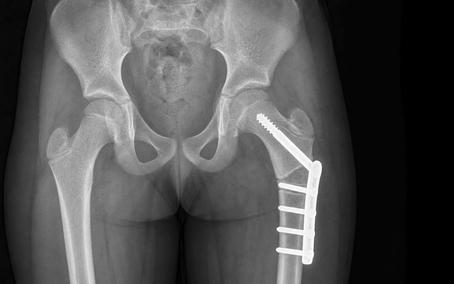

Preoperative planning. Obtain an AP pelvis, an abduction-internal-rotation (von Rosen) view to estimate congruent reduction and the varus needed, and a CT or clinical assessment of femoral anteversion. Calculate the neck-shaft angle and version preoperatively. Template the blade-plate angle (or plate) that will deliver the planned neck-shaft angle of 110-120 degrees, calculate the closing wedge for the varus correction, and decide the amount of shortening if reducing a high or dislocated hip.
Operative sequence
- Supine on a radiolucent table, a small bump under the operative hip, image intensifier available.
- A straight lateral longitudinal incision runs from the greater trochanter distally along the femoral shaft.
- Split the fascia lata and iliotibial band in line with the incision.
- Elevate vastus lateralis anteriorly off the lateral intermuscular septum (or split it) to expose the lateral femur subperiosteally.
- Incise and elevate the periosteum to expose the intertrochanteric or subtrochanteric region.
- Profunda femoris perforating arteries pierce the lateral intermuscular septum just behind the linea aspera — the principal bleeding hazard, lying immediately posterior to the osteotomy. Stay strictly subperiosteal and keep the posterior retractor flush on bone.
- The medial femoral circumflex artery, the dominant femoral head blood supply, ascends posteriorly along the neck — protected by staying at the osteotomy level and not stripping the posterosuperior neck.
- The sciatic nerve lies posterior to the hip and is at risk of stretch when a high dislocated cerebral palsy hip is reduced and derotated, and from posteriorly placed retractors.
- The femoral neurovascular bundle is anterior and is protected by subperiosteal lateral dissection and an anterior retractor placed on bone.
- Under image intensifier, place a guidewire up the femoral neck into the centre of the head in two planes — this sets the blade or screw trajectory. Stop short of subchondral bone and stay distal to the physis.
- Before any cut, insert two longitudinal K-wires straddling the planned osteotomy (one proximal, one distal) at an angle equal to the planned derotation, so that bringing them parallel after the cut confirms the rotation achieved.
- Following the guidewire, drive the seating chisel (for a blade plate) or insert the neck screw (for a locking plate or hip screw) along the planned trajectory under image guidance, stopping short of the joint and the physis.
- This is done BEFORE the osteotomy so the proximal fragment is controlled. Pre-drill and advance gently in osteopenic neuromuscular bone to avoid splitting or comminuting the fragment.
- Mark the intertrochanteric (or subtrochanteric) osteotomy level proximal to the lesser trochanter.
- Make a transverse osteotomy with an oscillating saw under constant cool irrigation, protecting the posterior cortex.
- If a closing wedge is planned for varus, resect the laterally based wedge as templated.
- For shortening in a high cerebral palsy hip, resect an additional cylindrical segment — decompressing the joint protects the sciatic nerve and the head blood supply when the hip is reduced.
- Internally rotate the distal fragment until the two K-wires are parallel — the planned derotation is achieved.
- Close the varus wedge by bringing the side plate onto the shaft. The varus is already built into the chosen blade-plate angle and the closing wedge, so bringing the plate onto the shaft delivers the correction.
- Confirm the corrected neck-shaft angle of 110-120 degrees on image — not less.
- Compress across the osteotomy and secure the side plate to the femoral shaft with bicortical (or locking) screws.
- Ensure enough bicortical screws distal to the osteotomy for a stable construct.
- Confirm stable fixation and acceptable correction on AP and lateral images, holding the reduction with the marker wires or a clamp until fixed.
- In older DDH and neuromuscular reconstruction, perform the planned pelvic osteotomy (Salter, Pemberton or Dega) and any open reduction or soft-tissue release through the appropriate exposure.
- Confirm a concentric, stable, contained reduction.
- Take final AP and abduction images.
- Confirm the head is concentrically reduced and contained, the neck-shaft angle is 110-120 degrees, anteversion is physiological, and there is no implant in the joint or across the physis.
- Repair vastus lateralis, then close fascia lata, subcutaneous tissue and skin in layers.
- In young children apply a hip spica for 4-6 weeks. Older children with rigid plate fixation may be managed without a spica depending on construct stability and compliance.
The perforating branches of the profunda femoris pierce the lateral intermuscular septum just behind the linea aspera, immediately posterior to the osteotomy plane. A posteriorly directed saw, an errant retractor hooked behind the femur, or uncontrolled distal exposure can lacerate a perforator and cause brisk, difficult-to-control bleeding that retracts into the septum. Keep strictly subperiosteal, protect the posterior cortex, keep the posterior retractor flush on bone, control the saw blade, and never blindly diathermy into the septum.
The fixed-angle device aimed up the neck can breach the articular surface or cross the proximal femoral physis, causing chondrolysis, growth arrest or trochanteric overgrowth. Use a guidewire under image intensifier in two planes before committing the chisel or blade, aim into the centre of the head stopping short of subchondral bone, and stay distal to the physis in the skeletally immature hip.
Place two longitudinal K-wires across the planned osteotomy set apart by the angle of anteversion you intend to remove. After the cut, internally rotate the distal fragment until the two wires are parallel — that is your derotation, reproducibly, not by eye.
Template the blade-plate angle so that when the side plate sits flush on the shaft the neck-shaft angle is your target of 110-120 degrees; the closing wedge delivers the rest. Confirm on the intensifier and do not add varus to "gain a bit more cover" — over-varus is the commonest error and causes abductor insufficiency.
For a high or dislocated cerebral palsy hip, resect an additional cylindrical segment. Decompressing the joint eases concentric reduction and protects the sciatic nerve and the femoral head blood supply when you reduce. Make the osteotomy transverse and cool the saw constantly to avoid thermal necrosis.


Aftercare & Complications
Rehabilitation | Timing | Immobilisation | Milestones | |--------|----------------|------------| | 0-6 weeks | Hip spica in young children; protected positioning if rigid fixation in older or compliant children | Neurovascular checks (especially after a high CP reduction); multimodal analgesia; wound care | | 6 weeks | Radiograph for callus and maintained position | Wean immobilisation if uniting | | 6-12 weeks | Protected, advancing | Progressive weight-bearing after radiographic union; abductor strengthening and gait re-education; range-of-motion work for the CP hip | | 6-12 months | None | Plan implant removal once united, particularly for prominent lateral implants in thin or neuromuscular children | Young children (under 8 years) remodel residual deformity, so undercorrection is tolerated. An adolescent has little remodelling reserve, so the on-table correction is final and must be planned precisely. Complications
- Incidence
- Common if not planned
- Recognition
- Persistent Trendelenburg gait and abductor lurch; neck-shaft angle less than 110 degrees on film; medialised trochanter
- Prevention and management
- Prevention: plan to 110-120 degrees, use the abduction film to size varus, never chase maximal coverage. Management: most improve with growth and remodelling in the young child and abductor strengthening; established symptomatic over-varus may need a valgus-producing revision osteotomy or trochanteric advancement
- Incidence
- Common (varus + shortening)
- Recognition
- Measured leg-length difference; pelvic obliquity; compensatory gait
- Prevention and management
- Prevention: limit shortening to what reduction needs; account for growth in the young. Management: shoe raise for a small discrepancy; contralateral epiphysiodesis or later lengthening for a significant projected discrepancy
- Incidence
- Higher when the femur is done alone in dysplasia
- Recognition
- Persistent uncovering; rising migration percentage on surveillance films; recurrent instability
- Prevention and management
- Prevention: add a pelvic osteotomy when the acetabulum is deficient; correct both version and angle. Management: revision with the missing pelvic procedure; repeat containment if the Perthes head is still at risk
- Incidence
- Frequent (thin or neuromuscular)
- Recognition
- Palpable or tender lateral hardware; bursitis over the plate; skin irritation
- Prevention and management
- Prevention: low-profile or locking plate; careful soft-tissue closure over the implant. Management: routine removal once united, typically 6-12 months postoperatively
- Incidence
- Uncommon with image control
- Recognition
- Blade or screw across the articular surface or physis on imaging; chondrolysis, growth arrest, trochanteric overgrowth
- Prevention and management
- Prevention: two-plane image-intensifier check before committing the device; stay short of subchondral bone and distal to the physis. Management: reposition the implant; manage growth arrest with later contralateral epiphysiodesis or corrective osteotomy
- Incidence
- Low (level-dependent)
- Recognition
- Sclerosis or collapse on follow-up films; pain, stiffness
- Prevention and management
- Prevention: keep at the osteotomy level, avoid posterosuperior neck stripping (medial femoral circumflex artery), avoid excessive forced reduction. Management: activity modification, containment if salvageable, later reconstruction
- Incidence
- Uncommon (intertrochanteric heals well)
- Recognition
- Persistent pain, motion at the osteotomy, lucency or lack of callus beyond 3-4 months
- Prevention and management
- Prevention: stable fixation, cancellous intertrochanteric level, preserve blood supply. Management: revision fixation with bone graft; address mechanical instability
- Incidence
- Intraoperative, level-dependent
- Recognition
- Brisk bleeding from the posterior plane that retracts into the lateral intermuscular septum
- Prevention and management
- Prevention: subperiosteal dissection, posterior retractor flush on bone, control the saw. Management: direct pressure, identify and ligate or clip the perforator; do not blindly diathermy into the septum
- Incidence
- Rare (high CP reduction)
- Recognition
- Postoperative foot drop or sensory deficit in the reduced, derotated limb
- Prevention and management
- Prevention: add adequate shortening rather than acute lengthening; gentle retraction. Management: relax the limb position, monitor, and explore if it does not recover
Viva & Exam Focus
VARUSVARUS — goals and pitfalls of the femoral osteotomy
CONTAINCONTAIN — when to add the pelvic side
Maximising varus to gain coverage drops the neck-shaft angle below roughly 110 degrees, medialises the trochanter, slackens the abductors and shortens the limb, producing a persistent Trendelenburg gait. Plan correction to a neck-shaft angle of 110-120 degrees and use the abduction-internal-rotation radiograph preoperatively to estimate the varus required for congruent reduction, then stop there.
The perforating branches of the profunda femoris pierce the lateral intermuscular septum just behind the linea aspera, immediately posterior to the osteotomy plane. A posteriorly directed saw, an errant retractor behind the femur, or uncontrolled distal exposure can lacerate a perforator and cause brisk, difficult-to-control bleeding that retracts into the septum. Stay subperiosteal, protect the posterior cortex and keep the retractor flush on bone.
The blade or fixed-angle screw aimed up the neck can breach the articular surface or cross the proximal femoral physis, causing chondrolysis, growth arrest or trochanteric overgrowth. Use a guidewire under image intensifier in two planes before committing the chisel or blade, aim into the centre of the head stopping short of subchondral bone, and stay distal to the physis in the skeletally immature hip.
In older DDH and neuromuscular subluxation the acetabulum is dysplastic and deficient, so a femoral osteotomy alone leaves an uncovered head that re-subluxates. Combine VDRO with a pelvic osteotomy (Salter, Dega or Pemberton) and, when needed, open reduction plus soft-tissue release. Femur-only correction is appropriate mainly in Perthes containment and in the very young, remodelling hip.
The femoral component of containment is rotational as much as angular. Leaving excessive anteversion (commonly greater than 50-60 degrees in CP and DDH) means the head still escapes anterolaterally despite a good neck-shaft angle. Measure version preoperatively (CT or a clinical Ryder test under image) and derotate to leave a physiological 10-15 degrees, marking rotation with longitudinal K-wires across the osteotomy before cutting.
A long-standing high CP dislocation acutely reduced and derotated places the sciatic nerve and femoral vessels under tension; the limb is suddenly lengthened and re-oriented. Add femoral shortening to decompress the joint and slacken the neurovascular structures, avoid acute large lengthening, and monitor distal perfusion and nerve function postoperatively.
Clinical Decision Scenarios
Practise clinical reasoning and management decisions out loud
“A 9-year-old boy with lateral pillar B Perthes disease in the fragmentation stage has a containable, spherical femoral head but increasing lateral extrusion. You are considering a femoral varus derotation osteotomy. How do you plan and perform it, and how do you avoid the common pitfalls?”
“A 7-year-old non-ambulant child with spastic quadriplegic cerebral palsy has a right hip migration percentage of 60% with early pain on transfers. Adductor tenotomy two years ago has not prevented progression. How do you manage this hip?”
“You have completed a femoral varus derotation osteotomy and fixed it with a blade plate. Talk me through how you intraoperatively confirm you have achieved the correct varus AND derotation, and how you avoid over-correction.”
Core concept
- VDRO corrects TWO deformities: varus (lowers an elevated neck-shaft angle of coxa valga) and derotation (reduces excessive femoral anteversion)
- Biomechanical aim: redirect the head into the acetabulum, improve containment and reduce the joint reaction force
- Performed at the intertrochanteric (preferred — cancellous, fast union) or subtrochanteric level
- Shortening is added to decompress the joint and ease reduction of a high or dislocated hip
- Targets: neck-shaft angle 110-120 degrees; anteversion 10-15 degrees
Indications
- DDH: aid concentric reduction; correct residual femoral deformity (with a pelvic osteotomy in the older child)
- Perthes: containment of a spherical, containable, at-risk head — best benefit in children over 8 years, lateral pillar B or B/C border
- Cerebral palsy: progressive subluxation (migration percentage greater than 40-50%) or dislocation
- Residual SCFE deformity: realignment for residual varus or retroversion
- Contraindicated: hip sepsis; hinge abduction in Perthes (a stiff, non-containable head)
When to add the pelvic side
- Older DDH (over 4 years) — the acetabulum will not remodel: add Salter, Pemberton or Dega
- CP reconstruction — almost always combined: VDRO plus Dega plus adductor or psoas release, with or without open reduction
- Femur-alone is mainly for Perthes containment and the very young, remodelling hip
- If the head stays uncovered on the abduction film, the acetabulum is deficient — correct it
Surgical anatomy
- Approach: lateral, subperiosteal exposure of the proximal femur (split fascia lata, elevate vastus lateralis)
- Profunda femoris perforators: behind the linea aspera, posterior to the osteotomy — the main bleeding hazard
- Medial femoral circumflex artery: posterosuperior neck — the head blood supply; do not strip it
- Sciatic nerve: posterior; at risk of stretch when a high CP hip is reduced or derotated
- Normal values: neck-shaft angle about 125-130 degrees; anteversion about 10-15 degrees
Operative technique — key steps
- 1. Plan from AP, the abduction view and a version measurement; template the blade-plate angle and wedge
- 2. Lateral subperiosteal exposure; protect the posterior cortex and perforators
- 3. Neck guidewire in two planes; place two rotation marker K-wires set at the planned version
- 4. Seat the fixed-angle device (blade or screw) BEFORE the osteotomy
- 5. Transverse intertrochanteric osteotomy; laterally based closing wedge for varus; add shortening if needed
- 6. Derotate until the marker wires are parallel; close the varus; fix the plate to the shaft
- 7. Confirm a neck-shaft angle of 110-120 degrees, physiological version, and the device clear of joint and physis
- 8. Add the planned pelvic osteotomy or open reduction; confirm a concentric, contained reduction
Danger zones
- Over-varus (less than 110 degrees): Trendelenburg gait, shortening, abductor insufficiency — the commonest error
- Profunda perforators behind the linea aspera: brisk bleeding from a posterior cut or errant retractor
- Joint or physeal penetration by the blade or screw: chondrolysis, growth arrest, trochanteric overgrowth
- Acute reduction of a high CP hip without shortening: sciatic nerve stretch and femoral head avascular necrosis
Complications
- Over-varus with abductor insufficiency and limb-length discrepancy — limit varus and shortening; plan to 110-120 degrees
- Under-correction or recurrent subluxation — add the pelvic procedure when the acetabulum is deficient
- Implant prominence — frequent in thin or neuromuscular children; routine removal once united
- Avascular necrosis, non-union (uncommon at the intertrochanteric level) and sciatic nerve palsy (after a high CP reduction)
Post-op and remodelling
- Spica for 4-6 weeks in young children; rigid plate fixation may permit spica-free management in older or compliant children
- Radiograph at 6 weeks; progressive weight-bearing after union (6-12 weeks); abductor strengthening
- Young child (under 8): remodels residual deformity — undercorrect deliberately
- Adolescent: little remodelling reserve — the on-table correction is final; plan precisely
- Plan implant removal after union (commonly 6-12 months)
Background & Evidence
Geometry the osteotomy manipulates. VDRO corrects both the coronal angle (varus) and the axial rotation (derotation) — correcting only one leaves residual uncovering. | Parameter | Normal (mature) | Pathological (DDH/CP) | VDRO target | |-----------|-----------------|------------------------|-------------| | Neck-shaft angle | About 125-130 degrees | Coxa valga, greater than 140 degrees | 110-120 degrees | | Femoral anteversion | About 10-15 degrees | Excessive, often greater than 50 degrees | 10-15 degrees | | Articulo-trochanteric distance | Trochanter tip about level with the head centre | Reduced (relative overgrowth) | Maintain; avoid over-varus | Osteotomy levels. The intertrochanteric level (between the greater and lesser trochanters) is the standard level for most paediatric VDRO — its cancellous bone heals rapidly. A subtrochanteric level (distal to the lesser trochanter) is used when more shortening or a particular fixation is planned; it is more diaphyseal and so unites more slowly. Fixation devices. A 90-degree fixed-angle blade plate is the classic AO paediatric device — the blade up the neck sets the varus and the side plate captures the shaft, so the chosen blade-plate angle dictates the final neck-shaft angle. A proximal femoral locking plate is anatomically pre-contoured, with locking screws into the neck and head setting the angle, and is useful in osteopenic neuromuscular bone. A paediatric hip screw or dynamic device is a fixed-angle screw construct used in larger children and adolescents. Why the thigh tolerates the cut. The proximal femur is enveloped by well-vascularised muscle, so intertrochanteric osteotomies have a high union rate. The trade-offs are abductor mechanics: varus medialises the trochanter and slackens the abductors, while derotation reorients the whole distal limb — both must be planned, not maximised. Epidemiology of the indications. DDH is the most common indication in the young child, where VDRO supports a concentric reduction. Perthes disease (Legg-Calve-Perthes) is an idiopathic avascular necrosis of the immature femoral head; the lateral pillar classification stratifies prognosis and the children over 8 years with lateral pillar B and B/C border hips are the group that benefits from containment. In cerebral palsy, progressive hip subluxation is driven by muscle imbalance and tracked by the Reimers migration percentage — the proportion of the femoral head lying lateral to Perkin's line, with a value greater than 30-33% regarded as abnormal and reconstruction commonly triggered at 40-50%. Key evidence. Herring (2004) showed that in children over 8 years with lateral pillar B or B/C border hips, surgical containment gave significantly better outcomes than non-operative treatment, with femoral varus osteotomy equivalent to an innominate (Salter) osteotomy. Joseph (2005) found that a femoral varus osteotomy shortened the disease course and better preserved head sphericity in containable Perthes hips, with the greatest benefit when performed in the avascular or early fragmentation stage. Reimers (1980) defined the migration percentage that now drives neuromuscular hip surveillance. Al-Ghadir (2009) showed that combined VDRO plus a San Diego (Dega) pelvic osteotomy outperformed VDRO alone in CP hips — a 25% revision rate after femur-only correction versus 0% after combined surgery. Hagglund (2014) showed that a population-based CP hip-surveillance programme over 20 years nearly eliminated painful dislocation and enabled timely reconstructive VDRO.
References
Legg-Calve-Perthes disease. Part II: Prospective multicenter study of the effect of treatment on outcome
- Prospective multicentre study of 451 affected hips; 345 hips followed to skeletal maturity, classified by the modified lateral pillar and Stulberg systems
- In children over 8 years at onset with lateral pillar B or B/C border hips, surgical treatment gave significantly better outcomes than non-operative treatment (p less than or equal to 0.05)
- Femoral varus osteotomy and innominate (Salter) osteotomy produced no significant difference in outcome — either containment method is acceptable
- Lateral pillar group C hips did poorly regardless of treatment; children 8 years or younger did well irrespective of treatment
How does a femoral varus osteotomy alter the natural evolution of Perthes' disease?
- Analysed 640 Perthes hips; 314 treated with femoral varus osteotomy compared with non-operated controls
- Varus osteotomy shortened disease duration, minimised femoral head extrusion at the most vulnerable stage, and reduced metaphyseal widening
- 34% of hips operated in the avascular necrosis stage bypassed the fragmentation stage altogether
- Sphericity of the femoral head was better preserved, with greatest benefit when surgery was performed in the avascular or early fragmentation stage
Combined femoral and pelvic osteotomies versus femoral osteotomy alone in the treatment of hip dysplasia in children with cerebral palsy
- 52 spastic CP hips: 36 treated with VDRO plus San Diego (Dega) pelvic osteotomy, 16 with VDRO alone; mean follow-up 4.4 years
- The combined procedure gave significantly greater improvement in centre-edge angle and acetabular index, and better pain relief
- 25% of the VDRO-alone group required revision surgery, versus 0% of the combined group
- No avascular necrosis, non-union or infection in either group
Prevention of dislocation of the hip in children with cerebral palsy: 20-year results of a population-based prevention programme
- Population-based CPUP surveillance register in southern Sweden followed for 20 years
- Hip dislocation fell from 9 children in the historical control group to 2 then 0 in successive surveillance cohorts (p less than 0.001)
- Of 689 children in the study groups, 91 (13%) underwent timely preventive reconstructive surgery (including VDRO)
- Every child who reached dislocation reported severe pain; four required salvage surgery
The stability of the hip in children: a radiological study of the results of muscle surgery in cerebral palsy
- Original description of the migration percentage (Reimers index) — the proportion of the femoral head lying lateral to Perkin's line
- Provides the radiological measure used worldwide to monitor neuromuscular hip displacement
- A migration percentage greater than 30-33% is regarded as abnormal; progressive rises commonly trigger reconstruction at 40-50%
- Forms the quantitative basis of modern hip surveillance programmes