Phalangeal and metacarpal fractures are the most common hand injuries. Phalangeal fractures are the least forgiving — one degree of malrotation equals about 5 mm of digital overlap at the fingertip, so rotation always requires correction. Fixation (K-wire, lag screw, mini-plate or intramedullary pin) is matched to the fracture pattern; Bennett's and Rolando are intra-articular thumb metacarpal base fractures requiring anatomical reduction.
- Rotation is the critical deformity in metacarpal and phalangeal fractures. One degree of rotational malunion causes 5 mm of digital overlap at the fingertip level — even small amounts cause symptomatic scissoring.
- Boxer's fracture (5th MCP neck): up to 40–70° of sagittal angulation may be acceptable (functional outcome good) but rotational deformity must always be corrected regardless of angulation.
- Phalangeal fractures are the least forgiving of all hand fractures: oblique or spiral patterns cause shortening and rotation; condylar fractures of the PIP joint require anatomical reduction to restore PIP function.
- Bennett's fracture (intra-articular base of thumb MC) — small volar fragment held by anterior oblique ligament while MC shaft displaces radially and proximally by APL pull. ORIF or percutaneous K-wire fixation required.
- “Test rotation clinically: ask the patient to flex all fingers toward the palm in tandem — all fingernails should be parallel and converge to the scaphoid tubercle. Any scissoring or crossing indicates malrotation.
- “Acceptable metacarpal shaft angulation: index 10°, middle 10°, ring 20°, small 30° (transverse fractures). For oblique and spiral fractures, shortening greater than 5 mm or any rotation is unacceptable.
- “Condylar fractures of the proximal phalanx (PIP joint): even 2 mm displacement leads to PIP joint incongruity and a poor outcome. Anatomical reduction via a lag screw is the gold standard.
- “Rolando fracture is a comminuted Bennett's fracture. If fragments are large: ORIF with lag screws or a mini-plate. If highly comminuted: external fixation, traction, or primary arthrodesis in older patients.
When & Why
Indication. Operative fixation of a metacarpal or phalangeal fracture is driven by three things — rotation, joint congruity, and unacceptable alignment — not by the radiograph in isolation. Rotation cannot be read reliably from a plain film and is the single non-negotiable indication for surgery. Metacarpal fractures — absolute indications - Any rotational deformity (clinically confirmed scissoring).
- Intra-articular fractures with displacement greater than 1–2 mm (MCP or CMC joint).
- Open fractures (debridement mandatory; fixation depends on contamination).
- Multiple metacarpal fractures with instability.
- Bennett's fracture (intra-articular base of the 1st MC with CMC joint involvement). Metacarpal fractures — relative indications - Angulation exceeding the acceptable limit for the specific ray (see table below).
- Shortening greater than 5 mm in oblique or spiral patterns.
- Failure of closed reduction to achieve acceptable alignment.
- Soft-tissue interposition preventing reduction.
- Boxer's fracture with rotation, or with greater than 50–70° of angulation depending on patient demand.
- Transverse angulation
- Up to 10°
- Oblique/spiral shortening
- Less than 3 mm
- Rotation
- Zero — any rotation unacceptable
- Rationale
- Rigid CMC joint — no compensation for angulation
- Transverse angulation
- Up to 10°
- Oblique/spiral shortening
- Less than 3 mm
- Rotation
- Zero — any rotation unacceptable
- Rationale
- Central pillar of the hand — limited CMC mobility to compensate
- Transverse angulation
- Up to 20°
- Oblique/spiral shortening
- Less than 5 mm
- Rotation
- Zero — any rotation unacceptable
- Rationale
- More CMC mobility (about 15°) allows some compensation
- Transverse angulation
- Up to 30° (neck up to 40–70°)
- Oblique/spiral shortening
- Less than 5 mm
- Rotation
- Zero — any rotation unacceptable
- Rationale
- Most CMC mobility (25–30°) compensates for angulation
Phalangeal fractures — operative indications (proximal and middle phalanx shaft) - Any rotational deformity.
- Angulation greater than 10° in the sagittal or coronal plane (phalangeal fractures are less tolerant).
- Shortening greater than 2–3 mm.
- Condylar fractures (PIP or DIP joint) with displacement greater than 1–2 mm.
- Unstable patterns (transverse with cortical comminution, oblique, spiral). Consent specifically for digital numbness or a painful neuroma (digital nerve), PIP stiffness (the commonest problem after phalangeal fractures), pin-site infection, non-union, residual rotational deformity needing a derotation osteotomy, and post-traumatic arthritis for intra-articular injuries. Setup. Supine, arm on a hand table, upper-arm tourniquet, regional (axillary/supraclavicular) or general anaesthesia. The image intensifier is essential for reduction and fixation; loupe magnification is used for articular and Bennett's work. Position the hand so the cascade can be assessed intra-operatively.
The Operation
The goal is to restore alignment, rotation and joint congruity with the least fixation compatible with early motion. Expose through the approach matched to the fracture, protect the digital neurovascular bundles and the extensor mechanism, reduce the fracture anatomically, confirm rotation clinically under anaesthesia, fix to suit the pattern, and splint in the intrinsic-plus position. The exposure is laid out in full as the first operative steps below — it is the heart of the procedure.
Operative sequence
- Supine, hand table, upper-arm tourniquet; exsanguinate and inflate.
- Image intensifier available for the whole case; loupes for condylar and Bennett's work.
- Plan the approach from the fracture pattern and the at-risk structures before cutting.
The exposure is chosen to give fracture access while protecting the neurovascular bundles and the extensor mechanism: - Dorsal longitudinal (metacarpal shaft) — over the dorsum between the extensor tendons; the digital neurovascular bundles (volar-lateral) are protected and out of field.
- Mid-lateral (phalanx) — an incision along Cleland's ligament at the axis of rotation, working between the dorsal and volar neurovascular bundles; safe, but gives limited access to volar structures.
- Volar zig-zag / Bruner (phalanx) — full access to the flexor sheath; the neurovascular bundles on each side must be protected.
- Wagner volar-radial (thumb CMC, Bennett's) — for the base of the thumb metacarpal; identify and protect the radial artery (deep/dorsal to APL and EPB) and the superficial branch of the radial nerve before the capsulotomy.
- The digital neurovascular bundle runs volar-lateral on each side of the digit, volar to the axis of rotation at MCP level; it is at risk with incisions too far volar or with aggressive retraction.
- The extensor mechanism overlies the phalanx dorsally — plates placed here lie directly beneath it and drive adhesions, so keep dissection tight and the periosteal stripping limited to the fracture zone.
- The extensor digitorum communis runs as the central slip to the middle phalanx base; lateral bands join to form the terminal tendon at the DIP.
- Reduce with bone reduction clamps, restoring length, angulation and rotation.
- Confirm rotation clinically — flex all digits together in a cascade: the fingernails must be parallel and the fingers must converge toward the scaphoid tubercle. Any scissoring is unacceptable.
- Re-check rotation after definitive fixation, not only after reduction.


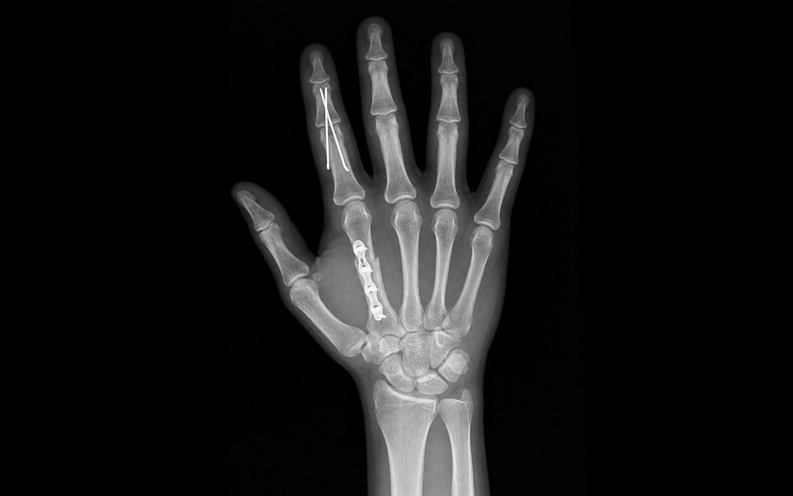
Choose the implant from the fracture geometry: - Transverse shaft — crossed K-wires or a mini-plate.
- Oblique or spiral shaft — lag screws (fracture length at least twice the shaft diameter).
- Comminuted shaft — mini-plate (bridging).
- Condylar / intra-articular — lag or headless compression screw.
- Bennett's — lag screw ORIF or percutaneous K-wire into the trapezium.
Crossed K-wire — metacarpal neck (Boxer's):
- Flex the MCP joint 90° to relax soft-tissue tension and aid reduction.
- Apply longitudinal traction and use the Jahss manoeuvre (pressure over the flexed proximal phalanx) to correct the apex-dorsal angulation.
- Introduce the first K-wire through the metacarpal head at about 45° to the shaft, crossing the fracture site.
- Introduce a second K-wire from the opposite side, crossing the first at the fracture.
- Confirm reduction fluoroscopically in AP, oblique and lateral views; bend and cut the wires outside the skin. Parallel K-wire — phalangeal shaft: two parallel wires through the distal fragment across the fracture into the proximal fragment give better rotational control than a single wire. K-wires give minimal rotational stability alone — rotation must be corrected before insertion. Avoid transfixing joints (causes stiffness); remove wires at 3–4 weeks.

Indications: spiral or oblique metacarpal shaft fractures (length at least twice the shaft diameter); condylar fractures of the PIP joint; Bennett's fracture. Technique — metacarpal spiral fracture:
- Expose through a dorsal longitudinal incision; protect the extensor tendon.
- Reduce with bone reduction clamps and confirm no rotation clinically.
- Drill the gliding hole in the near cortex (drill bit equal to the outer screw diameter).
- Drill the pilot hole in the far cortex (drill bit equal to the inner screw diameter).
- Measure, tap if required, and insert the cortical screw — the head compresses the near cortex to give the lag effect.
- Use a minimum of two lag screws for rotational stability (a single screw allows rotation). Technique — condylar fracture (PIP joint): mid-lateral or dorsal approach; lever the condylar fragment into anatomical position and check articular congruity directly; hold with a temporary K-wire; replace it with a 1.3 mm or 1.5 mm countersunk headless screw (Herbert technique). The screw must sit flush or sub-articular — a protruding head causes PIP impingement.

Indications: comminuted metacarpal fractures not suitable for lag screws alone; transverse metacarpal shaft fractures needing absolute stability (high-demand patient); Rolando fracture with large reconstructable fragments; revision after K-wire failure. Technique — dorsal mini-plate (metacarpal shaft):
- Longitudinal dorsal incision; reflect the extensor tendon or pass through the interval between EDC slips.
- Limited periosteal elevation over the fracture zone only.
- Reduce with reduction clamps.
- Apply a 2.0 mm or 2.4 mm straight plate to the dorsum with at least two screws each side of the fracture in cortical bone.
- Repair the periosteum and close the extensor interval; early active motion at 3–5 days. Phalangeal plates lie directly under the extensor mechanism — use the lowest-profile system available (1.0 mm or 1.3 mm) and commit to an early active mobilisation protocol.
- Anatomy of injury: the anterior oblique ligament (beak ligament) retains a small volar-ulnar fragment at the base of the 1st MC; APL pulls the metacarpal shaft proximally, radially and into supination, producing CMC subluxation.
- Approach: Wagner volar-radial incision at the thumb CMC joint.
- Protect the radial artery (deep/dorsal to APL and EPB) and the superficial branch of the radial nerve.
- Longitudinal capsulotomy; identify the volar fragment held by the AOL and reduce the thumb CMC joint (sometimes a dental pick is needed to elevate an impacted fragment).
- Fix: a 1.5 mm or 2.0 mm lag screw from the metacarpal shaft into the volar fragment, OR a percutaneous K-wire from the 1st MC into the trapezium (plus a second wire into the 2nd MC base for rotational control).
- Confirm anatomical reduction of the CMC joint fluoroscopically; repair the capsule and close in layers.
- Final clinical rotation check and fluoroscopic confirmation.
- Layered closure; release the tourniquet and confirm digit perfusion.
- Bury or trim K-wires; dress.
- Immobilise in the intrinsic-plus (safe) position — MCP joints 70–90° flexion, IP joints fully extended, wrist 20–30° extension.
- Bennett's fracture: thumb spica for 4 weeks, then mobilise.
One degree of malrotation at the fracture causes about 5 mm of digital overlap at the fingertip — a fracture that looks only mildly rotated on radiograph may cross dramatically in flexion. Assess rotation clinically under anaesthesia before and after fixation: flex all digits together and confirm the nails are parallel and converge to the scaphoid. Rotation cannot be read reliably from a plain film. For a spiral fracture use two lag screws, never one — a single screw allows rotation.
For a Bennett's fracture, identify and protect the radial artery (it lies deep and dorsal to APL and EPB in the snuffbox region) and the superficial branch of the radial nerve before the capsulotomy. These are the structures most often injured in this approach.
A spiral or oblique metacarpal fracture is ideal for lag-screw fixation only if the fracture length is at least twice the shaft diameter — and it always needs two screws. A single lag screw acts as a pivot and allows rotation; two screws give rotational stability.
A mini-plate on the dorsum of a phalanx lies directly beneath the extensor mechanism and drives adhesions and stiffness. Prefer a lag screw for phalangeal shaft fractures whenever possible; if a plate is required, use the lowest-profile implant and commit to early active motion (in one plating series, major complications occurred in 57% of patients).
Aftercare & Complications
Rehabilitation | Phase | Timing | Immobilisation | Therapy | |-------|--------|----------------|---------| | 1 | 0–3 weeks | Intrinsic-plus splint (K-wires removed at 3–4 weeks) | Wound care, elevation; gentle active ROM; buddy taping | | 2 | 3–6 weeks | Removable splint | Progressive active and active-assisted ROM; dynamic splinting if PIP stiffness develops | | 3 | 6–12 weeks | Splint for heavy tasks only | Strengthening (putty, grip); graded return to function | | 4 | 12+ weeks | None | Full activity; contact sport from 8–12 weeks once united | Intrinsic-plus (safe) position. MCP joints at 70–90° flexion (collateral ligaments under maximum tension, preventing MCP stiffness), IP joints at full extension (volar plate at maximum tension, preventing IP flexion contracture), wrist at 20–30° extension. Use it for any post-operative splint. Duration by fixation: K-wire fixation 3–4 weeks in an intrinsic-plus splint then mobilise; stable lag-screw or mini-plate fixation — early active motion at 3–5 days with buddy taping; Bennett's fracture — thumb spica 4 weeks then mobilise. Rehabilitation milestones - Week 0–3: wound care, elevation; intrinsic-plus splint between exercises; gentle active ROM 3–5 times per day; buddy taping to the adjacent digit.
- Week 3–6: remove K-wires at 3–4 weeks (confirm healing on radiograph); progressive active and active-assisted ROM; dynamic splinting if PIP stiffness develops; avoid strengthening until union.
- Week 6–12: progressive strengthening (putty, grip); return to light duties; sport or manual labour from 8–12 weeks depending on stability. Return to activity - Light work (keyboard, sedentary): 3–4 weeks.
- Medium work (non-dominant hand tasks): 4–6 weeks.
- Manual labour / gripping: 6–12 weeks.
- Contact sport: 8–12 weeks, with radiographic union before full loading. Boxer's fracture specific protocol - Buddy taping to the ring finger immediately; a metacarpal brace for comfort (not essential).
- Early active ROM at 1–2 weeks if acceptable alignment is maintained.
- Most patients regain full function by 6 weeks.
- A cosmetic dorsal knuckle prominence may persist — warn the patient pre-operatively. Complications
- Incidence
- 5–15% (commonest functional problem)
- Prevention
- Intra-operative rotation assessment under anaesthesia; confirm parallel nail cascade and convergence to scaphoid before finishing; two lag screws for spiral fractures
- Management
- Within 3 weeks — corrective closed manipulation under anaesthetic. Late malunion — derotation osteotomy (good results if within 6 months)
- Incidence
- 20–40% after phalangeal fractures
- Prevention
- Early active motion from 3–5 days; avoid transfixing joints with K-wires; low-profile implants; formal hand therapy
- Management
- Dynamic splinting, buddy taping, active-assisted ROM; manipulation under anaesthesia at 6 weeks if plateau; capsulotomy and tenolysis at 3–6 months if severe
- Incidence
- Up to 30% with dorsal plates on phalanges
- Prevention
- Prefer lag screw over plate; lowest-profile implant; careful paratenon repair; early active motion
- Management
- Tenolysis at 3–6 months once union is confirmed — under local anaesthetic so the patient can move actively to confirm excursion
- Incidence
- Less than 2% (uncommon)
- Prevention
- Adequate reduction and stable fixation; avoid excessive periosteal stripping; early mobilisation maintains biology; smoking cessation
- Management
- Revision ORIF with bone graft (iliac crest or distal radius); assess for infection
- Incidence
- 5–10% of percutaneous fixation
- Prevention
- Bury wires if left long-term; regular pin-site dressings; remove wires at 3–4 weeks
- Management
- Superficial — oral antibiotics (flucloxacillin) and dressings; deep infection or osteomyelitis — wire removal, IV antibiotics, debridement
- Incidence
- 10–20% after condylar or intra-articular fractures
- Prevention
- Anatomical articular reduction (step less than 2 mm); stable fixation allowing early motion; early mobilisation maintains cartilage nutrition
- Management
- Conservative — hand therapy, corticosteroid injection; surgical — PIP fusion (most reliable pain relief) or PIP arthroplasty
Viva & Exam Focus
ROTATORROTATOR — rotation assessment in the hand
FRACTUREFRACTURE — fixation principles for finger fractures
Clinical test: gently flex all digits toward the palm together — all fingernails should be parallel and the fingers should converge toward the scaphoid tubercle. The rule: 1° of malrotation at the fracture site causes 5 mm of digital overlap at the fingertip. Even subtle scissoring is unacceptable and requires surgical correction.
Metacarpal shaft (transverse): - Index: up to 10° - Middle: up to 10° - Ring: up to 20° - Small: up to 30° MCP neck (Boxer's): 5th ray tolerates 40–70° sagittal angulation, but rotation must be zero. Oblique/spiral: shortening greater than 5 mm is unacceptable in any ray.
Mechanism: the long lever arm of the finger amplifies small proximal rotational errors into large distal displacements. Implication: a fracture that looks only mildly rotated on radiograph may cross dramatically in flexion — always assess rotation clinically under anaesthesia. Correction: even 5–10° of rotational malunion warrants surgical correction.
Bennett's: two-part intra-articular thumb MC base fracture. The anterior oblique (beak) ligament retains the small volar-ulnar fragment; APL pulls the shaft radially and proximally. ORIF (lag screw) or percutaneous K-wire. Rolando: comminuted Bennett's (three or more parts). ORIF if fragments are large; external fixation or traction if highly comminuted. Prognosis is worse than Bennett's.
Why they are difficult: the bicondylar head of the proximal phalanx is complex — even 2 mm of articular displacement causes PIP incongruity and post-traumatic arthritis. Management: anatomical open reduction and lag (or headless) screw fixation under loupe magnification. Do not accept displacement. Early active motion to prevent PIP stiffness.
The problem: mini-plates on the dorsum of phalanges lie directly beneath the extensor mechanism — plate bulk drives adhesions and PIP/DIP stiffness (up to 30%, and 57% major complications in one plating series). Implication: prefer a lag screw over a plate for phalangeal shaft fractures; if a plate is required, use the lowest-profile implant and commit to early active motion.
Viva scenarios — finger fracture fixation
Practise clinical reasoning and management decisions out loud
“A 35-year-old carpenter sustains a spiral fracture of the 4th metacarpal shaft with 5mm of shortening confirmed on radiograph. There is no clinical rotation. Would you operate?”
“A 22-year-old university student presents with a Boxer's fracture of the 5th metacarpal neck with 55° of apex dorsal angulation. How do you manage this?”
“A 45-year-old physiotherapist sustains a Bennett's fracture of the thumb. What are your principles of management and how would you perform the ORIF?”
Critical numbers
- 1° malrotation = 5 mm finger overlap at the fingertip
- Index/middle MC shaft: up to 10° angulation acceptable
- Ring MC shaft: up to 20° angulation acceptable
- Small MC shaft (5th): up to 30° shaft, 40–70° MCP neck (Boxer's)
- Shortening: less than 5 mm acceptable (ring/small); less than 3 mm (index/middle)
- PIP condylar fracture: less than 2 mm displacement acceptable
- Lag screw: fracture length must be greater than twice the shaft diameter
Rotation assessment
- Flex all digits together — fingernails parallel, converge to scaphoid
- Any scissoring or crossing = malrotation, requires correction
- Test under anaesthesia before and after reduction
- Cannot assess rotation reliably from plain radiograph alone
- Correction mandatory regardless of angulation acceptability
Fixation choice by pattern
- Metacarpal neck (Boxer's): K-wire cross-pinning if rotation or greater than 70°
- Metacarpal shaft spiral: two lag screws (most stable for rotation)
- Metacarpal shaft transverse: mini-plate if unstable or unacceptable alignment
- Phalangeal shaft: lag screw preferred (low profile), plate if comminuted
- Condylar fracture (PIP): anatomical ORIF with headless compression screw
- Bennett's: lag screw ORIF or percutaneous K-wire into trapezium
Bennett's vs Rolando
- Bennett's: two-part intra-articular thumb CMC fracture
- AOL retains volar-ulnar fragment; APL deforms MC shaft
- Rolando: comminuted Bennett's (three or more parts)
- Both: anatomical reduction of the CMC joint required
- Bennett's ORIF: lag screw via Wagner approach
- Rolando: plate if three large fragments; external fixation or traction if highly comminuted
Immobilisation
- Intrinsic-plus position: MCP 70–90° flexion, IP fully extended
- K-wire fixation: 3–4 weeks then mobilise
- Lag screw or plate fixation: early active motion at 3–5 days
- Bennett's: thumb spica 4 weeks then mobilise
- Buddy taping to adjacent digit for guided motion after K-wire removal
Common complications
- Malrotation: most important functional complication
- PIP stiffness: most common after phalangeal fractures (20–40%)
- Extensor tendon adhesion: dorsal plates on phalanges (up to 30%)
- Non-union: rare in finger fractures due to good vascularity
- Post-traumatic arthritis: condylar PIP fractures (10–20%)
- Pin site infection: K-wire fixation (5–10%) — regular dressing care
Rehabilitation
- Early active motion at 3–5 days after lag screw or plate (stable fixation)
- Light work (sedentary): 3–4 weeks
- Manual labour / gripping: 6–12 weeks
- Dynamic splinting for PIP stiffness developing at 3 weeks
- Tenolysis at 3–6 months if extensor adhesion limits active range
References
- van Aaken 2015 (PMID 26559192): RCT — soft wrap non-inferior to cast for Boxer's up to 70°, no rotation
- Poolman 2005 (PMID 16034891): Cochrane — no single conservative regimen superior for 5th MC neck
- Kurzen/Fusetti 2006 (PMID 16612306): stiffness commonest complication after phalangeal plating (57% major complications)
- Barton 1979 (PMID 488787): phalangeal shaft fractures — accurate reduction critical, only 57% satisfactory
- Leclère 2012 (PMID 22438128): two-screw Bennett ORIF holds reduction in 96% at 7 years
Background & Evidence
Epidemiology & context. Metacarpal and phalangeal fractures are among the most common skeletal injuries, and the hand's long lever arms make small angular and rotational errors clinically significant. The metacarpal rays compensate for sagittal angulation through their CMC joints — the index and middle are essentially fixed, the ring has about 15° of flexion, and the small has 25–30° of flexion and rotation. This progressive ulnar CMC mobility is exactly why the small ray tolerates far more metacarpal shaft angulation than the index. Phalangeal fractures are the least forgiving hand fractures: the intrinsic tendons (lumbricals and interossei) volarly and the extensor mechanism dorsally create strong deforming forces — a proximal phalanx shaft fracture tends to apex volar, while a middle phalanx base fracture (FDS insertion) tends to apex dorsal. Bennett's vs Rolando — classification and pathomechanics. Both are intra-articular fractures of the thumb metacarpal base involving the trapezio-metacarpal joint. In a Bennett's fracture the anterior oblique (beak) ligament retains a small volar-ulnar fragment while the APL displaces the metacarpal shaft proximally, radially and into supination; a Rolando fracture is the comminuted (three-or-more-part) equivalent.
- Bennett's fracture
- Two-part intra-articular fracture of the thumb MC base
- Rolando fracture
- Comminuted (three or more parts) intra-articular fracture of the thumb MC base
- Bennett's fracture
- Retained by the anterior oblique (beak) ligament
- Rolando fracture
- Multiple fragments; ligament attachment variable
- Bennett's fracture
- APL pulls the shaft proximally, radially and into supination
- Rolando fracture
- Same APL pull on a comminuted base
- Bennett's fracture
- Anatomical CMC joint reduction
- Rolando fracture
- Anatomical CMC joint reduction
- Bennett's fracture
- Lag-screw ORIF (Wagner) or percutaneous K-wire into the trapezium
- Rolando fracture
- Mini-plate or lag screws if large fragments; external fixation or traction if highly comminuted
- Bennett's fracture
- Good with anatomical reduction (Leclère: 96% maintained at 7 years)
- Rolando fracture
- Worse than Bennett's regardless of treatment
Key evidence. The modern management of these fractures rests on a few pivotal studies. For Boxer's fractures, van Aaken's multicentre RCT showed that a soft wrap with buddy taping is non-inferior to reduction and casting for 5th metacarpal neck fractures with up to 70° of palmar angulation and no rotation — rotation, not angulation, drives the decision to operate. The Cochrane review (Poolman) found no single conservative regimen superior. For phalangeal plating, the Kurzen series documented a 57% major-complication rate (stiffness commonest) — the basis for preferring lag screws and lowest-profile implants. Barton's foundational phalangeal shaft series showed that only about 57% achieved a satisfactory result, underpinning the principle that phalangeal fractures are the least forgiving. For Bennett's fractures, Leclère's 7-year follow-up of two-screw ORIF showed reduction maintained in 96% with near-normal strength — while radiographic CMC arthritis was common but correlated poorly with sub-2 mm residual incongruity.
References
Soft wrap and buddy taping is non-inferior to reduction and casting for boxer's fracture with palmar angulation up to 70° and no rotation
- Prospective multicentre RCT of 68 patients with 5th metacarpal neck fractures (palmar angulation up to 70°, no rotational deformity)
- Soft wrap plus buddy taping was non-inferior to reduction and cast on the QuickDASH at 4 months (mean difference -10.4, within the pre-specified margin)
- No significant difference in pain, satisfaction, MCP mobility or grip; time off work was 11 days shorter with soft wrap
- Patients must accept loss of the dorsal knuckle prominence with non-reduction treatment
No single non-operative regimen is superior for closed fifth metacarpal neck fractures
- Systematic review of 5 RCTs (252 participants) comparing functional treatment with immobilisation for closed 5th metacarpal neck fractures
- No treatment modality was statistically superior for radiographic or clinical outcomes
- Trials were of limited quality and size; validated hand function was not reported as a primary outcome
- Supports minimising immobilisation in favour of early functional treatment
Stiffness is the most frequent complication after plate fixation of phalangeal fractures
- Retrospective review of 54 patients with 64 phalangeal fractures treated by open reduction and plate fixation
- One or more major complications occurred in 57% of patients (52% of fractures) despite early mobilisation
- Stiffness (composite ROM under 180°) was the single commonest complication (22 patients)
- Complication rate was independent of open vs closed injury, phalanx level, soft-tissue lesion or occupation
Outcomes of phalangeal shaft fractures depend on accurate reduction; comminuted fractures fare worse
- Follow-up series of 148 phalangeal shaft fractures (109 non-comminuted, 39 comminuted)
- Only about 57% achieved a satisfactory result in both groups
- Accurate reduction of non-comminuted fractures was emphasised as the key to improving outcomes
- Foundational outcomes data underpinning the principle that phalangeal fractures are the least forgiving hand fractures
Two-screw ORIF of Bennett fractures gives durable reduction and near-normal strength at 7 years
- 24 Bennett fractures treated with open reduction and screw fixation, mean follow-up 83 months
- Reduction was maintained in 96% when two lag screws were used
- Pinch and grip strength reached 92% and 89% of the contralateral side at 4 months
- No correlation between articular reduction accuracy (gap/step under 2 mm) and later CMC arthritis
Textbooks & references - Wolfe SW, Pederson WC, Kozin SH, Cohen MS, eds. Green's Operative Hand Surgery. 8th ed. Philadelphia: Elsevier, 2022.
- AO Foundation. AO Surgery Reference — Metacarpal and Phalangeal Fractures. Available at: surgeryreference.aofoundation.org