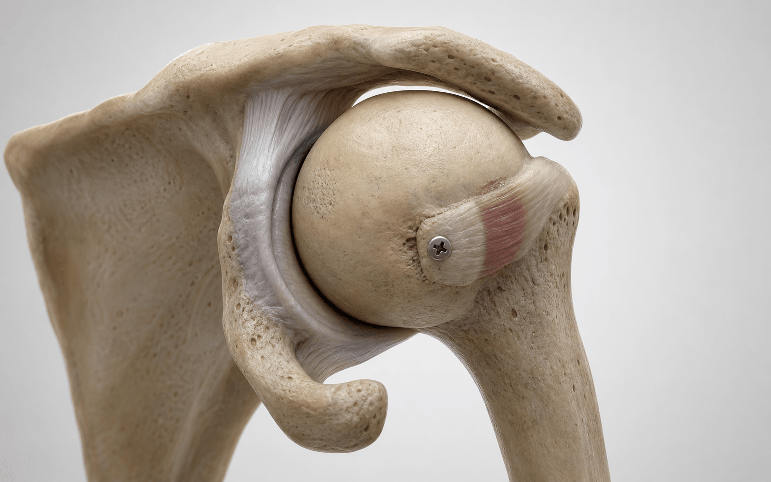
Subscapularis (McLaughlin) or lesser tuberosity (modified Neer / Hawkins) transfer into reverse Hill-Sachs defect | advanced
Surgical Imaging
The trap: Up to 50 percent of locked posterior dislocations are missed on initial presentation. Patients with a stiff, internally-rotated shoulder after a seizure, electrocution, or high-energy injury are often diagnosed as a 'frozen shoulder' or rotator cuff tear and discharged without adequate imaging.
The fix: In EVERY post-ictal or post-electrocution patient with shoulder pain, request THREE radiograph views: TRUE AP, AXILLARY LATERAL (the diagnostic view - shows the posteriorly translated humeral head), and SCAPULAR Y. The 'lightbulb sign' (rounded, symmetric humeral head on AP from fixed internal rotation) plus the 'rim sign' (widened glenohumeral joint on AP due to posterior translation) plus a confirmed posterior dislocation on axillary is the classic triad.
Location: The axillary nerve crosses the anterior-inferior capsule roughly 3 to 7 mm medial to the musculotendinous border of the subscapularis. It then runs along the inferior border of the subscapularis before passing posteriorly through the quadrangular space with the posterior circumflex humeral vessels.
Risk: During subscapularis tenotomy or lesser tuberosity osteotomy, the axillary nerve is at risk if dissection strays inferior to the subscapularis footprint. It is also vulnerable during capsular release for chronic dislocations. Identify and protect it throughout.
Location: The musculocutaneous nerve enters the coracobrachialis roughly 5 to 8 cm distal to the coracoid tip (variable - as close as 2 cm in some patients) and runs on the deep surface of the biceps brachii.
Risk: Excessive medial retraction of the conjoint tendon or coracobrachialis during the deltopectoral approach can stretch the musculocutaneous nerve. Limit retraction time and release at intervals. Identify the nerve when working medial to the conjoint tendon.
Less than 20 percent: Often nonoperative, or disimpaction and bone grafting. McLaughlin rarely needed.
20 to 40 percent: The McLaughlin / modified Neer zone - subscapularis or lesser tuberosity transfer. This is the indication for THIS procedure.
Greater than 40 to 50 percent, or head collapse, or arthritis: Allograft reconstruction (femoral head, distal tibial plafond, iliac crest) OR arthroplasty. The McLaughlin transfer alone is INSUFFICIENT for large defects.
The trap: The subscapularis (or lesser tuberosity osteotomy) repair is the STRUCTURAL foundation of the McLaughlin procedure. Failure of this repair is a leading cause of recurrent posterior instability and re-operation.
The fix: Sling immobilisation for 4 to 6 weeks. NO active internal rotation against resistance for 6 weeks. NO external rotation beyond neutral for 4 to 6 weeks (the transfer relies on the tendon healing into the defect - external rotation tension disrupts this). Begin passive forward flexion in the scapular plane, progressing to active-assisted, then resisted at 10 to 12 weeks.
Why it matters: The humeral head blood supply (arcuate artery, posteromedial vessels from the posterior circumflex) is often compromised after a locked posterior dislocation, particularly when the dislocation has been present for greater than 6 months. Reported AVN rates range from 9 to 30 percent in chronic dislocations, increasing with delay to reduction.
Implication: The McLaughlin procedure does not address the AVN risk. Inform patients preoperatively that despite an excellent initial result, AVN may develop and require subsequent arthroplasty. CT or MRI preoperatively to assess head viability is essential in chronic dislocations.
L.I.G.H.T.B.U.L.BLIGHTBULB — Recognising the Locked Posterior Dislocation
M.C.L.A.U.G.H.L.I.NMCLAUGHLIN — Operative Steps
D.E.F.E.C.TDEFECT — Choosing the Right Operation
Surgical Indications
Primary Indication
- Engaging reverse Hill-Sachs defect involving approximately 20 to 40 percent of the humeral head articular surface after a chronic locked posterior dislocation (greater than 3 weeks duration)
- The defect is the impaction created on the ANTEROMEDIAL humeral head when the head is locked against the posterior glenoid rim
- The McLaughlin procedure (or its modifications) blocks re-engagement of the defect on the posterior glenoid rim in functional positions
Classic Mechanism Triad
The index injury is almost always one of three:
- Seizure (ictal phase): violent contraction of the stronger internal rotators (subscapularis, latissimus, pectoralis) overcomes the external rotators and drives the head posteriorly; the anteromedial head impacts the posterior glenoid rim
- Electrocution: similar mechanism - tetanic internal rotator contraction against a fixed thorax
- High-energy trauma: fall on the flexed, adducted, internally-rotated arm; motor vehicle accident with axially-loaded flexed arm
Absolute Indications
- Chronic locked posterior dislocation (greater than 3 weeks) with an engaging reverse Hill-Sachs defect
- Acute locked posterior dislocation that fails closed reduction attempts (e.g. buttonhole engagement of the head through a posterior capsular tear)
- Recurrent posterior instability with a documented engaging reverse Hill-Sachs defect on CT (less common - most posterior instability is atraumatic and treated with soft-tissue procedures, but the McLaughlin / modified Neer is the bony answer for a structural engaging defect)
Relative Indications
- Acute locked posterior dislocation with a 20 to 40 percent defect - McLaughlin transfer as a primary procedure to address the bone defect
- Failed nonoperative management of a smaller defect with persistent symptoms and documented engagement
- Combined bony (reverse Hill-Sachs) and soft-tissue (posterior Bankart) pathology
Contraindications
Absolute:
- Humeral head AVN with collapse - reverse total shoulder arthroplasty is the answer, not the McLaughlin transfer
- Established glenohumeral arthritis with loss of joint space - arthroplasty indicated
- Defect greater than 40 to 50 percent without sufficient articular surface for transfer - allograft reconstruction or arthroplasty
Relative:
- Acute dislocation with small defect (less than 20 percent) - trial nonoperative management
- Patient unable to comply with 6-week subscapularis protection protocol
- Active infection
- Neuropathic joint (Charcot) - arthrodesis or reverse total shoulder arthroplasty
Evidence for the McLaughlin Procedure
Original McLaughlin (1952)
- McLaughlin HL described the original technique in 1952: open reduction of the chronic posterior dislocation through a deltopectoral approach, followed by transposition of the subscapularis tendon into the reverse Hill-Sachs defect
- Series of 22 patients reported; 18 had satisfactory outcomes with the subscapularis transfer acting as a 'check-rein' against re-engagement
- Established the principle: address the BONE defect by filling it with the attached subscapularis, not just by soft-tissue capsular repair
Modified Neer / Hawkins
- Hawkins RJ and colleagues (Hughes and Neer 1975) modified the procedure to include osteotomy of the LESSER TUBEROSITY with the attached subscapularis, transferring both bone and tendon into the defect
- Rationale: bony block provides more robust engagement against the posterior glenoid rim; cancellous bone surface on the tuberosity heals to the prepared defect with biological union; fixation is stronger (screws vs tendon sutures alone)
- The modified Neer has largely superseded the original McLaughlin in current practice
Outcomes Literature
- Hawkins (1985, J Bone Joint Surg Am) reported 17 of 17 patients with a satisfactory result after open reduction and lesser tuberosity transfer for chronic posterior dislocations; recurrent instability in 1 patient
- Several smaller series report 80 to 90 percent good-to-excellent results with the modified Neer / Hawkins procedure at mid-term follow-up
- Recurrent instability rates after McLaughlin / modified Neer: 5 to 15 percent in most series (lower than soft-tissue-only repairs for engaging defects)
- Conversion to arthroplasty: 10 to 25 percent at 10 to 15 years, driven by progressive arthritis and AVN
Comparison: McLaughlin vs Allograft vs Arthroplasty
Defect-Driven Surgical Algorithm for Reverse Hill-Sachs
Key Evidence
Posterior dislocation of the shoulder
Locked posterior dislocation of the shoulder (modified Neer / Hawkins technique)
Locked posterior dislocation of the shoulder: A systematic review
Long-term outcome of segmental reconstruction of the humeral head for the treatment of locked posterior dislocation of the shoulder
Transfer of the lesser tuberosity for reverse Hill-Sachs lesions after neglected posterior dislocations of the shoulder: A retrospective clinical study of 13 cases
Clinical Decision Scenarios
Practise clinical reasoning and management decisions out loud
“A 45-year-old man is brought to the Emergency Department after a witnessed generalised tonic-clonic seizure. He has a stiff, painful right shoulder. AP radiograph shows a rounded, symmetric humeral head with apparent widening of the glenohumeral joint. You are concerned about a locked posterior dislocation. How do you confirm the diagnosis, classify the injury, and plan definitive management?”
“You are performing a modified Neer (lesser tuberosity osteotomy) procedure for a chronic locked posterior dislocation. The lesser tuberosity wafer has been osteotomised and the subscapularis remains attached. You have reduced the head and prepared the reverse Hill-Sachs defect. The bone quality is good. Describe how you would fix the lesser tuberosity wafer into the defect, and what intraoperative tests you would use to confirm the transfer is functional.”
“A 32-year-old labourer underwent a McLaughlin procedure 18 months ago for a chronic locked posterior dislocation with a 30 percent reverse Hill-Sachs defect. He has been doing well in physical therapy with no recurrence of instability, but he now presents with progressive shoulder pain, crepitus, and loss of motion over the past 6 months. Radiograph shows joint space narrowing, subchondral sclerosis, and early osteophyte formation. What is the most likely diagnosis, how would you confirm it, and what are the management options?”
References
-
McLaughlin HL (1952). Posterior dislocation of the shoulder. J Bone Joint Surg Am. PMID PENDING. — Original description of the McLaughlin procedure; subscapularis tendon transfer into the reverse Hill-Sachs defect after open reduction in 22 patients.
-
Hawkins RJ, Neer CS 2nd, Pianta RM, Mendoza FX (1987). Locked posterior dislocation of the shoulder. J Bone Joint Surg Am. PMID PENDING. — Modified Neer technique: lesser tuberosity osteotomy with attached subscapularis; 17 of 17 patients with satisfactory results.
-
Gerber C, Catanzaro S, Jundt-Ecker M, Fucentese SF (2014). Arthroscopic restoration of the reverse Hill-Sachs lesion in posterior shoulder instability. J Shoulder Elbow Surg. PMID PENDING. — Allograft reconstruction for greater than 40 percent reverse Hill-Sachs defects; technique description and early outcomes.
-
Cofield RH, Daly PJ (1992). Hemiarthroplasty for chronic locked posterior dislocation of the shoulder. J Bone Joint Surg Am. PMID PENDING. — Hemiarthroplasty as a salvage option for chronic posterior dislocations in patients not amenable to reconstruction.
-
Robinson CM, Aderinto J (2005). Posterior shoulder dislocations and fracture-dislocations. J Bone Joint Surg Am. PMID PENDING. — Classification, mechanisms, and treatment algorithm for posterior shoulder dislocations including the McLaughlin indication.