Open reduction and internal fixation of a displaced medial humeral epicondyle apophyseal avulsion | intermediate
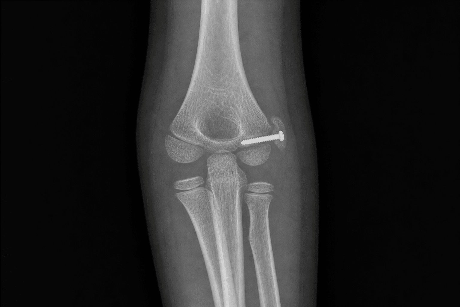
Surgical Imaging
The trap: After elbow dislocation the medial epicondyle can be drawn into the joint; on the AP film it sits near the trochlear region and is mistaken for the trochlear ossification center or a normal variant.
The fix: CRITOE helps. The medial epicondyle ossifies BEFORE the trochlea, so a "trochlea" that appears too early, or a medial epicondyle absent from its normal position, is the incarcerated fragment. An incarcerated fragment is an ABSOLUTE indication for ORIF. Confirm with a true lateral and CT if doubt remains, then extract and fix it.
Location: The ulnar nerve lies posterior to the medial epicondyle in the cubital tunnel, immediately deep to the surgical field. It can be dragged distally with the fragment or, rarely, incarcerated within the joint alongside it.
Risk: Direct injury during dissection, traction, or post-operative compression over hardware. Pre-injury ulnar symptoms are common with this fracture.
Protection: Identify it first, protect it with a vessel loop, decompress the cubital tunnel, and consider anterior transposition when there is pre-operative neuropathy or the fragment was incarcerated.
The trap: applying the traditional greater-than-5-mm displacement rule as an absolute indication to operate.
The fix: displacement alone is CONTESTED. The Kamath systematic review found no evidence that ORIF of a displaced fragment improves outcome over nonoperative care, and Farsetti and Josefsson showed good long-term function with fibrous union. Operate for incarceration, nerve dysfunction, or instability, not for displacement alone.
The trap: apparent fragment position is unreliable when the elbow is splinted in flexion, and the fragment may be confused with the trochlea, the lateral condyle, or a normal ossification center.
The fix: obtain true AP and lateral views (and a comparison view of the opposite elbow in subtle cases). Actively exclude an associated elbow dislocation, a lateral condyle fracture, and confirm the radiocapitellar line. Use the CRITOE order to locate the medial epicondyle.
Why it matters: the medial epicondyle bears the ulnar collateral ligament origin. An incompletely restored fragment leaves valgus laxity that is disabling for an overhead thrower.
The fix: in high-demand throwing athletes keep a lower threshold for anatomic reduction and rigid fixation to restore UCL tension, and counsel on a delayed, progressive return to throwing.
The trap: the paediatric elbow stiffens readily after this injury, especially when associated with a dislocation, and prolonged immobilisation compounds the loss of motion.
The fix: use stable fixation that allows early motion, immobilise briefly, and begin guided range-of-motion exercises. Watch for heterotopic ossification, the risk of which rises with a concomitant elbow dislocation or a head injury.
M.E.D.I.A.LMEDIAL - The Fracture at a Glance
O.P.E.R.A.SOPERAS - When to Operate
S.C.R.E.WSCREW - Fixation Principles
Surgical Indications
Absolute Indications
- Incarcerated (intra-articular) fragment that does not reduce after closed reduction of the elbow - the fragment blocks motion and will not unite in a functional position
- Ulnar nerve dysfunction - especially a nerve incarcerated within the joint or a persistent symptomatic neuropathy
- Open fracture
- Vascular injury requiring exploration (rare)
- An irreducible elbow dislocation in which the fragment is interposed and prevents reduction
Relative Indications (the contested ground)
- Significant displacement - the traditional thresholds of greater than 5 mm, greater than 10 mm, and greater than 15 mm are widely debated and are not supported as standalone indications by modern evidence
- High-demand overhead-throwing athletes in whom valgus stability is critical to function
- Objective valgus instability on stress testing, indicating incompetence of the ulnar collateral ligament origin
- A fragment that lies within the cubital tunnel and irritates the ulnar nerve
Contraindications
Absolute:
- A fracture without incarceration, nerve compromise, open injury, or demonstrated instability - these do well nonoperatively
Relative:
- Gross swelling or fracture blisters that delay safe surgery
- A near-skeletal-maturity child with a chronic, asymptomatic fibrous nonunion
The Displacement Controversy
Traditional teaching (Smith; Hines) recommended ORIF for displacement greater than 5 mm, with some authors proposing thresholds of greater than 10 mm or greater than 15 mm, citing theoretical risks of nonunion, valgus instability, and ulnar nerve problems.
Modern evidence challenges this position:
- The Kamath systematic review (2009) found no conclusive evidence that operative treatment of displaced medial epicondyle fractures improves outcome over nonoperative care, and highlighted the lack of high-quality prospective studies.
- Farsetti and colleagues (2001) reported excellent long-term elbow function in patients treated nonoperatively despite residual displacement and frequent fibrous union.
- Josefsson and Danielsson (1986) found that adults reviewed long after childhood nonoperative treatment were largely asymptomatic, even when the fragment had gone on to fibrous union.
- Louahem and colleagues (2010) showed in a multicentre series that long-term outcome was driven by associated elbow injuries and ulnar nerve involvement rather than by the degree of fragment displacement.
The defensible modern position is to operate for absolute indications (incarceration, ulnar nerve dysfunction, open injury, instability), to treat the remainder nonoperatively, and to individualise for the high-demand throwing athlete in whom the ulnar collateral ligament origin must be restored.
Non-Operative Treatment
- A long-arm splint or cast at 90 degrees of flexion for 1 to 3 weeks, followed by early active motion.
- Acceptable for minimally displaced fractures and for displaced fractures that carry no absolute indication.
- Fibrous union is common and is usually asymptomatic; it is not by itself an indication for surgery.
- Counsel the family to expect a minor loss of terminal extension and a small residual cosmetic bump at the medial epicondyle.
Evidence at a Glance - Operative versus Non-Operative
Operative versus Non-Operative Management
Clinical Decision Scenarios
Practise clinical reasoning and management decisions out loud
“A 12-year-old boy fell onto his outstretched hand. Radiographs show a medial epicondyle fracture with the fragment displaced approximately 8 mm but lying outside the joint, no elbow dislocation, and a normal ulnar nerve examination. How do you manage him?”
“A 13-year-old presents after an elbow dislocation that has been reduced in the emergency department. The post-reduction AP radiograph shows what appears to be a trochlear ossification center, but the medial epicondyle is not visible in its normal position. The child has ulnar-distribution paraesthesia. What is going on and how do you manage it?”
“You are fixing a displaced medial epicondyle fracture in a 14-year-old elite junior baseball pitcher. What specific considerations apply, and how do your fixation and rehabilitation differ from those of a non-throwing child?”