Adult Monteggia Fracture-Dislocation — ORIF

Shoulder & ElbowAdvancedCore Procedure

Adult Monteggia Fracture-Dislocation — ORIF

Surgical technique for open reduction and internal fixation of adult Monteggia fracture-dislocation — Bado classification, plate fixation of the proximal ulna with indirect radial-head reduction, management of associated radial-head and coronoid fractures, complications including posterior interosseous nerve palsy and recurrent instability

High-yield overview

Open reduction and plate fixation of the proximal ulna with indirect radial-head reduction | advanced

Surgical Imaging

Adult Monteggia ulna plating
Adult Monteggia injury: the proximal ulna fracture is plated to restore length and alignment, which reduces the dislocated radial head.Credit: AI-generated medical illustration · OrthoVellum
Critical Danger Structures and Exam Traps
Missed Radial-Head Dislocation — The Classic Trap

The trap: The ulna fracture is obvious and distracting; the radial-head dislocation is subtle or missed entirely on initial radiographs, particularly in Bado Type I (anterior dislocation) where the radial head may appear nearly reduced on the AP view.

The fix: ALWAYS check the radiocapitellar line on every view — a line drawn along the radius must pass through the centre of the capitulum. Obtain dedicated elbow AP and lateral radiographs in addition to the forearm films. If any doubt, obtain a CT of the elbow. A missed Monteggia lesion treated as an isolated ulna fracture leads to chronic radial-head dislocation, progressive pain, proximal radioulnar joint instability, and limited forearm rotation.

Malreduction of the Ulna — Persistent Radial-Head Dislocation

The trap: After plate fixation of the ulna, the radial head remains dislocated. The surgeon accepts this and proceeds to radial-head fixation or excision without re-examining the ulna.

The fix: The most common cause of persistent radial-head dislocation is residual malreduction of the ulna — typically inadequate restoration of ulnar length or failure to correct the proximal dorsal angulation (the sag). Remove the plate, re-reduce the ulna anatomically, and re-fix. Only after the ulna is definitively anatomic should you accept persistent radial-head displacement as being caused by annular-ligament or capsular interposition requiring open clearance.

Posterior Interosseous Nerve (PIN) Palsy

Location: The PIN branches from the radial nerve in the radial groove, passes anterior to the lateral epicondyle, then enters the supinator muscle through the arcade of Frohse. It supplies all wrist and finger extensors except ECRB (which is supplied by the radial nerve proximal to the PIN).

Risk: The PIN is at risk during posterior or Kocher approaches to the radial head/neck, during reduction manoeuvres of a dislocated radial head, and from postoperative swelling or callus around the proximal radius. In Bado Type III (lateral dislocation) the nerve may be directly injured by the displaced radial head. Document finger and thumb extension preoperatively. Most PIN palsies are neurapraxic and recover within 3-6 months.

Bado Type II — Associated Radial Head and Coronoid Fractures

The trap: Treating a Bado Type II as an isolated ulna fracture without identifying the associated radial-head fracture, coronoid fracture, or lateral collateral ligament injury. This leads to unstable fixation, persistent subluxation, and early post-traumatic arthritis.

The fix: Bado Type II almost always has associated injuries. CT of the elbow is mandatory. The Jupiter sub-classification helps: IIA (radial head fracture), IIB (coronoid fracture), IIC (both), IID (olecranon fracture), IIE (associated lateral collateral ligament disruption). Each associated injury must be addressed at surgery — radial-head fixation or replacement, coronoid fixation, lateral collateral ligament repair.

Inadequate Plate Length and Fixation

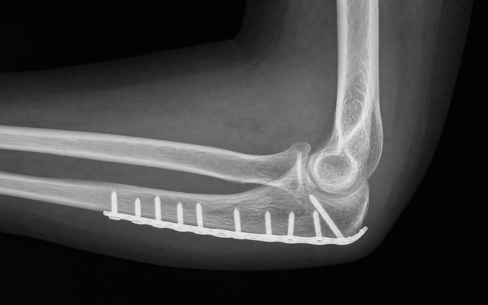
The trap: Using a short plate (fewer than 6 cortices proximal and distal to the fracture) or a plate that does not bridge the olecranon-coronoid apex, leading to fixation failure, loss of reduction, and nonunion.

The fix: Use a 3.5 mm LCDCP or pre-contoured proximal ulna locking plate with at least 3 screws (6 cortices) proximal and 3 screws (6 cortices) distal to the fracture. In proximal fractures, the plate should extend distal enough to capture intact cortex. In olecranon fractures (Bado Type IID), supplement with tension-band or additional plate fixation if needed.

Recurrent Instability After Fixation

The trap: Postoperative radial-head instability attributed to hardware removal or inadequate fixation, when the real cause is an unrecognised Essex-Lopresti lesion (interosseous membrane disruption with distal radioulnar joint instability) or an unaddressed coronoid fracture.

The fix: Before wound closure, assess forearm rotation and radial-head stability in all planes. Check the distal radioulnar joint (DRUJ) for instability — a positive piano-key sign or ballotable ulnar head suggests an Essex-Lopresti lesion. If the coronoid is fractured, fix it to restore the anterior buttress of the elbow. If the DRUJ is unstable, consider interosseous membrane repair or temporary K-wire fixation of the DRUJ.

Mnemonic

B.A.D.O.BADO — Monteggia Classification

Mnemonic

M.O.N.T.Y.MONTY — Key Surgical Principles in Adult Monteggia ORIF

Mnemonic

P.I.N.PIN — Posterior Interosseous Nerve in Monteggia

Surgical Indications

Absolute Indications

  • All adult Monteggia fracture-dislocations (Bado Types I-IV) — non-operative treatment in adults has unacceptably high rates of redislocation, malunion, and chronic instability
  • Irreducible radial head after attempted closed reduction, suggesting interposition of the annular ligament, capsule, or fracture fragment
  • Open Monteggia fractures — require urgent debridement and fixation regardless of Bado type
  • Monteggia fracture with associated neurovascular injury — PIN palsy with clinical evidence of entrapment, or vascular injury

Relative Indications

  • Bado Type II with associated radial-head fracture — requires radial-head fixation or replacement in addition to ulna plating
  • Bado Type II with coronoid fracture involving greater than 50% of coronoid height — requires coronoid fixation for elbow stability
  • Chronic unreduced Monteggia (presenting greater than 6 weeks after injury) — requires more complex reconstruction: ulna osteotomy, plate fixation, and annular ligament reconstruction (Bell-Tawse procedure)
  • Pathological fracture of the proximal ulna with radial-head dislocation in metastatic disease

Contraindications

Absolute:

  • None for the injury itself — virtually all adult Monteggia fractures require surgical fixation

Relative:

  • Severe medical comorbidity precluding general or regional anaesthesia
  • Active infection at the surgical site (defer until resolved)
  • Patient non-ambulatory with end-stage disease where the functional demands do not justify surgical risk

Evidence for Non-Operative Treatment

Paediatric Exception

Non-operative treatment is the standard for most paediatric Monteggia fractures because the annular ligament has good healing potential in children, the ulna remodels with growth, and closed reduction with casting is successful in a majority of cases. This paediatric principle does NOT apply to adults.

Adult Evidence Against Non-Operative Treatment

  • In adults, the proximal ulna has minimal remodelling potential and the annular ligament does not heal in a reduced position without anatomic ulnar fixation
  • Non-operative management of adult Monteggia fractures results in high rates of redislocation of the radial head, painful proximal radioulnar joint instability, restricted forearm rotation, and early post-traumatic arthritis
  • Multiple case series report poor outcomes with non-operative treatment in adults: persistent dislocation, malunion of the ulna with secondary cubitus valgus or varus, and progressive disability
  • The standard of care in adults is ORIF of the ulna for all Bado types

Evidence for Surgery

Plate Fixation of the Ulna — The Definitive Treatment

The established surgical principle across all Bado types in adults is that anatomic reduction and stable plate fixation of the ulna fracture restores the normal proximal radioulnar joint anatomy and allows indirect reduction of the radial head. This was established by series in the 1980s-2000s showing reliable radial-head reduction after ulna plating in greater than 90% of cases.

Key surgical evidence:

  • Anatomic plate fixation of the ulna with 3.5 mm LCDCP or locking compression plate achieves indirect reduction of the radial head in greater than 90% of Bado I and III cases and 80-85% of Bado II cases
  • Persistent radial-head dislocation after ulna fixation is most commonly caused by residual ulnar malreduction — re-reduce and re-plate the ulna before accepting persistent displacement
  • In Bado Type II, associated injuries to the radial head and coronoid must be addressed to achieve a stable elbow

Bado Type II and the Jupiter Sub-Classification

Jupiter et al. sub-classified Bado Type II based on associated injuries, which guides the operative plan:

IIA
Pattern
Ulna fracture at olecranon level
Associated Injuries
Radial head fracture
IIB
Pattern
Ulna fracture at coronoid level
Associated Injuries
Anterior coronoid fracture
IIC
Pattern
Ulna fracture at olecranon + coronoid
Associated Injuries
Radial head AND coronoid fractures
IID
Pattern
Transverse olecranon fracture
Associated Injuries
Both column olecranon fracture
IIE
Pattern
Any Type II
Associated Injuries
Lateral collateral ligament complex disruption

Key Evidence

Evidence

Unstable fracture-dislocations of the forearm. The Monteggia and Galeazzi lesions

Level IV
Reckling FW, Cordell LDArch Surg
Clinical implication: Established plate fixation of the ulna as the foundation of adult Monteggia treatment.
Source: Arch Surg. 1968;96(6):999-1007
Evidence

Monteggia fractures in adults

Level IV
Ring D, Jupiter JB, Simpson NSJ Bone Joint Surg Am
Clinical implication: Accuracy of ulnar reduction is the single most important surgical determinant of outcome in adult Monteggia fractures.
Source: J Bone Joint Surg Am. 1998;80(12):1733-44
Evidence

The posterior Monteggia lesion

Level IV
Jupiter JB, Leibovic SJ, Ribbans W, Wilk RMJ Orthop Trauma
Clinical implication: Posterior (Bado Type II) Monteggia lesions are complex injuries with frequent associated fractures of the radial head and coronoid — thorough preoperative CT and operative treatment of all associated injuries is essential.
Source: J Orthop Trauma. 1991;5(4):395-402
Evidence

Monteggia fractures and Monteggia-like-lesions: a systematic review

Level III
Weber MM, Rosteius T, Schildhauer TA, Königshausen M, Rausch VArch Orthop Trauma Surg
Clinical implication: Bado Type II Monteggia fractures require meticulous attention to associated injuries to optimise outcomes.
Source: Arch Orthop Trauma Surg. 2023;143(7):4085-4093
Evidence

Surgical Management of Complex Adult Monteggia Fractures

Level IV
Xiao RC, Chan JJ, Cirino CM, Kim JMJ Hand Surg Am
Clinical implication: Complex Monteggia fractures require a systematic approach: fix the ulna anatomically first, then address all associated injuries at the index procedure.
Source: J Hand Surg Am. 2021;46(11):1006-1015

Clinical Decision Scenarios

Practise clinical reasoning and management decisions out loud

Viva scenarioAdvanced
Clinical prompt

A 35-year-old man falls directly onto the point of his flexed right elbow. Radiographs show a comminuted olecranon fracture with posterior angulation and a posteriorly dislocated radial head. The radial head appears to have a single large fracture fragment. How do you classify this injury and what is your operative plan?

Practical approach
This is a Bado Type II Monteggia fracture-dislocation, specifically a Jupiter Type IIA pattern (olecranon-level fracture with associated radial-head fracture). The radial head is dislocated posteriorly and fractured. **Classification**: Bado Type II — posterior dislocation of the radial head with posterior angulation of the ulna fracture at the olecranon level, plus an associated radial-head fracture (Jupiter IIA). I would request a CT of the elbow to further define the radial-head fracture pattern, assess the coronoid for a fracture, and characterise the olecranon comminution before proceeding to surgery. **Operative plan**: I would use a posterior approach to the ulna and a Kocher lateral approach to the radial head (prep the entire arm preoperatively). First, I would fix the ulna with a 3.5 mm LCDCP or proximal ulna locking plate, restoring the olecranon articular surface, length, and the proximal dorsal angulation. I would then assess the radial head on fluoroscopy. If the radial head remains dislocated after anatomic ulna fixation, I would proceed to the Kocher approach, inspect the radiocapitellar joint for interposed tissue, reduce the radial head, and fix the radial-head fracture with headless compression screws. If the radial-head fracture is Mason Type III (comminuted, greater than 3 fragments) and not fixable, I would replace it with a modular metallic prosthesis. Before closure, I would check elbow valgus stability, forearm rotation through the full arc, and the distal radioulnar joint. **Post-operative**: posterior splint for 10-14 days, then active-assisted elbow mobilisation from week 2, forearm rotation exercises as tolerated. Formal physiotherapy from week 4. Weight-bearing progression from week 6 based on radiographic union.
Viva scenarioAdvanced
Clinical prompt

A 28-year-old woman presents with forearm pain and limited rotation 4 months after a fall. Radiographs show a healed proximal ulna fracture with residual anterior angulation and a chronically anteriorly dislocated radial head. She was treated in a cast initially for an 'isolated ulna fracture'. How do you manage this?

Practical approach
This patient has a chronic unreduced Bado Type I Monteggia fracture-dislocation. The original injury was misdiagnosed as an isolated ulna fracture — the radial-head dislocation was missed. The ulna has healed in malunion with anterior angulation, and the radial head has been dislocated for 4 months. The annular ligament will have scarred and shortened, making closed or simple open reduction impossible. **Workup**: I would obtain dedicated elbow radiographs (AP, lateral, radiocapitellar views) and a CT scan to assess the olecranon-coronoid relationship, the degree of ulna malunion, and the position of the radial head. I would check for proximal radioulnar joint arthritic changes and assess forearm rotation clinically. I would also document PIN function. **Surgical plan**: I would perform a combined procedure. First, through a posterior approach to the ulna, I would perform an osteotomy at the site of the malunion (just distal to the previous fracture line) and correct the ulnar alignment, length, and sag, fixing the osteotomy with a long 3.5 mm plate. This corrects the bony architecture. Second, through a Kocher lateral approach, I would open the radiocapitellar joint, reduce the radial head, and reconstruct the annular ligament using a strip of triceps fascia or flexor carpi ulnaris as a graft (Bell-Tawse procedure). The new annular ligament must be tensioned with the radial head reduced and the forearm in neutral rotation. **Post-operative**: long-arm cast with the elbow at 90 degrees and the forearm in neutral for 4-6 weeks to protect the annular ligament reconstruction, followed by a graduated mobilisation programme. Functional outcomes are inferior to acute Monteggia fixation, and I would counsel her that she is likely to have some permanent restriction of forearm rotation, particularly supination. **Viva point**: this case illustrates the critical importance of ALWAYS checking the radiocapitellar line on every forearm injury. A missed Monteggia lesion treated as an isolated ulna fracture is a recurrent exam trap.
Viva scenarioStandard
Clinical prompt

You have just completed plate fixation of a Bado Type I Monteggia fracture in a 40-year-old man. The ulna is beautifully plated. On the post-fixation lateral fluoroscopy, the radial head is still anteriorly dislocated. The radiocapitellar line is disrupted. What do you do?

Practical approach
This is the critical decision point in Monteggia ORIF. The radial head is still dislocated despite what appears to be a well-plated ulna. The most common mistake at this juncture is to assume the annular ligament is interposed and proceed directly to open reduction of the radial head. The correct first step is to re-examine the ulna. **Step 1 — Re-check the ulna**: Remove the distal plate screws and re-assess the ulnar reduction on fluoroscopy. I would carefully check: (a) Is there residual anterior angulation that I missed? (b) Is the ulnar length fully restored? (c) Is the proximal dorsal angulation (sag) correct? In my experience, a subtle residual malreduction — even 5-10 degrees of residual angulation — can be enough to prevent the radial head from reducing. I would re-reduce the ulna, re-plate, and re-check the radial head on fluoroscopy. **Step 2 — If the ulna is definitively anatomic and the radial head remains dislocated**: only now do I accept that the cause is soft-tissue interposition (annular ligament, capsule, or rarely a small fracture fragment). I would proceed to a Kocher lateral approach, open the radiocapitellar joint, identify and remove the interposed tissue, and manually reduce the radial head. I would confirm concentric reduction on fluoroscopy and check forearm rotation. **Step 3 — Before closing**: I would assess forearm rotation through a full arc with the radial head reduced to confirm stability. If the radial head subluxates during rotation, I would check for additional instability (IOM disruption, DRUJ instability, LCL injury). This systematic approach — ulna first, then radial head — is the most important surgical principle in adult Monteggia ORIF and is a frequent exam topic.
Exam day cheat sheet
Adult Monteggia Fracture-Dislocation ORIF — Exam Day Summary

References

Evidence

Unstable fracture-dislocations of the forearm. The Monteggia and Galeazzi lesions

Level IV
Reckling FW, Cordell LDArch Surg
Clinical implication: Established the foundational principle of adult Monteggia treatment: fix the ulna and the radial head reduces.
Source: Arch Surg. 1968;96(6):999-1007
Evidence

Monteggia fractures in adults

Level IV
Ring D, Jupiter JB, Simpson NSJ Bone Joint Surg Am
Clinical implication: Accuracy of ulnar reduction is the single most important determinant of outcome.
Source: J Bone Joint Surg Am. 1998;80(12):1733-44
Evidence

The posterior Monteggia lesion

Level IV
Jupiter JB, Leibovic SJ, Ribbans W, Wilk RMJ Orthop Trauma
Clinical implication: The Jupiter sub-classification guides the operative plan: identify all associated injuries on preoperative CT and address each.
Source: J Orthop Trauma. 1991;5(4):395-402
Evidence

Monteggia fractures and Monteggia-like-lesions: a systematic review

Level III
Weber MM, Rosteius T, Schildhauer TA, Königshausen M, Rausch VArch Orthop Trauma Surg
Clinical implication: Bado Type II requires meticulous attention to associated injuries to optimise outcomes.
Source: Arch Orthop Trauma Surg. 2023;143(7):4085-4093
Evidence

Surgical Management of Complex Adult Monteggia Fractures

Level IV
Xiao RC, Chan JJ, Cirino CM, Kim JMJ Hand Surg Am
Clinical implication: Consider extensor-sparing approach when Monteggia involves the olecranon to preserve the triceps mechanism.
Source: J Hand Surg Am. 2021;46(11):1006-1015
Editorially reviewed — transparent references and correction processPublished by OrthoVellum Medical Education TeamEditorial boardMethodologyReview policy
Educational disclosure

Educational content is reviewed for source visibility, editorial coherence, and correction readiness.

No individual clinician credential is claimed unless a named person is shown.

Verify before clinical use; this is not medical advice or a substitute for local guidance.