Nail bed and fingertip injury repair — trephination, nail-bed repair, Seymour fracture and fingertip reconstruction
- The germinal matrix (under the proximal nail fold) produces about 90 percent of the nail plate. Any germinal matrix injury that is not anatomically repaired causes a permanent nail deformity — a split nail, a ridged nail, or failure of the nail to grow.
- Subungual haematoma: the old rule of removing the nail when the haematoma exceeds 50 percent is superseded (Roser and Gellman). If the nail plate AND the nail margin are intact, trephination alone gives equivalent results regardless of haematoma size. Remove the plate to inspect and repair the bed only when the nail fold is disrupted, the plate is avulsed or displaced, or a laceration is evident.
- A Seymour fracture is an open physeal fracture of the distal phalanx in a child with nail-fold disruption — it is NOT a simple mallet finger. It requires irrigation, debridement and nail-bed repair; missed cases develop osteomyelitis.
- Replace the nail plate after repair as a biological dressing and splint: it maintains the nail-fold space, provides a template for nail regrowth, and reduces pain. If the plate is unusable, a silicone sheet or the foil from a suture packet suffices.
When & Why
Indication. A nail bed or fingertip injury needing surgical care — a crush or entrapment mechanism with a subungual haematoma, a nail-fold laceration or avulsion, an open distal phalanx fracture, or a fingertip amputation. The decision to operate rests on nail-fold integrity (not on haematoma size) and on the Allen zone of any amputation. When to trephinate only (conservative).
- The nail plate and nail margin are structurally intact — the haematoma size is NOT a contraindication to trephination when the nail is intact.
- No displaced or open distal phalanx fracture (radiograph the distal phalanx to confirm).
- Performed within 48 hours while the haematoma is still liquefied; later, the clot may be too organised to drain. When to remove the nail plate and repair the bed.
- Any disruption of the proximal nail fold — the plate avulsed or displaced from the fold.
- A visible nail-bed laceration or avulsion.
- A Seymour fracture (a child with a physeal fracture and nail-fold disruption).
- The nail plate avulsed entirely from its bed.
- The classic teaching adds "a subungual haematoma greater than 50 percent" — but with an intact nail and nail margin the evidence supports trephination alone irrespective of size; explore on nail-fold integrity, not on haematoma size. Fingertip reconstruction.
- Allen Zone II or greater: exposed bone requires flap coverage.
- Zone I may be managed conservatively, particularly in children, who show remarkable healing.
- A composite graft (replacing the amputated tip) is an option if available within about 6 hours and the patient is cooperative. The reconstruction options at a glance. Match the flap to the zone and the pattern of loss:
Volar V-to-Y flap, 5 to 8 mm of advancement, sensate glabrous skin. The default for Zone II dorsal-oblique or transverse amputations.
Two lateral V-Y flaps. Better suited to volar-oblique Zone II amputations; bilateral sensate cover.
Based on the proper digital artery with the nerve in the pedicle — 10 to 15 mm of advancement and excellent sensation, but more demanding.
Two-stage flap from the dorsum of the adjacent digit, divided at 3 weeks. For Zone III; initially insensate and risks PIP stiffness from immobilisation.
Two-stage glabrous flap for index or middle Zone III to IV injuries; risks PIP flexion contracture — avoid in patients older than about 50 years.
Reserved for cases where preserving length is impossible; sacrifices fingertip length and risks a hook nail.
Consent specifically for residual nail deformity (ridging or a split nail) even after a perfect repair, fingertip numbness or cold intolerance, infection, and — for flap reconstructions — donor-site morbidity, stiffness and a two-stage pathway. The decision at a glance.
- Allen zone
- Zone I equivalent
- Recommended management
- Trephination under digital block alone; no nail removal needed even for a large haematoma (Roser and Gellman)
- Expected outcome
- Excellent — nail regrows normally
- Allen zone
- Zone I to II
- Recommended management
- Remove the plate, inspect and repair the bed with 6-0/7-0 absorbable suture, replace the nail as a splint
- Expected outcome
- Good with anatomical repair — some ridging possible
- Allen zone
- Zone I to II
- Recommended management
- Remove the nail, repair under loupe magnification, replace the nail as a splint
- Expected outcome
- Good — depends on completeness of germinal matrix repair
- Allen zone
- Zone I
- Recommended management
- Conservative in children; composite graft or semi-occlusive dressing in adults
- Expected outcome
- Excellent in children; good in adults with conservative care
- Allen zone
- Zone II
- Recommended management
- V-Y advancement (Atasoy or Kutler) or homodigital island flap
- Expected outcome
- Good — maintains length, sensate skin
- Allen zone
- Open Zone II equivalent
- Recommended management
- Irrigation and debridement, nail-bed repair, fracture reduction, antibiotics
- Expected outcome
- Good if treated; osteomyelitis if missed
Setup. Supine with the hand on a hand table. Most nail-bed work is done under a digital block (2 percent lignocaine, or ropivacaine for longer duration); a ring or Penrose tourniquet at the finger base gives a bloodless field. Loupe magnification (minimum 2.5×, ideally 3.5–4.5×) is essential — identifying and repairing a 1 mm germinal matrix defect is the whole game. Always obtain a radiograph of the distal phalanx first to exclude a fracture.
The Operation
The goal depends on the injury. For a simple subungual haematoma with an intact nail, decompression is the whole operation. For a lacerated nail bed, the operation is: atraumatically remove the nail plate to expose the bed, inspect every laceration under loupe magnification, repair the germinal matrix first and the sterile matrix second, then replace the plate as a biological splint. For a Seymour fracture the same exposure becomes the route to irrigate, debride and reduce an open physeal fracture.
Nail-bed repair — operative sequence
- Confirm the injury pattern and obtain a radiograph of the distal phalanx to exclude a fracture.
- Digital block (no tourniquet needed for trephination; a ring or Penrose tourniquet for repair).
- Loupe magnification on, fine instruments ready (needle driver, iris scissors, fine forceps).
- If the nail plate and nail margin are intact — even with a large haematoma — trephinate only: a heated paper clip, electrocautery tip, or an 18-gauge needle rotated gently over the centre of the discoloration. Allow spontaneous decompression; do not force the instrument through.
- Remove the nail plate to inspect and repair the bed when the nail fold is disrupted, the plate is avulsed or displaced, or a laceration is evident. This decision is made on nail-fold integrity, not on haematoma size.
- Begin distally: slide a flat periosteal (Freer) elevator under the distal edge of the nail plate between plate and bed.
- Advance proximally, gently separating the nail-bed adhesions, then advance the elevator into the proximal nail fold to free the germinal matrix attachments.
- Extract the nail plate intact and preserve it — clean it with saline and keep it in saline-soaked gauze for later replacement.
- Visualise the entire nail bed — the proximal germinal matrix, the lunula and the sterile matrix.
- Identify every laceration and note any missing tissue (avulsion), comminution or contamination.
- Irrigate the wound thoroughly with saline.
- The germinal matrix is the highest priority — approximate it anatomically with a 7-0 absorbable suture (chromic or Vicryl) under loupe magnification.
- Accept NO gap: even a 1 to 2 mm germinal matrix defect causes a permanent split or ridged nail.
- Do NOT use braided non-absorbable suture — the knots trap debris, cause granulomas and deform the nail.
- Repair the sterile matrix with a 6-0 absorbable suture, interrupted as needed for full approximation.
- Ensure no gap remains — even a 1 mm sterile matrix gap may cause non-adherence (onycholysis).
- Tissue adhesive alternative: 2-octylcyanoacrylate (Dermabond) is an evidence-based option for clean lacerations — a randomised trial (Strauss 2008) showed equivalent cosmesis and function at roughly one-third of the operative time. Suture remains preferred for complex or comminuted matrix injuries.
- Fenestrate the cleaned nail plate (two small drainage holes) and trim it to fit if needed.
- Slide it gently under the proximal nail fold and secure it with 4-0 nylon through the fenestrations into the lateral nail folds.
- The plate acts as a biological dressing, a splint maintaining the nail-fold space, and a template for regrowth.
- If the plate is destroyed, substitute sterile foil from a suture packet, a silicone sheet (Mepitel), or petroleum-impregnated gauze rolled into the nail-fold space.
- A non-adherent primary layer (Mepitel or paraffin gauze) directly on the repair, an absorptive secondary layer, and a light compressive bandage — not tight, to avoid digital vascular compromise.
- Elevate the hand above heart level for the first 48 hours.
- Leave the replaced nail plate in situ for 6 to 8 weeks; the securing 4-0 nylon suture is removed at 2 to 3 weeks. Full nail regrowth takes 3 to 4 months.

The germinal matrix lies under the proximal nail fold and is not visible until the nail plate is removed. Every significant crush to the proximal nail must be assumed to have injured it. Remove the plate, inspect directly, and repair every germinal matrix laceration anatomically with 7-0 absorbable suture under loupe magnification — no gaps. A missed or poorly repaired germinal matrix injury produces a permanent split nail, and prevention at the index operation is far superior to revision.
Use a heated paper clip, an electrocautery tip, or an 18-gauge needle rotated gently over the thinnest area of the haematoma (usually its centre). Let it decompress spontaneously — do not force the instrument through — and gently express the rest. No suture is needed; the wound self-seals.
In a viva, acknowledge the classic threshold of a subungual haematoma greater than 50 percent as a historical indication for nail removal. Then state that the modern evidence (Roser and Gellman) shows trephination is equivalent when the nail and nail margin are intact, and that you explore based on nail-fold integrity rather than haematoma size alone.
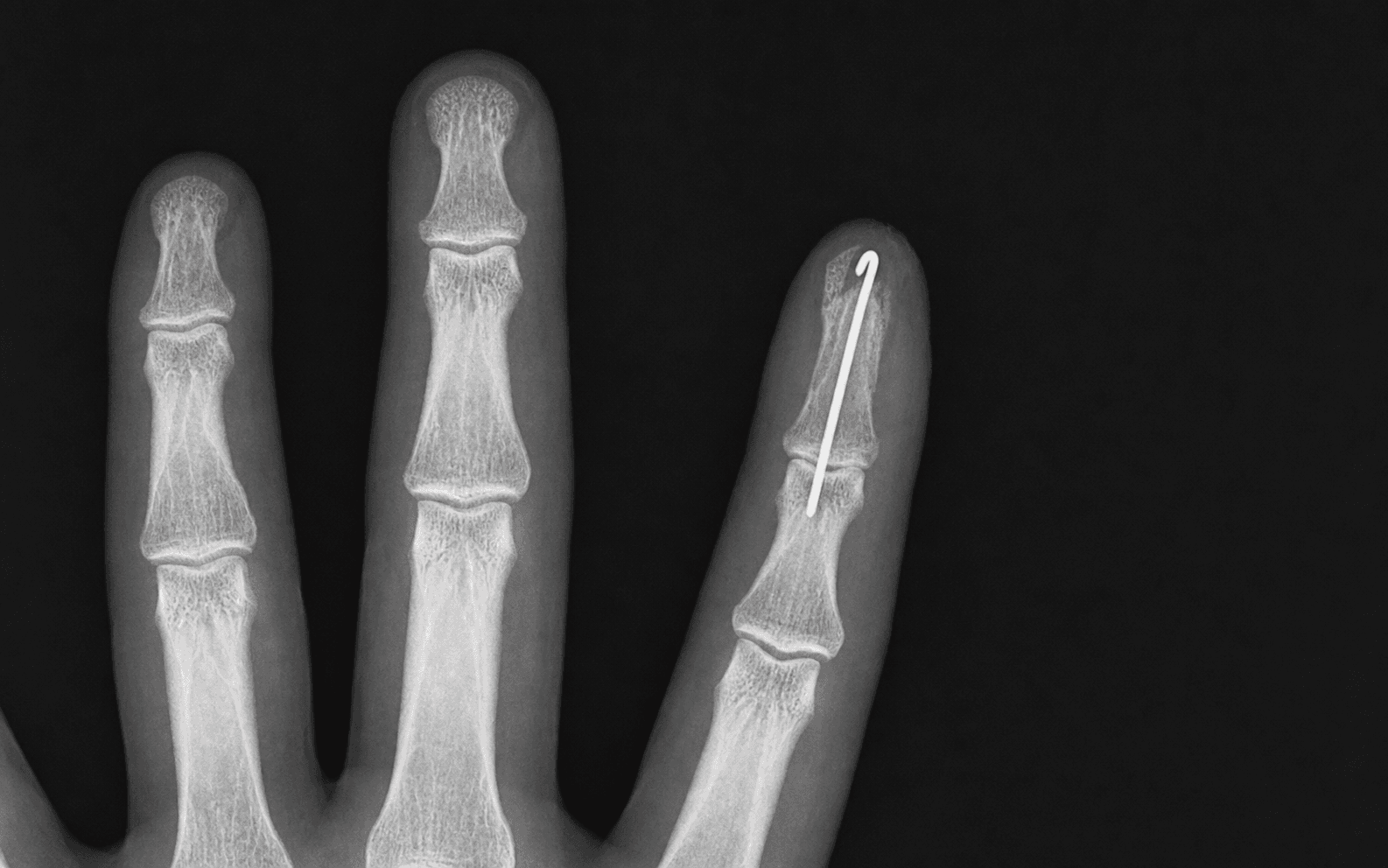
Seymour fracture — a specific operative protocol A Seymour fracture is an open physeal fracture (Salter-Harris I or II) of the distal phalanx in a child, with the nail plate avulsed from or lodged under the nail fold. It is NOT a simple mallet finger, and the nail-fold biofilm makes it an antibiotic-resistant open fracture — missed cases develop osteomyelitis, physeal arrest and digital shortening.

Aftercare & Complications
Rehabilitation | Phase | Timing | Immobilisation & therapy | |-------|--------|---------------------------| | 1 | 0 to 2 weeks | Non-adherent dressing, finger-tip protector; elevation above the heart for 48 hours; contaminated wounds get 5 days of oral antibiotics, clean lacerations need none | | 2 | 2 to 3 weeks | Remove the securing 4-0 nylon suture; wound check, assess for infection, haematoma and nail-plate position | | 3 | 6 to 8 weeks | The replaced nail plate is shed as the new nail emerges from under the proximal fold; begin scar care | | 4 | 3 to 4 months | Full nail regrowth (the fingernail grows about 3 mm per month); final nail-appearance assessment | After a flap reconstruction, splint in slight flexion for 7 to 10 days, begin active range of motion at 10 to 14 days, and start scar management (silicone gel, massage) from 3 to 4 weeks. A cross-finger flap is divided at 3 weeks under local anaesthesia, with hand therapy from division to address the expected PIP stiffness — full recovery takes 8 to 12 weeks. Counselling key points. Warn the patient that some ridging or a split nail may occur even with a perfect repair, that the nail looks abnormal until full regrowth at 3 to 4 months, that fingertip sensitivity and cold intolerance are common for 6 to 12 months after a flap, and that children heal better than adults. Complications
- How it arises
- A longitudinal scar in the germinal matrix — the nail grows in two halves — when a germinal matrix laceration was not repaired anatomically at the primary operation
- Prevention
- Remove the nail plate at the index procedure to inspect the germinal matrix; repair all lacerations with 7-0 absorbable suture under loupe, no gaps
- Management
- Late revision: excise the scar, mobilise the matrix edges and re-approximate. Results are variable; prevention is far superior
- How it arises
- Loss of distal phalanx bone support — the nail curves over the shortened tip when bone loss exceeds about 50 percent of the distal phalanx without soft-tissue support
- Prevention
- Preserve bone length; provide an advancement flap to support the nail when bone loss is greater than 50 percent
- Management
- Nail-fold elevation with a dermal graft, a fingertip advancement flap, or terminal nail ablation if severe
- How it arises
- A sterile matrix scar or loss — the nail grows but does not adhere distally, lifting off and collecting debris
- Prevention
- Repair the sterile matrix at the primary operation with 6-0 absorbable suture; replace the nail plate to maintain contact during healing
- Management
- Conservative if mild; surgical scar excision and repair, or nail-bed grafting from a toe for a large defect. Results variable
- How it arises
- Contamination at the time of injury (crush, bite, machinery), inadequate irrigation, or a retained foreign body under the nail fold
- Prevention
- Thorough irrigation at the primary operation; remove all contaminated tissue; antibiotics for contaminated wounds and open fractures
- Management
- Superficial — oral antibiotics and dressing changes; deep paronychia — formal drainage, remove the plate if under the fold, culture; osteomyelitis — IV antibiotics and debridement
- How it arises
- Proper digital nerve damage from the original injury or from flap dissection; a cross-finger flap is initially insensate
- Prevention
- Preserve the digital nerves during flap dissection; include the nerve in the homodigital island flap pedicle for the best sensation
- Management
- Most flaps re-innervate over 6 to 18 months; a neuroma is managed with desensitisation, steroid injection or excision
- How it arises
- An open physeal fracture not formally debrided — Staphylococcus aureus colonises the fracture haematoma; the nail-fold biofilm resists antibiotics
- Prevention
- Correctly diagnose a Seymour fracture in every child with an apparent mallet deformity and nail-fold disruption; irrigate, debride and give antibiotics
- Management
- IV antibiotics (flucloxacillin or cephalosporin) and surgical debridement; physeal growth arrest and digital shortening are late sequelae
Viva & Exam Focus
NAILNAIL — nail bed repair principles
ALLENALLEN — fingertip amputation zone management
Clinical Decision Scenarios
Practise clinical reasoning and management decisions out loud
“A 9-year-old child presents after catching a finger in a door. There is a mallet deformity of the ring finger DIP joint and the nail plate appears displaced proximally from the nail fold. What is your diagnosis and management?”
“A 28-year-old chef sustains a fingertip amputation of the dominant index finger at the level of the distal third of the distal phalanx (Allen Zone II). There is 8 mm of pulp loss and the bone is exposed. The amputated tip is not available. What are your reconstructive options?”
“You review a patient at 4 months after nail-bed repair for a crush injury. She has a split nail deformity — her nail grows in two halves with a longitudinal ridge. How do you explain what has happened and what can be done?”
Nail unit anatomy
- Germinal matrix: under the proximal nail fold, produces 90 percent of the nail plate
- Sterile matrix: distal nail bed, produces 10 percent, anchors the plate
- Lunula: the white crescent — the visible distal edge of the germinal matrix
- The nail grows about 3 mm per month — full regrowth in 3 to 4 months
- Hyponychium: the seal between the nail and the fingertip pulp
Subungual haematoma decision
- Nail plate AND margin intact: trephinate only, any haematoma size (Roser and Gellman)
- Nail fold disrupted, plate avulsed/displaced, or a laceration seen: remove the nail, repair the bed
- Quote the 50 percent rule — but base the decision on nail-fold integrity, not haematoma size
- Trephinate with a heated paper clip, electrocautery or an 18-gauge needle
- Never trephinate a Seymour fracture — it needs formal debridement
- Always radiograph the distal phalanx to exclude a fracture
Allen classification
- Zone I: distal to bone — conservative (children) or a composite graft
- Zone II: bone exposed — V-Y advancement or a homodigital island flap
- Zone III: loss to DIP level — cross-finger flap or a thenar flap
- Zone IV: proximal to the DIP — replantation or formal amputation
- In every zone: if the nail bed is involved, repair it
Nail-bed repair technique
- Remove the nail plate gently with a Freer elevator (distal to proximal)
- Loupe magnification is essential (minimum 2.5×, ideally 3.5×)
- 6-0 or 7-0 absorbable suture (chromic or Vicryl) — never braided non-absorbable
- 2-octylcyanoacrylate (Dermabond) is an equivalent, faster alternative (Strauss RCT)
- Repair the germinal matrix first with 7-0 suture
- Replace the fenestrated nail plate as a biological splint, secured with 4-0 nylon
- Alternative splint: aluminium foil from a suture packet, trimmed to size
Seymour fracture
- Child plus apparent mallet deformity plus nail-fold disruption equals a Seymour fracture
- Lateral radiograph: a Salter-Harris I or II physeal fracture of the distal phalanx
- It is an open fracture through the nail bed — NOT a simple mallet finger
- Treatment: irrigation, debridement, nail-bed repair, fracture reduction, antibiotics
- A missed diagnosis leads to osteomyelitis, physeal growth arrest and digital shortening
Nail deformity prevention
- Split nail: repair the germinal matrix anatomically at the primary procedure
- Hook nail: preserve bone length; use a flap for support if bone loss exceeds 50 percent
- Non-adherence: repair the sterile matrix; replace the nail plate to maintain contact
- No nail growth: complete germinal matrix destruction — consider nail ablation
- For every deformity: prevention at the index surgery is far superior to revision
Flap summary
- V-Y Atasoy (volar): Zone II dorsal/transverse, 5 to 8 mm, sensate
- V-Y Kutler (bilateral lateral): Zone II volar-oblique pattern
- Homodigital island: Zone II, greater advancement 10 to 15 mm, nerve included
- Cross-finger flap: Zone III, two-stage, PIP stiffness risk, initially insensate
- Thenar flap: Zone III index/middle, younger patients, two-stage
Background & Evidence
Nail unit anatomy. The nail plate is a hard keratinised structure produced by the matrix, growing continuously at about 3 mm per month (a fingernail), attached on its undersurface to the nail bed and enclosed proximally by the nail fold. The germinal matrix lies under the proximal nail fold (it is NOT visible without removing the nail plate), produces about 90 percent of the nail plate, and the lunula (the white crescent at the base of the visible nail) marks its distal extent; injury here causes a permanent deformity — even a 1 to 2 mm defect gives a split nail or ridging. The sterile matrix extends from the distal lunula to the hyponychium, produces about 10 percent of the plate, and anchors the plate to the bed — injury causes onycholysis if not repaired. The proximal (eponychial) nail fold maintains the space critical for the direction of nail regrowth; its avulsion disrupts the germinal matrix (the Seymour fracture equivalent in children). The hyponychium is the thickened epidermis sealing the nail plate to the fingertip pulp.

- Definition
- Distal to bone — skin and pulp loss only, no bone exposed
- Typical management
- Conservative (dressings alone heal well in children); a composite graft if the tip is available
- Definition
- Distal phalanx exposed at the tip, proximal nail bed preserved
- Typical management
- V-Y advancement (Atasoy or Kutler), homodigital island flap, or shortening and primary closure
- Definition
- Loss to the DIP joint level — extensive tissue loss
- Typical management
- Cross-finger flap (two-stage), thenar flap (index or middle, younger patient), or pollicisation for the thumb
- Definition
- Loss proximal to the DIP joint — extensive soft-tissue and skeletal loss
- Typical management
- Replantation if the digit is available; otherwise formal amputation or complex reconstruction
Key evidence. Roser and Gellman (1999) studied 53 fingers in 52 children with a subungual haematoma and an INTACT nail and nail margin, allocated to nail removal plus formal nail-bed repair versus simple trephination or observation; outcomes were equivalent regardless of haematoma size, fracture, mechanism or age, and the operative group cost roughly four times as much. This is why an intact nail with an intact nail fold is simply decompressed. Strauss and colleagues (2008) randomised 40 nail-bed lacerations to 2-octylcyanoacrylate versus 6-0 chromic suture and found equivalent cosmesis, pain and function with the adhesive in roughly one-third of the time (9.5 versus 27.8 minutes), establishing tissue adhesive as a legitimate faster alternative. The Seymour fracture was defined by Seymour (1966) as an open juxta-epiphyseal distal phalanx injury with the nail plate displaced from the eponychial fold and matrix interposed in the fracture; Krusche-Mandl and colleagues (2013) confirmed that with timely debridement, reduction and antibiotic cover, 23 of 24 patients regained full motion with no infections. The Cochrane review (Capstick and Giele, 2014) found the overall evidence sparse — routine prophylactic antibiotics after simple repair were not clearly beneficial, and a non-adherent silicone dressing aided atraumatic dressing changes in children.
References
Comparison of nail bed repair versus nail trephination for subungual hematomas in children
- 53 fingers in 52 children with a subungual haematoma and an INTACT nail and nail margin, allocated to nail removal plus formal nail-bed repair (26 fingers) versus simple decompression by trephination or observation (27 fingers)
- Mean follow-up over 2 years; outcomes were equivalent between groups regardless of haematoma size, presence of fracture, mechanism or age
- Only transient, self-resolving nail abnormalities in each group (3 operative, 1 non-operative)
- Cost was approximately 4-fold higher in the operative group (mean USD 1,263 versus USD 283)
A prospective, randomized, controlled trial of 2-octylcyanoacrylate versus suture repair for nail bed injuries
- 40 patients with acute nail-bed lacerations randomised to 2-octylcyanoacrylate (Dermabond, 18 patients) versus 6-0 chromic suture repair (22 patients)
- Tissue adhesive was markedly faster: mean 9.5 minutes versus 27.8 minutes for suture (p less than 0.0003)
- No difference in physician-rated cosmesis, patient-perceived cosmesis, pain or function at 1, 3 and 6 months
- Establishes tissue adhesive as an efficient, equivalent alternative for nail-bed laceration repair
Juxta-epiphysial fracture of the terminal phalanx of the finger
- Original description of the juxta-epiphyseal (physeal) fracture of the distal phalanx in children with an associated nail-bed laceration and ungual subluxation — the eponymous Seymour fracture
- Characterised by an apparent mallet posture with the nail plate displaced from beneath the eponychial fold
- Established that the interposed nail matrix blocks reduction and that the injury behaves as an open fracture
- Recognised the requirement for formal treatment rather than simple splinting
Seymour fractures: retrospective analysis and therapeutic considerations
- 24 skeletally immature patients (mean age 8.5 years) with Seymour fractures, mean follow-up 10 years
- 9 treated non-operatively; 9 by debridement, open reduction and fixation; 5 needed an additional trans-DIP K-wire for instability
- All surgically treated patients received perioperative cephalosporin; 23 of 24 regained full motion with no infections and no residual flexion deformity
- Minor long-term nail or physeal growth disturbance in a minority, rarely cosmetically relevant
Interventions for treating fingertip entrapment injuries in children (Cochrane systematic review)
- Systematic review of 2 RCTs (191 children) on fingertip entrapment injury management
- Antibiotic RCT after surgical repair: infection in 1 of 66 (antibiotic) versus 1 of 69 (no antibiotic) — no significant difference; both infected children had partial amputations
- Silicone-net versus paraffin-gauze dressing: similar healing time and complications; silicone was less adherent and less distressing at the first dressing change
- Overall low-quality evidence — no RCT recorded fingertip function, nail growth or nail deformity