Patella ORIF — Tension Band and Screw Fixation

TraumaIntermediateCore Procedure

Patella ORIF — Tension Band and Screw Fixation

Open reduction and internal fixation of displaced transverse patellar fractures using modified anterior tension band wiring or cannulated screw tension band constructs — indications, midline anterior approach, articular reduction, fixation techniques, complications and rehabilitation

High-yield overview

Open reduction internal fixation of displaced transverse patellar fractures | intermediate

Surgical Imaging

Transverse patella fracture fixed with tension-band wiring
Transverse patella fracture fixed with tension-band wiring — anteroposterior and lateral radiographs showing two K-wires and a figure-of-eight cerclage.Credit: Hellerhoff via Wikimedia Commons (CC BY-SA 3.0)
Critical Danger Structures and Exam Traps
Articular Step-off Threshold

The rule: Any patellar fracture with articular step-off greater than 2 mm or gap greater than 3 mm requires anatomic reduction and internal fixation. Non-operative management is reserved for fractures with step-off less than 2 mm and an intact extensor mechanism.

The risk: Malreduction greater than 2 mm increases contact pressure on the remaining articular surface and accelerates post-traumatic arthritis. Intra-operative confirmation of the retropatellar surface under direct vision is mandatory — fluoroscopy alone misses subtle steps.

Extensor Mechanism Continuity

The test: Active knee extension against gravity must be assessed before deciding on operative versus non-operative care. A patient who can perform a straight-leg raise has an intact extensor mechanism even if the fracture is displaced on radiographs.

The trap: Radiographic displacement alone does not dictate surgery. A patient with a displaced fracture but preserved active extension can sometimes be treated non-operatively in a cylinder cast; conversely, a minimally displaced fracture with loss of extension requires exploration and repair.

K-wire and Wire Prominence

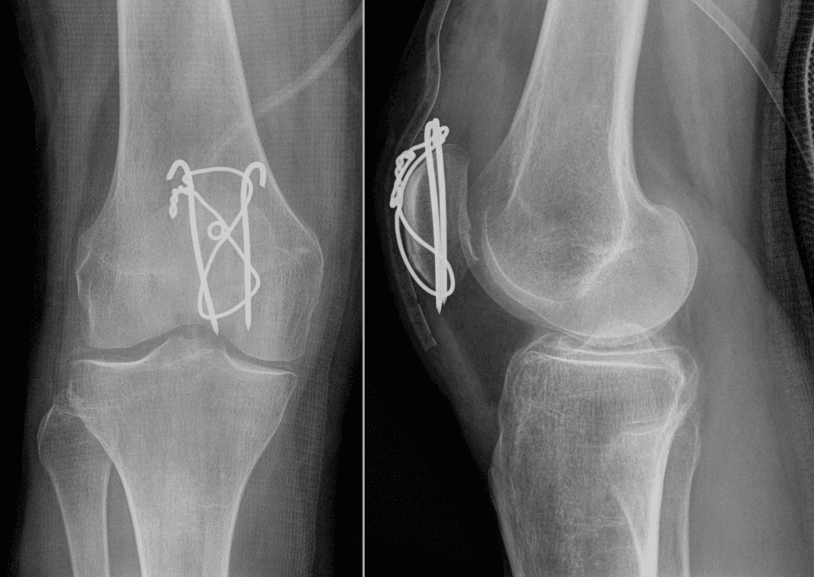
The problem: Prominent K-wire ends and tension-band wire knots are the most common cause of re-operation after patellar ORIF. Up to 50% of patients require hardware removal for irritation or skin breakdown.

The fix: Cut K-wires short and bend the ends into the quadriceps or patellar tendon. Use cannulated screws with the tension-band wire passed through the screw heads when soft-tissue coverage is marginal. Consider low-profile plate constructs in thin patients.

Comminuted Inferior Pole Fractures

The challenge: The inferior pole is frequently comminuted in high-energy injuries. Small fragments cannot hold screws or wires and risk fixation failure or patellar tendon avulsion.

The solution: Perform partial patellectomy of the comminuted pole and reattach the patellar tendon directly to the remaining patella with heavy non-absorbable transosseous sutures or suture anchors. Preserve at least 50% of patellar height to maintain extensor mechanism leverage.

Quadriceps and Patellar Tendon Integrity

The assessment: Always examine the entire extensor mechanism. A transverse patellar fracture may coexist with a quadriceps tendon rupture proximally or a patellar tendon rupture distally — these must be repaired simultaneously.

The consequence: Missing a concomitant tendon rupture leads to early fixation failure when the patient attempts active extension. Palpate the quadriceps tendon above the patella and the patellar tendon below for gaps or tenderness before proceeding to the operating theatre.

Open Fractures and Soft-Tissue Injury

The principle: Up to 10% of patellar fractures are open. The subcutaneous location of the patella means even closed fractures can have significant skin contusion or degloving.

The management: Open fractures require urgent irrigation and debridement within 6 hours. Delayed definitive fixation with temporary spanning external fixation or a spanning cast is appropriate until soft-tissue swelling subsides. Do not place definitive hardware through compromised skin.

Mnemonic

T.E.N.S.I.O.NTENSION — Modified Anterior Tension-Band Principle

Mnemonic

P.A.T.E.L.L.APATELLA — Operative Decision Algorithm

Mnemonic

R.E.D.U.C.EREDUCE — Intra-operative Reduction and Fixation Checklist

Surgical Indications

Absolute Indications

  • Articular step-off greater than 2 mm or fracture gap greater than 3 mm on radiographs or CT
  • Loss of active knee extension (inability to perform straight-leg raise)
  • Open patellar fracture requiring debridement and stabilisation
  • Displaced fracture with significant displacement of the extensor mechanism
  • Associated ipsilateral injuries requiring surgical stabilisation (tibial plateau, distal femur)

Relative Indications

  • Minimally displaced fracture (step-off less than 2 mm) with preserved active extension but patient preference for early mobilisation
  • Athletes or high-demand patients where anatomic reduction may improve long-term function
  • Comminuted fractures where partial patellectomy and tendon reattachment can restore a functional extensor mechanism

Contraindications

Absolute:

  • Minimally displaced fracture with intact active extension and acceptable alignment for non-operative management
  • Patient medically unfit for surgery
  • Chronic non-union with established post-traumatic arthritis where salvage (patellectomy or arthroplasty) is more appropriate

Relative:

  • Severe soft-tissue compromise requiring delayed definitive fixation
  • Low-demand elderly patient with comorbidities where non-operative care in extension bracing is acceptable
  • Vertical fracture patterns better suited to lag screw fixation alone

Evidence for Operative versus Non-Operative Treatment

Non-Operative Management

  • Indicated for fractures with articular step-off less than 2 mm and an intact extensor mechanism
  • Cylinder cast or hinged knee brace locked in extension for 4-6 weeks followed by progressive mobilisation
  • Good to excellent results in 70-85% of appropriately selected minimally displaced fractures
  • Risk of late displacement if patient compliance with extension bracing is poor

Operative Treatment Outcomes

  • Anatomic reduction and stable fixation allows early mobilisation and reduces the risk of post-traumatic arthritis
  • Modified anterior tension-band wiring achieves union rates greater than 90% in transverse fractures
  • Cannulated screw tension-band constructs show lower rates of hardware prominence and re-operation compared with K-wire constructs in several series
  • Partial patellectomy for comminuted poles restores extension with acceptable functional scores when at least 50% of patellar height is preserved

Key Evidence

Evidence

Long-term results after operative treatment of patellar fractures

Level III
Bostrom AActa Orthop Scand
Clinical implication: Anatomic reduction remains the cornerstone of modern patellar fracture surgery; malreduction greater than 2 mm is associated with inferior long-term results.
Source: Acta Orthop Scand Suppl 1972;143:1-80
Evidence

Biomechanical comparison of tension-band wiring versus cannulated screw fixation for transverse patellar fractures

Level II
Carpenter JE, Kasman RA, Patel N, et al.J Orthop Trauma
Clinical implication: Cannulated screw tension-band constructs are a biomechanically sound alternative to K-wire constructs and may reduce hardware prominence in thin patients.
Source: J Orthop Trauma 1997;11(5):351-6
Evidence

Partial patellectomy for comminuted patellar fractures

Level III
Saltzman CL, Goulet JA, McClellan RT, et al.J Bone Joint Surg Am
Clinical implication: Partial patellectomy is a valid salvage option for severely comminuted poles that cannot be reconstructed; aim to retain at least half the patella.
Source: J Bone Joint Surg Am 1990;72(9):1279-85
Evidence

Hardware removal after patellar tension-band fixation

Level IV
Smith ST, Cramer KE, Karges DE, et al.J Orthop Trauma
Clinical implication: Counsel patients pre-operatively about the high likelihood of hardware removal; use low-profile constructs when soft-tissue coverage is marginal.
Source: J Orthop Trauma 1997;11(3):183-7

Clinical Decision Scenarios

Practise clinical reasoning and management decisions out loud

Viva scenarioStandard
Clinical prompt

A 42-year-old labourer sustains a displaced transverse patellar fracture after a fall from scaffolding. He is unable to perform a straight-leg raise. Radiographs show 4 mm of articular step-off. How do you manage this patient?

Practical approach
This patient has an absolute indication for operative fixation: articular step-off greater than 2 mm and loss of active extension confirming extensor mechanism disruption. **Pre-operative plan**: CT scan to assess comminution and plan implant placement. Consent for ORIF with possible partial patellectomy if the inferior pole is irreconstructible. Regional anaesthesia with femoral block for post-operative analgesia. **Surgical approach**: Midline anterior longitudinal incision. Full-thickness flaps. Evacuate haematoma and visualise the retropatellar surface through the fracture or a small arthrotomy. Achieve anatomic reduction with pointed clamps. Place two parallel K-wires in the anterior half of the patella. Pass a figure-of-eight 18-gauge wire through the quadriceps and patellar tendons. Tension the wire with the knee in extension, then confirm compression with knee flexion to 90 degrees. Obtain AP, lateral, and sunrise fluoroscopic views. **Alternative construct**: If the patient has thin soft tissues or high demand, use 4.5 mm cannulated screws with the tension-band wire passed through the screw heads. **Post-operative**: Hinged brace locked in extension, touch weight-bearing for 2 weeks. Immediate quadriceps setting. Unlock brace at 6 weeks if radiographic healing is progressing. Return to heavy labour at 4-6 months.
Viva scenarioAdvanced
Clinical prompt

A 35-year-old female athlete presents with a comminuted inferior pole patellar fracture after a dashboard injury. The inferior pole is in multiple small fragments. She has loss of active extension. Describe your operative plan.

Practical approach
This is a challenging fracture because the comminuted inferior pole cannot reliably hold screws or wires. The goal is to restore a functional extensor mechanism while preserving as much patellar height as possible. **Assessment**: CT scan to quantify comminution and remaining bone stock. If the inferior pole fragments are too small for reconstruction, plan for partial patellectomy. **Surgical technique**: Midline approach. Identify the comminuted inferior pole. Excise the small, non-reconstructible fragments. Freshen the remaining inferior patellar surface. Reattach the patellar tendon to the remaining patella using heavy non-absorbable transosseous sutures passed through three parallel drill holes in the coronal plane, or with two suture anchors. Ensure at least 50% of the original patellar height remains. If the remaining patella is too small, consider total patellectomy with tendon imbrication as a salvage. **Tension-band alternative**: If a single large inferior pole fragment exists, it can sometimes be fixed with a cannulated screw and incorporated into a tension-band construct. This is preferable to excision when technically feasible. **Rehabilitation**: Longer period of protected weight-bearing (8-12 weeks) because tendon-to-bone healing is slower than bone-to-bone. Brace locked in extension. Gradual active flexion once the repair is clinically stable.
Viva scenarioStandard
Clinical prompt

A 28-year-old man underwent patellar ORIF with K-wire tension band for a transverse fracture 9 months ago. He has united but complains of severe anterior knee pain when kneeling and a palpable prominence over the superior pole. Radiographs show the fracture is healed. How do you manage him?

Practical approach
This patient has symptomatic hardware prominence, the most common reason for re-operation after patellar tension-band fixation. The K-wire ends and wire knot are irritating the skin and subcutaneous tissues, particularly during kneeling. **Assessment**: Confirm radiographic union. Examine for signs of infection. Assess range of motion and extensor strength. Counsel that hardware removal is appropriate once union is confirmed. **Surgical plan**: Removal of hardware under local or regional anaesthesia. Make small incisions over the prominent wire ends. Cut the K-wires short and remove them. Remove the tension-band wire. Inspect the fracture site to ensure it remains stable. Close in layers. No post-operative bracing is required if the fracture is solidly united. **Prevention for future cases**: Use cannulated screw tension-band constructs in patients with thin soft-tissue envelopes. Bury K-wire ends deep in the quadriceps tendon. Consider low-profile plate constructs in high-risk patients. **Outcome**: Most patients experience significant relief after hardware removal. Some residual anterior knee discomfort from the original injury may persist, but the mechanical irritation from prominent hardware is eliminated.
Exam day cheat sheet
Patella ORIF — Tension Band and Screw Fixation — Exam Day Summary

References

Evidence

Long-term results after operative treatment of patellar fractures

Level III
Bostrom AActa Orthop Scand
Source: Acta Orthop Scand Suppl 1972;143:1-80
Evidence

Biomechanical comparison of tension-band wiring versus cannulated screw fixation for transverse patellar fractures

Level II
Carpenter JE, Kasman RA, Patel N, et al.J Orthop Trauma
Source: J Orthop Trauma 1997;11(5):351-6
Evidence

Partial patellectomy for comminuted patellar fractures

Level III
Saltzman CL, Goulet JA, McClellan RT, et al.J Bone Joint Surg Am
Source: J Bone Joint Surg Am 1990;72(9):1279-85
Evidence

Hardware removal after patellar tension-band fixation

Level IV
Smith ST, Cramer KE, Karges DE, et al.J Orthop Trauma
Source: J Orthop Trauma 1997;11(3):183-7
Editorially reviewed — transparent references and correction processPublished by OrthoVellum Medical Education TeamEditorial boardMethodologyReview policy
Educational disclosure

Educational content is reviewed for source visibility, editorial coherence, and correction readiness.

No individual clinician credential is claimed unless a named person is shown.

Verify before clinical use; this is not medical advice or a substitute for local guidance.