Femoral varus and/or Salter pelvic osteotomy to contain the deformable necrotic femoral head | intermediate
- CONTAINMENT principle: keep the soft, plastic necrotic femoral head deeply seated within the acetabulum so it reossifies as a sphere — a 'ball in a mould'. The acetabulum acts as the mould only while the head is biologically plastic (initial/fragmentation stages).
- AGE AT ONSET is the single most powerful prognostic factor — children older than 8 years (chronological age) at onset with lateral pillar B or B-C are the group shown by Herring to benefit most from surgical containment; younger children often do well without surgery.
- Lateral pillar (Herring) grade and the 'head at risk' signs (Gage sign, lateral calcification, lateral subluxation, horizontal physis, metaphyseal cysts) guide the decision to operate — lateral pillar C does poorly regardless of treatment.
- Containment is only effective EARLY — in the initial or fragmentation stage while the head is still mouldable and reducible. Once the head is healed/deformed or hinge abduction has developed, containment osteotomy is contraindicated and a salvage/realignment procedure is needed.
When & Why
The containment principle. During the necrosis and fragmentation stages of Legg-Calvé-Perthes disease, the femoral head is biologically plastic and deformable. If it is kept deeply seated within the spherical acetabulum, the acetabulum acts as a mould and the head reossifies as a sphere — the 'ball in a mould' concept. If the head is allowed to extrude or sublux laterally, it deforms into a non-spherical shape and becomes incongruent, leading to early osteoarthritis. The biological principle exploited by surgery is that the plastic head will remodel to the shape of whatever it is moulded against. The decision-making framework. The aim of all treatment is containment — keeping the femoral head within the acetabulum during the plastic phase so it remodels spherically and congruently. Not every Perthes hip needs surgery; the decision integrates age, lateral pillar grade, range of motion, and head-at-risk signs.

Prognostic factors (decision drivers):
- Age at onset — the single most important factor. In the Herring study no treatment effect was seen below a chronological age of 8 years (or a skeletal age of 6 years), so younger children usually do well non-operatively; children older than 8 years at onset remodel poorly and are more likely to benefit from surgery.
- Lateral pillar grade — A (good regardless), B/B-C (benefits from surgery if older), C (poor regardless).
- Head-at-risk signs — especially lateral subluxation — favour intervention.
- Loss of containment / extrusion and progressive loss of abduction.
- Hinge abduction — if present, varus containment is contraindicated. Non-operative containment — largely abandoned:
- Abduction bracing / casting (e.g. Petrie casts, Scottish-Rite orthosis) was historically used to hold the hip abducted and contained.
- High-quality evidence (including the Herring multicentre study) showed bracing is ineffective at altering outcome and is poorly tolerated — it is no longer recommended as a containment strategy.
- Modern non-operative management focuses on symptomatic care: activity modification, physiotherapy to maintain range of motion (especially abduction and internal rotation), analgesia, and observation — appropriate for the younger child and lateral pillar A. Indications for surgical containment:
- Best-supported indication — chronological age older than 8 years at onset with lateral pillar B or B/C — this is the group with demonstrated benefit from operative containment (femoral or pelvic osteotomy) in the Herring multicentre prospective study. (Wiig used a chronological-age threshold of 6 years with greater than 50% head necrosis to recommend femoral varus osteotomy.)
- Progressive subluxation / extrusion of the head (head-at-risk signs) with loss of containment.
- Failure to maintain containment with conservative measures in an at-risk hip.
- Lateral pillar B/B-C in the early (initial/fragmentation) stage while the head is still reducible and congruent in abduction. Contraindications to containment (varus/Salter):
- Healed or late reossification stage — head no longer plastic, deformity fixed.
- Hinge abduction confirmed on dynamic arthrogram — abduction worsens congruity (needs valgus osteotomy or shelf).
- Uncontainable head (markedly enlarged coxa magna that cannot be seated) — consider lateral shelf / Chiari salvage.
- Stiff, incongruent hip with fixed deformity. Prerequisite assessment before surgery:
- Dynamic arthrogram under anaesthesia — the key planning step. Confirms the head reduces and is congruent in abduction and internal rotation and excludes hinge abduction.
- Restore range of motion first if the hip is stiff (traction, adductor release / physiotherapy) — a hip must abduct before it can be contained.
- Lateral pillar A
- Observe — excellent
- Lateral pillar B / B-C
- Observe — usually good
- Lateral pillar C
- Guarded; observe, surgery limited benefit
- Lateral pillar A
- Observe
- Lateral pillar B / B-C
- Consider surgery if at-risk signs (Wiig: VO if greater than 50% necrosis)
- Lateral pillar C
- Poor regardless; symptomatic care
- Lateral pillar A
- Observe
- Lateral pillar B / B-C
- SURGERY — clearest benefit
- Lateral pillar C
- Poor regardless; counsel realistically
- Lateral pillar A
- N/A
- Lateral pillar B / B-C
- Varus contraindicated — valgus/shelf
- Lateral pillar C
- Salvage (shelf / Chiari)
The Herring lateral pillar classification (A / B / B-C / C) is the strongest radiographic predictor and is assessed in the FRAGMENTATION stage on the AP radiograph — not at presentation. Assessing it too early (initial stage) or too late (reossification) misgrades the hip and misdirects the decision to operate.
The Operation
The goal is to keep the deformable femoral head seated within the acetabular mould so it reossifies as a sphere. Containment can be achieved on the femoral side (redirect the head into the socket by reorienting the proximal femur), the pelvic side (redirect the acetabulum to cover the head), or both. Salvage procedures (shelf, Chiari) are used when the head is too large or uncontainable, or when hinge abduction is present. Every case is opened with a dynamic arthrogram under anaesthesia to confirm the head reduces and is congruent in abduction and to exclude hinge abduction before any bone is cut.


- Mechanism
- Reorients proximal femur to seat the head deeper
- Best indication
- Older child, pillar B/B-C, good abduction, congruent in abduction
- Key drawback
- Limb shortening; relative trochanteric overgrowth; coxa vara if over-corrected
- Mechanism
- Redirects acetabulum to cover the anterolateral head
- Best indication
- Containment needed without wanting femoral shortening; anterolateral deficiency
- Key drawback
- Increases joint pressure; needs mobile, congruent hip; slight limb lengthening
- Mechanism
- Both reorientations for severe uncontainment
- Best indication
- Older child, severe extrusion, single redirection insufficient
- Key drawback
- Larger procedure; more morbidity; technically demanding
- Mechanism
- Augments lateral coverage with a bone shelf
- Best indication
- Large head / older child / mild hinge; salvage of lateral deficiency
- Key drawback
- Non-anatomic; coverage by fibrocartilage not articular cartilage
- Mechanism
- Medialises hip, creates capsular interposition coverage
- Best indication
- Salvage of incongruent / uncontainable head
- Key drawback
- Non-anatomic; medialises joint; salvage only
- Mechanism
- Moves congruent medial segment under the load; opens out hinge
- Best indication
- Hinge abduction — head congruent in adduction
- Key drawback
- Worsens abductor lever arm differently; for specific deformity only
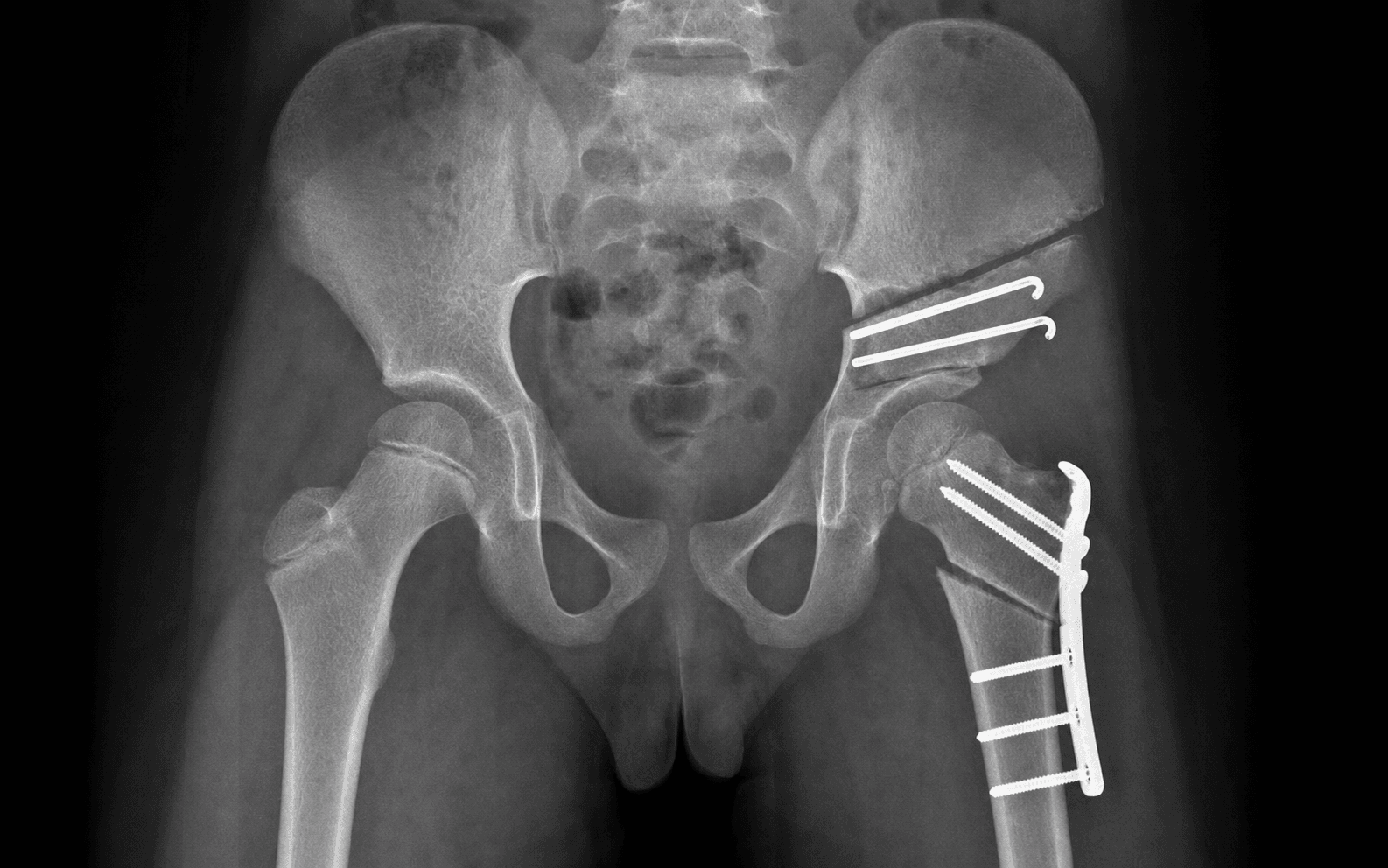
Femoral varus derotation osteotomy (VDRO) — the default containment operation
- Supine on a radiolucent table with the image intensifier available throughout.
- Position the whole femur so it can be imaged in AP and lateral without repositioning.
- General anaesthesia; examine and document hip range of motion (abduction, internal rotation) before draping.
- A straight lateral incision along the proximal femur, centred on the intended intertrochanteric/subtrochanteric osteotomy level.
- Incise fascia lata in line; split vastus lateralis off the lateral intermuscular septum and retract it anteriorly to expose the lateral shaft and trochanteric region.
- Stay subperiosteal on bone to protect the perforating branches of the profunda femoris as they pierce the septum.
- Clear the lateral cortex at the intertrochanteric/subtrochanteric region subperiosteally.
- Insert the blade of a pre-contoured paediatric blade plate (or a proximal femoral locking plate) along the planned proximal-fragment trajectory, checked on the image intensifier, to set the neck-shaft angle.
- Make the transverse intertrochanteric osteotomy below the greater trochanter.
- Bring the distal fragment into varus (and derotate to neutralise excessive anteversion), seating the femoral head more deeply into the acetabulum and improving anterolateral cover.
- Aim for a neck-shaft angle of about 105-110 degrees — enough varus to contain the head without producing a permanent Trendelenburg gait, which the older child cannot remodel.
- Secure the plate to the distal fragment; confirm reduction, correction and fixation on AP and lateral imaging.
- Resect only a wedge sufficient to obtain bone contact and the desired varus without excessive shortening.
- Layered closure over a drain.
- Apply a hip spica or a protected-weight-bearing regime depending on fixation rigidity and the child's age; plate removal is commonly undertaken at 6-12 months once united.
- Excessive varus — produces a persistent Trendelenburg gait and abductor weakness; the older child cannot remodel it. Limit to a neck-shaft angle around 105-110 degrees.
- Limb-length discrepancy — varus and wedge resection shorten the limb; document and counsel; usually 1-2 cm and tolerated.
- Relative trochanteric overgrowth — varus relatively elevates the greater trochanter, shortening the abductor lever arm.
If the lateral head levers on the acetabular rim and the medial joint gaps open on abduction (hinge abduction), a varus or Salter containment procedure is contraindicated — both rely on the abducted position, which is precisely the incongruent position. Confirm congruity with a dynamic arthrogram in abduction BEFORE choosing varus. Hinge abduction is managed with a valgus osteotomy or shelf.
Femoral varus osteotomy shortens the limb and can aggravate a Trendelenburg gait via relative trochanteric overgrowth. A Salter osteotomy lengthens the limb slightly and avoids femoral shortening — relevant to limb-length planning when choosing between the two.
Salter innominate (pelvic redirectional) osteotomy — the pelvic alternative
- Supine with a sandbag under the ipsilateral buttock.
- Bikini incision from below the iliac crest toward the groin; expose the iliac wing subperiosteally down to the sciatic notch and the anterior inferior iliac spine (AIIS).
- A straight, complete cut through the innominate bone from the sciatic notch to the AIIS, just above the acetabulum.
- Complete the cut fully to the notch so the distal fragment can hinge on the symphysis pubis; an incomplete posterior cut risks the sciatic nerve and gives an inadequate hinge.
- Rotate the distal fragment (bearing the acetabulum) anterolaterally to cover the extruded femoral head.
- Hold the opened osteotomy with a tricortical iliac crest wedge graft and two threaded Kirschner wires across the osteotomy into the distal fragment.
- Layered closure; image to confirm wire position and head cover.
- Single hip spica for approximately 6 weeks, then mobilise; remove the threaded wires once healed.
- Increased joint reaction force — redirection raises contact pressure; contraindicated in a stiff or incongruent hip.
- Incomplete posterior osteotomy / sciatic notch — risk to the sciatic nerve and inadequate hinge; complete the cut to the notch.
- Pin migration / graft displacement — secure two-pin fixation and protected weight-bearing.
Salvage and hinge-abduction procedures. When the head is too large to contain anatomically, or hinge abduction is present, containment is abandoned in favour of realignment or augmentation: a shelf acetabuloplasty augments lateral coverage of a large head with a bone shelf; a Chiari osteotomy medialises and salvages an incongruent/uncontainable hip by capsular metaplasia; and a valgus (± extension) osteotomy is the procedure of choice for hinge abduction, because the head is congruent in adduction — bringing that congruent medial portion under load relieves the hinge and improves abduction and gait.
Aftercare & Complications
Post-operative care by procedure:
- Femoral osteotomy — hip spica or protected weight-bearing depending on fixation rigidity; plate removal commonly at 6-12 months once united.
- Salter osteotomy — single hip spica for approximately 6 weeks, then mobilise; remove threaded wires once healed.
- Rehabilitation — regain abduction and internal rotation; physiotherapy; monitor for trochanteric overgrowth and leg-length discrepancy to maturity. Complications
- Frequency / context
- More common with lateral pillar C and older age
- Recognition
- Continued collapse and asphericity on serial radiographs; persistent stiffness
- Prevention and management
- Prevention: correct case selection (congruent, reducible, early stage); avoid containment in healed/hinge hips. Management: salvage realignment (valgus/shelf/Chiari) at maturity; counsel re eventual arthritis
- Frequency / context
- Common natural sequela of LCPD
- Recognition
- Enlarged, broadened femoral head wider than the acetabulum on radiograph
- Prevention and management
- Prevention: early containment maintains sphericity. Management: usually tolerated if congruent; if uncontainable consider shelf augmentation
- Frequency / context
- Physeal damage and premature growth arrest
- Recognition
- Short, broad femoral neck; high-riding greater trochanter on radiograph
- Prevention and management
- Prevention: minimise physeal insult. Management: trochanteric transfer/epiphysiodesis for relative overgrowth; neck-lengthening osteotomy in selected cases
- Frequency / context
- Relative overgrowth after early physeal arrest or excessive varus
- Recognition
- Abductor weakness, positive Trendelenburg, trochanter above centre of head
- Prevention and management
- Prevention: avoid excessive varus; consider greater trochanteric apophysiodesis in young child at risk. Management: distal/lateral trochanteric transfer to restore abductor lever arm
- Frequency / context
- Femoral varus shortening, growth disturbance
- Recognition
- Measured/clinical leg-length difference; pelvic obliquity
- Prevention and management
- Prevention: limit varus/wedge resection; Salter avoids shortening. Management: shoe raise; contralateral epiphysiodesis timed to maturity if significant
- Frequency / context
- Excessive varus or trochanteric overgrowth
- Recognition
- Abductor lurch, positive Trendelenburg test
- Prevention and management
- Prevention: neck-shaft angle around 105-110 degrees, not more varus. Management: abductor strengthening; trochanteric transfer if structural
- Frequency / context
- Pre-existing stiffness, prolonged immobilisation
- Recognition
- Reduced abduction and internal rotation post-op
- Prevention and management
- Prevention: restore motion before surgery; avoid over-immobilisation. Management: physiotherapy; rarely arthrolysis
- Frequency / context
- Long-term outcome of aspherical incongruent hip (Stulberg IV-V)
- Recognition
- Pain, stiffness, joint space narrowing in young adulthood
- Prevention and management
- Prevention: achieve Stulberg I-III by good containment. Management: joint-preserving osteotomy in young adult; eventual arthroplasty
- Frequency / context
- Technique-dependent
- Recognition
- Pain, failure to unite, lateral hip prominence/bursitis
- Prevention and management
- Prevention: stable fixation, adequate bone contact. Management: implant removal once united; revision fixation/grafting for non-union
Viva & Exam Focus
C.O.N.T.A.I.NCONTAIN — principles of Perthes containment
GLSHMGLSHM — Catterall head-at-risk signs
Critical decision points and exam traps
The trap: treating all Perthes hips the same. Children under 6 years (skeletal age) have such good remodelling potential that most do well with symptomatic treatment alone. The fix: reserve surgical containment for the older child (chronological age older than 8 years at onset) with lateral pillar B/B-C. This is the group with proven benefit from the Herring multicentre study. Younger children rarely need surgery.
The trap: performing a containment osteotomy in the late reossification or healed stage. The head is no longer plastic and cannot remodel into the acetabular mould. The fix: containment is an EARLY-stage intervention (initial or fragmentation). The head must be reducible and congruent in abduction. Late deformity needs salvage, not containment.
Definition: an enlarged, deformed lateral head segment levers (hinges) on the lateral acetabular rim during abduction, causing lateral gapping medially and worsening congruity. Why it matters: a varus or Salter containment procedure relies on abduction to contain the head — in hinge abduction this is harmful. Confirm congruity with a dynamic arthrogram in abduction BEFORE choosing varus. Hinge abduction needs a valgus osteotomy or shelf.
Evidence: in the Herring lateral pillar C group, outcomes are poor irrespective of operative or non-operative treatment, and across all age groups. Implication: do not promise that surgery will rescue a pillar C hip. Counsel realistically. The clearest surgical benefit is the older child with pillar B / B-C.
Catterall head-at-risk signs: Gage sign (V-shaped lucency lateral epiphysis/metaphysis), lateral calcification of the epiphysis, lateral subluxation of the head, horizontal physis, and diffuse metaphyseal reaction/cysts. Implication: presence of head-at-risk signs (especially lateral subluxation) shifts the balance towards containment surgery, particularly in the older child.
Perthes: painless or mild limp, restricted abduction and internal rotation, child typically 4-8 years, afebrile, AVN of epiphysis on imaging. SCFE: older/heavier child (10-16 years), externally rotated limb, obligatory external rotation on hip flexion. Septic hip: febrile, refuses to weight-bear, raised CRP/WCC — an emergency requiring aspiration/washout, never an elective containment problem.
Clinical Decision Scenarios
Practise clinical reasoning and management decisions out loud
“A 9-year-old boy presents with a 4-month history of a painless limp and reduced hip abduction. Radiographs show fragmentation of the right proximal femoral epiphysis with the lateral pillar reduced to about 40% of the contralateral height. How do you assess and manage him?”
“What is the containment principle in Perthes disease, and how do femoral and pelvic osteotomies achieve it? When would you choose one over the other?”
“During examination under anaesthesia and arthrography of an older child with Perthes, you find that the femoral head is congruent in adduction but levers on the lateral acetabular rim and gaps medially when you abduct the hip. What is this phenomenon, why does it contraindicate your planned varus osteotomy, and what would you do instead?”
Disease and staging
- LCPD = idiopathic AVN of the proximal femoral epiphysis; age 4-8 years, male predominance about 4-5:1, bilateral 10-15% (usually asynchronous)
- Waldenstrom stages: 1 initial/necrosis, 2 fragmentation, 3 reossification, 4 remodelling/healed
- Lateral pillar assessed in the FRAGMENTATION stage on AP radiograph
- Synchronous symmetrical bilateral disease — screen for epiphyseal dysplasia / hypothyroidism
Classifications
- Lateral pillar (Herring): A = no height loss (good); B = over 50% maintained; B/C border about 50%; C = under 50% (poor) — strongest radiographic predictor
- Catterall I-IV = extent of epiphyseal involvement; source of head-at-risk signs
- Catterall head-at-risk: Gage sign, lateral calcification, lateral subluxation, horizontal physis, metaphyseal cysts
- Stulberg I-V (at maturity) predicts OA: I-II spherical congruent (good), III aspherical congruent (moderate), IV-V incongruent (high OA risk)
The containment principle
- Keep the plastic necrotic head seated in the acetabular mould so it reossifies spherically — 'ball in a mould'
- Effective ONLY in early (initial/fragmentation) stages while the head is plastic and reducible
- Requires a head that reduces and is congruent in abduction — confirm on dynamic arthrogram
- Goal is a Stulberg I-III (congruent) hip; avoid Stulberg IV-V incongruency
Indications for surgery
- Best-supported: chronological age OVER 8 years at onset with lateral pillar B or B/C (Herring study); Wiig recommends femoral varus osteotomy over 6 years with greater than 50% head necrosis
- Progressive subluxation/extrusion with head-at-risk signs
- Lateral pillar A: observe (good regardless). Lateral pillar C: poor regardless — counsel realistically
- Bracing/abduction casting is NO LONGER recommended — ineffective at altering outcome
Surgical options
- Femoral varus derotation osteotomy: neck-shaft angle around 105-110 degrees; shortens limb; risk of trochanteric overgrowth/Trendelenburg
- Salter innominate osteotomy: redirects acetabulum anterolaterally; slight limb lengthening; needs congruent mobile hip
- Combined femoral + pelvic: severe extrusion when single redirection insufficient
- Salvage: shelf acetabuloplasty (large head / lateral deficiency), Chiari (incongruent/uncontainable hip)
- Valgus osteotomy: for HINGE ABDUCTION (head congruent in adduction)
Contraindications to containment (varus/Salter)
- Healed / late reossification stage — head shape fixed
- Hinge abduction confirmed on arthrogram — needs valgus or shelf instead
- Uncontainable enlarged head (coxa magna) — consider shelf / Chiari
- Stiff, incongruent hip with fixed deformity
Complications
- Coxa magna, coxa breva/short neck, greater trochanteric overgrowth, limb-length discrepancy
- Persistent Trendelenburg / abductor weakness — avoid excessive varus
- Progressive AVN and head deformity (especially pillar C)
- Premature osteoarthritis in aspherical incongruent (Stulberg IV-V) hips
- Osteotomy non-union, implant prominence, pin migration
Key exam points
- Age at onset is the single most powerful prognostic factor — older than 8 years (chronological) at onset does worse
- Lateral pillar C does poorly regardless of treatment, all ages
- ALWAYS do a dynamic arthrogram before containment — confirm reducibility/congruity and exclude hinge abduction
- Hinge abduction = valgus osteotomy (head congruent in adduction), NOT varus
- Salter lengthens the limb; femoral varus shortens it — relevant to limb-length planning
Background & Evidence
Epidemiology and pathoanatomy. Legg-Calvé-Perthes disease (LCPD) is an idiopathic avascular necrosis of the proximal femoral epiphysis in the growing child. Interruption of blood supply (predominantly via the lateral epiphyseal vessels of the medial femoral circumflex artery) leads to a self-limiting but staged sequence of necrosis, revascularisation, collapse, and repair. The vulnerable window is the period of necrosis and fragmentation, during which the softened, plastic epiphysis can deform under load. Typical age is 4-8 years with a male predominance of about 4-5:1; disease is bilateral in up to 10-15% (usually asynchronous — synchronous bilateral symmetrical disease should prompt consideration of epiphyseal dysplasia or hypothyroidism). Presentation is a painless or mildly painful limp, restricted abduction and internal rotation, and occasionally referred knee pain.
- Name
- Initial / necrosis
- Radiographic features
- Smaller, sclerotic epiphysis; medial joint space widening; subchondral fracture (crescent sign)
- Name
- Fragmentation
- Radiographic features
- Epiphysis fragments into segments; this is when the lateral pillar is assessed
- Name
- Reossification
- Radiographic features
- New bone fills the fragmented areas from medial to lateral
- Name
- Remodelling / healed
- Radiographic features
- Final head shape established; remodelling continues to skeletal maturity
Key timing principle: containment surgery is effective only in stages 1-2 while the head is plastic and reducible. By stages 3-4 the final shape is largely set.
- Lateral pillar height
- No loss of height
- Prognosis
- Good outcome regardless of treatment
- Lateral pillar height
- More than 50% height maintained
- Prognosis
- Outcome depends on age — benefits from containment if older
- Lateral pillar height
- About 50%, narrow/poorly ossified
- Prognosis
- Intermediate; older children benefit from surgery
- Lateral pillar height
- Less than 50% height maintained
- Prognosis
- Poor outcome regardless of treatment
- Description
- Spherical head, normal
- OA risk
- Minimal
- Description
- Spherical head, with coxa magna/short neck/steep acetabulum
- OA risk
- Low
- Description
- Aspherical (ovoid) but congruent
- OA risk
- Moderate (late OA in middle age)
- Description
- Flat head, congruent (flat acetabulum)
- OA risk
- Higher
- Description
- Flat head, incongruent
- OA risk
- Highest — early severe OA
The Stulberg classification predicts long-term osteoarthritis: Class I-II (spherical congruent) good, III (aspherical congruent) intermediate, IV-V (aspherical incongruent) high OA risk. A round head in a matching socket, or a flat head in a flat socket ('aspherical congruency'), both do better than an incongruent hip — so the goal of containment is a Stulberg I-III hip.
Determinants of outcome. The long-term outcome of Perthes is determined chiefly by the final shape and congruence of the femoral head (Stulberg class). Treatment aims to achieve a spherical or aspherical-congruent (Stulberg I-III) hip and to avoid an incongruent (IV-V) hip, which leads to early osteoarthritis. The most important prognostic factors are age at onset, lateral pillar grade, final head sphericity and congruence, head-at-risk signs (especially lateral subluxation/extrusion), and range of motion maintained throughout the disease. Femoral vs Salter — outcome comparison. Comparative series show no consistent difference in final sphericity or congruence (Stulberg outcome) between femoral and Salter osteotomy when the hip is congruent and reducible — both achieve containment. In the most-cited direct comparison (Kitakoji et al., 2005), the two procedures produced equivalent head sphericity and hip congruity, but the Salter group had a neck-shaft angle, acetabular coverage, and articular-trochanteric distance closer to normal, and avoided the surgical scarring and coxa vara seen after femoral osteotomy. Choice is guided by deformity pattern, surgeon experience, and limb-length considerations.
- Under 8 years at onset
- Good
- Over 8 years at onset
- Good
- Treatment implication
- No surgical benefit — observe
- Under 8 years at onset
- Good regardless
- Over 8 years at onset
- Better with surgery
- Treatment implication
- Operate if older than 8 years
- Under 8 years at onset
- Generally good
- Over 8 years at onset
- Better with surgery
- Treatment implication
- Operate if older than 8 years
- Under 8 years at onset
- Poor regardless
- Over 8 years at onset
- Poor regardless
- Treatment implication
- Surgery does not reliably improve outcome
Long-term and natural history. Many patients function well into adulthood; significant osteoarthritis typically presents in the 5th-6th decade, correlating with Stulberg class. Stulberg I-II carries minimal long-term OA risk; Stulberg III moderate OA in middle age; Stulberg IV-V a high risk of early, severe OA requiring joint-preserving osteotomy or arthroplasty. Counselling should be realistic: containment improves the odds of a congruent hip but cannot guarantee a normal hip, especially in older children and lateral pillar C.
References
Legg-Calvé-Perthes disease. Part II: prospective multicenter study of the effect of treatment on outcome
- Large controlled prospective multicentre study — 438 patients, 451 hips, all aged 6.0-12.0 years at onset, none previously treated; 345 hips followed to skeletal maturity.
- No difference in outcome between no treatment, bracing, and range-of-motion therapy — bracing confers no benefit.
- No significant difference between femoral varus osteotomy and innominate (Salter) osteotomy.
- In lateral pillar B and B/C-border hips in children OVER 8.0 years at onset, surgery gave significantly better outcomes than non-operative care (p less than or equal to 0.05).
- Lateral pillar group (p less than 0.0001) and age at onset (p = 0.0001) were the two strongest prognostic factors; group C and group B under 8 years showed no treatment effect.
The lateral pillar classification of Legg-Calvé-Perthes disease
- 93 hips in 86 patients classified during the fragmentation stage by lateral-pillar radiolucency and followed to maturity (Stulberg outcome).
- Group A had a uniformly good outcome (100% Stulberg I-II).
- Group B did well in children under 9 years at onset but poorly when older than 9 years; group C frequently became aspherical regardless of age.
- Inter-observer agreement was 78%; the lateral pillar group was a stronger determinant of outcome than age at onset.
The natural history of Legg-Calvé-Perthes disease
- Long-term follow-up (30-40 years) of two cohorts (88 and 68 patients) with radiographs from onset to maturity.
- Defined five classes of residual deformity at maturity, each with a characteristic long-term course.
- Spherical congruency (Class I-II) — no arthritis develops.
- Aspherical congruency (Class III-IV) — mild-to-moderate arthritis in late adulthood.
- Aspherical incongruency (Class V) — severe arthritis before age 50.
Prognostic factors and outcome of treatment in Perthes' disease: a prospective study of 368 patients with five-year follow-up
- Nationwide prospective study, 28 Norwegian hospitals, 368 unilateral cases, 97% follow-up at 5 years.
- Strongest predictor of outcome was femoral-head involvement greater than 50% (OR 7.76), followed by age at diagnosis and lateral pillar group.
- In children over 6 years at diagnosis with more than 50% head necrosis, proximal femoral varus osteotomy gave significantly better outcomes than orthosis or physiotherapy (p = 0.001).
- No difference between physiotherapy and orthosis groups, and no treatment effect in children under 6 years; the authors concluded the abduction orthosis should be abandoned.
Which is a better method for Perthes' disease: femoral varus or Salter osteotomy?
- Comparative series at skeletal maturity — 46 femoral varus osteotomies versus 30 Salter innominate osteotomies.
- No significant difference in femoral-head sphericity or hip congruity between the two procedures.
- Acetabular head coverage, neck-shaft angle, and articular-trochanteric distance were closer to normal after Salter osteotomy.
- Femoral varus osteotomy left more residual coxa vara, trochanteric prominence, and surgical scarring.
Further reading:
- Catterall A (1971). The natural history of Legg-Calvé-Perthes disease. J Bone Joint Surg Br 53(1):37-53. — Original Catterall classification and the head-at-risk signs guiding prognosis.
- Salter RB (1984). The present status of surgical treatment for Legg-Perthes disease. J Bone Joint Surg Am. — Rationale and technique for innominate osteotomy as a containment procedure.
- Joseph B, Nair NS, Narasimha Rao K, et al. Optimal timing for containment surgery for Perthes disease. J Pediatr Orthop. — Evidence that containment is effective only in the early (avascular/fragmentation) stages.