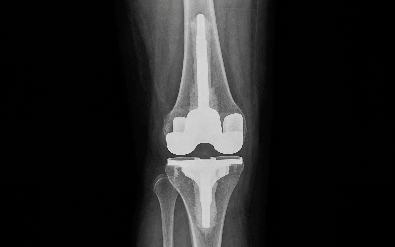
Medial parapatellar arthrotomy through the previous incision, with extensile exposure options when needed (quadriceps snip, V-Y turndown, or tibial tubercle osteotomy for the stiff knee) · Adult reconstruction
- Failed primary TKR requiring femoral component revision for aseptic loosening (AORI Type 2-3), polyethylene wear with femoral osteolysis, component malposition, instability, or infection (staged revision).
- “Know the AORI femur classification — tested in vivas with radiographs. Type 1 intact metaphysis (standard components), Type 2 damaged metaphysis (augments), Type 3 severe deficiency (large augments, sleeves, allograft, custom). Femoral Type 3 is rare compared with tibial Type 3.
- “Understand the constraint ladder thoroughly: PS to CCK to RHK to fixed hinge, with a specific indication for each level. CCK for MCL insufficiency or varus/valgus laxity greater than 10mm. RHK for severe instability or unbalanceable gaps. Each step up needs longer stems to transfer load from metaphysis to diaphysis.
- “Femoral component rotation is a critical exam topic. Three methods: transepicondylar axis (gold standard, hard to identify), 3 degrees external to the posterior condylar axis (easiest, variable in valgus), and Whiteside line (AP axis) perpendicular to it (most reliable in revision with bone loss). Internal rotation causes patellar maltracking and medial polyethylene wear.
- “Gap balancing in revision: the extension gap is set by the tibial cut and distal femoral resection; the flexion gap by the posterior femoral cuts and posterior condylar offset. Gaps should be equal within 2mm. Tight extension: recut the tibia or downsize the femur. Tight flexion: add a posterior augment or increase the tibial slope. If you cannot balance the gaps, use constraint (CCK).
When & Why
Indications for femoral component revision - Aseptic loosening with AORI Type 2-3 bone loss
- Polyethylene wear with femoral osteolysis
- Component malposition (rotation, flexion, sizing)
- Instability not correctable with a polyethylene exchange alone
- Periprosthetic fracture with a loose component
- Staged revision for periprosthetic joint infection Pre-operative planning - Review long-leg alignment films for the mechanical axis
- CT scan to assess component rotation and bone loss
- Measure the femoral canal diameter for stem sizing
- Assign the AORI classification from the radiographs (see Background & Evidence)
- Confirm the implants are available: augments, stems, and a range of constraint options
- Exclude infection — CRP, ESR, and joint aspiration with synovial WCC and differential (occult infection in 7-10 percent of presumed aseptic revisions) Setup and positioning Supine on a radiolucent table with a leg holder or lateral post, upper-thigh tourniquet. Regional or general anaesthesia with DVT prophylaxis initiated pre-operatively. Decide the incision, the exposure, and the extensile options before draping. Choosing the constraint level — the key planning decision Select the least constraint that achieves a stable knee; each step up the ladder requires longer stems to transfer load from the compromised metaphysis to the diaphysis.
- Indication
- PCL deficient, collaterals intact, balanced gaps
- Stem requirement
- Short stem 50-75mm
- Indication
- MCL insufficiency, varus/valgus laxity greater than 10mm, moderate gap mismatch
- Stem requirement
- Stems required 75-100mm for load transfer
- Indication
- Severe instability, collateral deficiency, gaps that cannot be balanced
- Stem requirement
- Long stems mandatory 100-150mm
- Indication
- Salvage only, an alternative to arthrodesis
- Stem requirement
- Long stems mandatory
The Operation
The goal is to remove the failed femoral component with minimal bone loss, reconstruct the femoral defects, restore the joint line and femoral rotation, balance the flexion and extension gaps, and implant a stable, well-fixed revision construct. The workhorse exposure is the medial parapatellar arthrotomy through the previous incision, with extensile options (quadriceps snip, V-Y turndown, tibial tubercle osteotomy) reserved for the stiff knee. Exposure and component removal are the heart of the operation and are laid out step by step below.
Operative sequence
- Identify and use the previous scar (the most lateral of multiple scars).
- Develop full-thickness medial and lateral skin flaps.
- Perform a medial parapatellar arthrotomy extending 5-6cm proximal to the superior pole of the patella through the quadriceps tendon.
- Excise hypertrophic synovium, scar tissue and adhesions.
- Attempt patellar eversion with the knee flexed.
In a stiff knee with less than 70 degrees of flexion, release the lateral gutter before attempting eversion and score adhesions in the suprapatellar pouch. If the patella will not evert safely, a quadriceps snip (45 degrees from the lateral border of the VMO, proximally) adds 10-15 degrees of flexion; a V-Y turndown is for severe stiffness (risk of avulsion); a tibial tubercle osteotomy is the last resort for an ankylosed knee.
- Patellar tendon avulsion during forced eversion in a stiff knee
- Patella fracture through thin or osteopenic bone
- Quadriceps tendon rupture with aggressive manipulation
- MCL damage during medial dissection
- Remove the polyethylene insert first to gain access to the femoral component.
- Flex the knee to 90 degrees and use curved osteotomes or dedicated extraction instruments to disengage the locking mechanism.
- Deliver the insert anteriorly.
- Inspect for the wear pattern (anterior, posterior, medial, lateral), backside wear from micromotion, and delamination.
Document the wear pattern systematically — asymmetric wear suggests instability or malalignment; anterior wear in a PS design suggests cam-post impingement or excessive posterior slope; medial wear suggests varus malalignment or MCL insufficiency. Send explanted components for analysis and photograph them for medicolegal documentation.
- Damaging femoral component surfaces if planning a selective revision
- Losing locking-mechanism fragments into the joint
- Scratching well-fixed components during extraction
- Use thin flexible osteotomes to gently test femoral component stability around the periphery (distal, anterior and posterior chamfers).
- Assess for micromotion, tilting, or gross loosening.
- Compare with pre-operative radiographs showing radiolucent lines, osteolysis or subsidence.
- If the component is well-fixed and well-positioned, consider retaining it and revising only the tibial side.
Indications: malalignment (greater than 3 degrees varus/valgus or greater than 10 degrees rotation), flexion-extension mismatch, osteolysis needing access, polyethylene wear into the component, need for higher constraint, or instability not correctable by tibial revision alone. If retaining it, clean the surfaces thoroughly and check for damage or metallosis.
- Creating iatrogenic bone defects while assessing a stable component
- Propagating occult fracture lines during manipulation
- Missing subtle loosening on clinical exam
- Use thin flexible osteotomes to disrupt the cement-bone or bone-implant interface.
- Work systematically: anterior flange first, then the medial and lateral condyles, then the posterior condyles.
- Use Gigli saws for well-fixed cementless components.
- Apply a slap hammer to the extraction device attached to the femoral component.
- Remove the component in flexion with controlled force.
This is the most challenging step; a well-fixed cemented component may take 30-45 minutes to remove safely. For an ingrown cementless component an oscillating saw or a Gigli saw passed under the component is safer than osteotomes (less bone loss). Create small windows in the anterior flange if needed to access cement. Preserve bone stock — patience is critical. Consider ultrasonic cement removal for a well-fixed cement mantle.
- Supracondylar or condylar fracture (the most common intraoperative complication)
- Posterior cortex perforation with vascular injury
- MCL or LCL avulsion from the epicondyle during extraction
- Excessive bone loss from aggressive osteotome use
- Fracture propagation in osteopenic or osteolytic bone
- Remove all cement, fibrous membrane and debris down to viable bleeding bone.
- Use narrow osteotomes, curettes, rongeurs, a high-speed burr and ultrasonic cement-removal devices.
- Work distal to proximal, anterior to posterior.
- Preserve the cortical shell and condylar bone stock.
- Clear cement from the intercondylar notch and posterior condyles.
Complete cement removal is essential to assess the true bone defects and achieve fixation of the revision component. Use a headlight and magnification; pulsatile lavage after gross cement removal. A cement restrictor deep in the femoral canal may need a flexible drill or osteotome. Send 5-6 tissue samples for culture and histology even in presumed aseptic cases (occult infection in 7-10 percent).
- Posterior cortex perforation during posterior cement removal
- Condylar fracture through weakened or osteolytic bone
- Thermal injury from the high-speed burr (continuous irrigation essential)
- Converting contained defects into uncontained ones by aggressive curettage
- Assess the femoral bone defects systematically with the AORI classification once all cement is removed (Type 1 intact metaphysis; Type 2 damaged metaphysis; Type 3 severe deficiency — see Background & Evidence).
- Map the defects on the distal and posterior condyles, measuring depth and extent.
- Categorize each defect as cavitary (contained) or segmental (uncontained).
Distal uncontained defects arise from loosening or osteolysis; posterior notching from a malpositioned component or osteolysis. Uncontained defects greater than 5mm need metal augments (screw-fixed where possible); contained defects less than 5mm can use cement or impaction grafting. Check the posterior condyles carefully — they are often underestimated.
- Underestimating defect depth, leading to component undersizing
- Missing posterior condyle defects (difficult to visualise)
- Misclassifying an uncontained defect as contained
- For a stemmed revision component, prepare the femoral canal with flexible reamers starting 1-2mm larger than the canal diameter.
- Ream in 1mm increments to cortical chatter.
- For press-fit stems ream to the exact stem diameter; for cemented stems over-ream 2mm.
- Aim for 4-6cm of diaphyseal contact (press-fit) or 8-10cm (cemented).
- Check alignment with an intramedullary rod.
Short stems (50-75mm) for AORI 1-2 give rotational control and reduce stress shielding; long stems (100-150mm) for AORI 3 or a deficient metaphysis give fixation and load transfer. Press-fit is preferred (better long-term fixation). Offset stems are available for canal deformity or malunion. Start small — you can always ream larger but cannot make the canal smaller. Fluoroscopy helps confirm alignment.
- Anterior cortical perforation (the femur has an anterior bow in the sagittal plane)
- Spiral fracture propagation in osteopenic bone
- Creating a false passage in a deformed or sclerotic canal
- Weakening the supracondylar region (fracture risk)
- Use an intramedullary alignment rod (via the stem trial) or an extramedullary guide.
- Set the resection perpendicular to the mechanical axis in the coronal plane (5-7 degrees valgus to the anatomic axis), at 3 degrees external rotation and 0 degrees flexion in the sagittal plane.
- Resect the minimum bone needed for a flat surface — typically 2-4mm beyond the primary resection.
- Use a distal femoral cutting guide.
Perpendicular to the mechanical axis (coronal), neutral flexion-extension (sagittal), minimal bone resection. Use the intact condyle as the reference (usually lateral in varus knees, medial in valgus knees). With massive asymmetric bone loss, use an asymmetric resection or augments. Avoid notching the anterior cortex (supracondylar fracture risk). The resection level sets the extension gap — balance it against the flexion gap later.
- Excessive resection compromising metaphyseal bone stock
- Creating varus or valgus malalignment
- Anterior cortex notching (supracondylar fracture risk)
- Resecting into an uncontained defect and losing peripheral support
- Set femoral component rotation using the posterior condylar axis (3 degrees external rotation), the transepicondylar axis (gold standard), or Whiteside line (the AP axis) — most reliable in revision with posterior bone loss (see Background & Evidence for the reference axes).
- Make the anterior, posterior and chamfer cuts with the appropriate cutting guides.
- Size the femoral component to avoid anterior notching and to optimize posterior condylar offset.
- Remove posterior osteophytes.
Internal rotation causes patellar maltracking and medial polyethylene wear; external rotation causes lateral polyethylene wear and PCL tightness. Use the transepicondylar axis if the landmarks are visible, otherwise 3 degrees external to the posterior condylar axis; in revision with bone loss Whiteside line is most reliable. Avoid oversizing (notching, overstuffing) and undersizing (instability, poor coverage). Restoring posterior condylar offset maintains the flexion gap.
- Anterior cortex notching (a stress riser for supracondylar fracture)
- Malrotation causing patellar maltracking and instability
- Asymmetric posterior resection creating a flexion-gap imbalance
- Damage to collateral ligament origins during the chamfer cuts
- Based on the AORI classification, reconstruct defects with metal augments.
- Distal femoral defects: use distal augment blocks (5-15mm) secured with screws where possible, fully supported peripherally by host bone.
- Posterior condyle defects: use posterior augment wedges.
- Size augments to fill the defect completely.
- Trial to confirm stability and joint-line restoration.
Metal augments are modular blocks or wedges that fill specific defects; they must be fully supported by host bone at the periphery (no cantilever), with screw fixation into host bone preferred. They are cemented to both bone and prosthesis at final implantation. Modular systems offer built-in augmentation. Avoid structural allograft for femoral defects where possible (high resorption rate).
- Undersizing augments, leading to subsidence or collapse
- Screw penetration into neurovascular structures
- Incomplete seating creating interface gaps
- An augment not fully supported, leading to failure
- Insert the femoral trial with stem (if used) and augments, the tibial trial, and select the polyethylene thickness.
- Reduce the knee and assess through a full range of motion.
- Check the extension gap (0 degrees) versus the flexion gap (90 degrees) — they should be equal within 2mm.
- Assess medial-lateral stability with varus/valgus stress (maximum 5mm opening).
- Check patellar tracking. Target 0-110 degrees of motion.
The extension gap is set by the tibial and distal femoral cuts; the flexion gap by the posterior femoral cuts and condylar offset. Tight extension: recut the tibia or downsize the femur. Tight flexion: add a posterior augment or increase the tibial slope. If the gaps differ by greater than 2mm, revise the cuts or use constraint (CCK). Assess the joint line — it should be 2-3cm distal to the medial epicondyle.
- Overstuffing the extension gap (limited extension, anterior knee pain)
- Overstuffing the flexion gap (limited flexion, PCL impingement if retained)
- Accepting instability (early failure and dislocation)
- Joint-line elevation greater than 8mm (extensor-mechanism dysfunction, instability)
- From the trial assessment, select the constraint level (see When & Why for the full ladder).
- PS if the PCL is deficient but the collaterals are intact with good bone stock and balanced gaps.
- CCK if the MCL or LCL is incompetent, or there is a flexion-extension gap imbalance with moderate instability.
- RHK for severe instability, collateral deficiency, or unbalanceable gaps.
- Fixed hinge for salvage only.
CCK has a taller post and wider box than PS and gives 3-7 degrees of varus/valgus constraint; RHK allows axial rotation but prevents varus/valgus tilting. Higher constraint needs longer stems for load transfer. Use the least constraint that achieves stability.
- Under-constraining (early instability and failure)
- Over-constraining (interface stress, loosening, reduced range of motion)
- Using an RHK without a stem (high failure rate from metaphyseal stress)
- Missing an indication for constraint (recurrent instability)
- Prepare the bone surfaces with pulsatile lavage (6-9L) and dry thoroughly with suction and swabs.
- Mix high-viscosity antibiotic-loaded bone cement (1g vancomycin plus 3.6g tobramycin per 40g cement).
- Apply cement to the bone and implant with a cement gun.
- For hybrid fixation, press-fit the stem then cement the metaphyseal component; for fully cemented fixation, cement the stem and component as one construct.
Clean dry bone, pressurization, cement-gun application, component insertion during the dough phase, and removal of excess before polymerization. Hybrid fixation (press-fit stem, cemented metaphyseal component) combines biological fixation with immediate stability. Use antibiotic cement in all revisions (even aseptic) to reduce infection risk. Apply cement to the augments and the bone-augment interface.
- Cement extrusion posteriorly into neurovascular structures
- Air entrapment creating weak spots in the cement mantle
- Premature polymerization before the component is seated
- Inadequate cement penetration into cancellous bone
- Insert the stem first if using a hybrid technique.
- Apply cement to the femoral bone surfaces and the undersurface of the component.
- Insert the femoral component with firm steady pressure, ensuring correct rotation (aligned with the transepicondylar axis), AP position (no anterior notching, adequate posterior coverage) and seating.
- Pressurize the cement, remove excess, and hold until polymerized.
- Select and insert the final polyethylene after the cement has set.
Rotation 3 degrees external to the posterior condylar axis or parallel to the transepicondylar axis; flexion neutral (0 degrees or slight flexion acceptable, avoid extension); sizing to maximize coverage without notching and to restore condylar offset. Polyethylene thickness: minimum 10mm for PS, 12-15mm for CCK (taller post), 15-20mm for RHK. Lock the insert securely and confirm it cannot be removed by traction.
- Malrotation (internal rotation causes patellar problems and wear)
- Anterior notching (supracondylar fracture risk)
- Incomplete seating leaving gaps under the component
- Polyethylene insert not fully locked (catastrophic failure)
- Final check: range of motion 0-110 degrees minimum, stable through the arc, no lift-off with varus/valgus stress, patellar tracking central.
- Lavage to remove all cement and debris.
- Achieve hemostasis; insert a drain if there is significant dead space.
- Close the capsule and extensor mechanism with strong interrupted sutures (No.2 or No.5) in flexion.
- Close in layers and apply a compression dressing with the knee in extension.
Tranexamic acid 1g IV at induction and at closure; ice and elevation; TED stockings and chemical thromboprophylaxis (LMWH or DOAC) for 35 days; IV antibiotics 24h; early mobilization on day 1 with physiotherapy, weight-bearing as tolerated with a frame; CPM if available (0-60 degrees advancing to 0-90 degrees by week 2); remove the drain at 24-48h; radiographs AP/lateral/skyline to check alignment, component position and the absence of fracture; discharge day 4-7 to rehabilitation; follow up at 6 weeks, 3 months, 1 year, then annually.
- Wound dehiscence (higher risk with extensile exposures)
- Deep infection (5-10 percent risk in revision surgery)
- Supracondylar fracture during early mobilization
- Extensor-mechanism failure or rupture
- Persistent instability or stiffness requiring re-revision
- Neurovascular complications (DVT, PE, nerve palsy)
Aftercare & Complications
Rehabilitation follows the post-operative protocol in Step 15: early mobilization, weight-bearing as tolerated, CPM, and 35 days of thromboprophylaxis. Protected rehabilitation is guided by the fixation achieved and by any intra-operative fracture. Most patients are discharged to rehabilitation at 4-7 days and followed up at 6 weeks, 3 months, 1 year, then annually. Complications
- Recognition
- Intra-op: visible fracture during extraction. Post-op: sudden pain, deformity, inability to weight-bear
- Prevention
- Gentle component removal, preserve bone stock, identify osteopenic bone pre-op
- Management
- Intra-op stable: continue with a long stem bypassing 2 diameters. Unstable: ORIF plus stem. Post-op: the Lewis-Rorabeck classification guides treatment
- Recognition
- Acute: fever, wound drainage, erythema. Chronic: persistent pain, loosening. Labs: raised CRP and ESR, positive synovial WCC
- Prevention
- Antibiotic prophylaxis, laminar flow, antibiotic cement, minimal operative time, send intra-op cultures
- Management
- Acute (less than 3 weeks): DAIR. Chronic: two-stage revision with an antibiotic spacer for 6-12 weeks. Suppressive antibiotics if unfit for surgery
- Recognition
- Progressive pain, radiolucent lines greater than 2mm, subsidence on serial radiographs
- Prevention
- Press-fit stems, adequate constraint, proper alignment, avoid stress shielding
- Management
- Re-revision with AORI assessment, longer stems for load transfer, metaphyseal sleeves if bone loss progresses
- Recognition
- Early: polyethylene dissociation or dislocation. Late: giving way, recurrent effusions, abnormal laxity on exam
- Prevention
- Appropriate constraint selection, gap balancing, adequate stem fixation
- Management
- Higher constraint level (CCK to RHK), address alignment, revision with stems if metaphyseal failure
- Recognition
- Limited range of motion despite rehabilitation, pain at end range, palpable scar tissue
- Prevention
- Proper gap balancing, early mobilization, CPM, avoid joint-line elevation greater than 8mm
- Management
- MUA within 12 weeks. Arthroscopic or open arthrolysis if greater than 12 weeks. Revision if component malposition is identified
- Recognition
- Patellar tendon rupture: loss of active extension, palpable gap. Quadriceps rupture: similar presentation
- Prevention
- Avoid forced eversion, protect the mechanism during closure, use a TTO instead of aggressive exposure
- Management
- Acute: primary repair with augmentation. Chronic: allograft reconstruction. Protect for 6-12 weeks with a brace
- Recognition
- Peroneal palsy: foot-drop, sensory changes lateral leg. Vascular: pulseless limb, expanding haematoma, compartment signs
- Prevention
- Careful lateral release, avoid lengthening greater than 3cm, identify vessels with posterior work
- Management
- Peroneal palsy: AFO, most recover. Vascular: emergency vascular repair. Compartment syndrome: fasciotomy of all 4 compartments
Viva & Exam Focus
FEMURFEMUR — AORI femoral bone-loss classification
GAPSGAPS — gap-balancing algorithm in revision TKA
Posterior to the distal femur in the popliteal fossa, separated from the posterior capsule by a fat pad. At risk during posterior cement removal and posterior capsular release.
Anterior to the femur in Hunter canal, becoming the popliteal vessels at the adductor hiatus. At risk with anterior cortical perforation during reaming.
Wraps around the fibular neck laterally, 1-2cm distal to the fibular head. At risk with valgus correction, lateral release, or leg lengthening greater than 3cm.
Superficial and deep fibres from the medial epicondyle to the proximal tibia. At risk during medial dissection, epicondylar osteotomy, or aggressive cement removal.
From the lateral epicondyle to the fibular head. At risk during lateral releases, lateral condyle augment placement, or extension of a lateral approach.
Clinical Decision Scenarios
Practise clinical reasoning and management decisions out loud
“A 72-year-old woman presents 12 years after primary TKA with progressive pain and instability. Radiographs show a loose femoral component with AORI Type 2B bone loss (both condyles affected). How do you approach this revision?”
“During femoral component removal you create a supracondylar fracture that extends into the diaphysis. The fracture is displaced with a separate butterfly fragment. How do you manage this intraoperatively?”
“You are revising a TKA in a patient with rheumatoid arthritis. Intra-operatively, after balancing the gaps with trials, you find greater than 15mm of varus-valgus laxity in extension despite using a PS design. What is your approach?”
Key indications
- Aseptic loosening with AORI Type 2-3 bone loss
- Polyethylene wear with femoral osteolysis needing access
- Component malposition (rotation greater than 10 degrees, flexion, sizing errors)
- Instability not correctable with a polyethylene exchange alone
AORI femoral classification
- Type 1: intact metaphysis — standard revision stems acceptable
- Type 2A: single condyle damaged — augments to that condyle
- Type 2B: both condyles damaged — multiple augments needed
- Type 3: severe deficiency — sleeves, cones, or allograft
Constraint ladder
- PS: PCL deficient, collaterals intact, balanced gaps
- CCK: MCL insufficiency, varus/valgus laxity greater than 10mm
- RHK: severe instability, unbalanceable gaps, collateral deficiency
- Higher constraint requires longer stems for load transfer
Femoral rotation methods
- Transepicondylar axis: gold standard, 0 degrees to this line
- Posterior condylar axis: 3 degrees external rotation (easiest)
- Whiteside line (AP axis): perpendicular — most reliable in revision
- Internal rotation causes patellar maltracking and medial wear
Gap balancing
- Extension gap: set by the tibial and distal femoral cuts
- Flexion gap: set by the posterior cuts and condylar offset
- Gaps should be equal within 2mm
- Tight extension: recut tibia or downsize femur; tight flexion: posterior augment or more tibial slope
Danger zones
- Popliteal vessels — posterior to the distal femur
- Femoral vessels — risk with anterior cortical perforation
- Common peroneal nerve — at the fibular neck, risk with valgus correction
- MCL and LCL — during dissection and component removal
Critical numbers
- Joint line 2-3cm distal to the medial epicondyle
- Joint-line elevation greater than 8mm causes extensor dysfunction
- Stem must bypass a fracture by 2 cortical diameters (8-10cm)
- Occult infection in 7-10 percent of presumed aseptic revisions
Background & Evidence
Epidemiology and modes of failure. Revision TKA is performed far less often than primary TKA but carries a higher re-operation rate and lower survivorship. Across the major registries — the National Joint Registry (NJR, England/Wales/NI), the American Joint Replacement Registry (AJRR), the Australian Orthopaedic Association National Joint Replacement Registry (AOANJRR), the Swedish Knee Arthroplasty Register and the New Zealand Joint Registry — the dominant modes of failure after revision are infection, instability and aseptic loosening. This is why meticulous gap balancing, appropriate constraint and sound metaphyseal fixation are the principles the whole operation is built around.
- Metaphysis
- Intact metaphyseal bone
- Reconstruction
- Standard revision stems (PS or CCK)
- Metaphysis
- Damaged single condyle
- Reconstruction
- Augments to the affected condyle (5-15mm)
- Metaphysis
- Both condyles damaged
- Reconstruction
- Multiple augments plus a stemmed component
- Metaphysis
- Severe metaphyseal deficiency
- Reconstruction
- Large augments, metaphyseal sleeves or cones, allograft, or a custom implant
- Setting
- 0 degrees (gold standard)
- Reliability in revision
- Best landmark but hard to identify with bone loss
- Setting
- 3 degrees external rotation
- Reliability in revision
- Easiest but unreliable in valgus or deficient condyles
- Setting
- Perpendicular to this line
- Reliability in revision
- Most reliable in revision with posterior bone loss
- Setting
- Match the femur to a balanced tibial cut
- Reliability in revision
- Useful once the tibia is balanced
Guidelines, registries and global practice. Registry survivorship of revision TKA (implant survival to re-revision): national joint registries consistently report higher re-revision and lower survivorship for revision TKA than for primary TKA, identifying infection, instability and aseptic loosening as the dominant modes of failure. There is global consensus across AAOS (US), BOA/BASK (UK) and EFORT/European practice on the constraint-ladder principle — use the least constraint that achieves stability, and lengthen stems as constraint and bone loss increase to transfer load to the diaphysis. Hybrid fixation (cemented metaphysis with a press-fit diaphyseal stem) and fully cemented stems are both accepted; the choice is guided by bone quality and canal geometry rather than a single national standard. Diagnosis of periprosthetic joint infection before revision follows internationally harmonised criteria (Musculoskeletal Infection Society / International Consensus Meeting and EBJIS definitions), combining serum CRP/ESR, synovial white-cell count and differential, culture and histology; routine intra-operative cultures (5-6 samples) are recommended even in presumed aseptic revision. AAOS and NICE both endorse risk-stratified chemical and mechanical VTE prophylaxis after major knee arthroplasty, and routine peri-operative tranexamic acid to reduce blood loss and transfusion.
References
Further reading 1. Whiteside LA, Arima J. The anteroposterior axis for femoral rotational alignment in valgus total knee arthroplasty. Clin Orthop Relat Res. 1995;(321):168-172. PMID: 7497664. 2. Fehring TK, Odum S, Griffin WL, Mason JB, Nadaud M. Early failures in total knee arthroplasty. Clin Orthop Relat Res. 2001;(392):315-318. 3. Morgan HD, Battista V, Leopold SS. Constraint in primary total knee arthroplasty. J Am Acad Orthop Surg. 2005;13(8):515-524. 4. Parvizi J, Zmistowski B, Berbari EF, et al. New definition for periprosthetic joint infection: from the Workgroup of the Musculoskeletal Infection Society. Clin Orthop Relat Res. 2011;469(11):2992-2994. 5. Rorabeck CH, Taylor JW. Classification of periprosthetic fractures complicating total knee arthroplasty. Orthop Clin North Am. 1999;30(2):209-214.
Bone loss with revision TKA: defect classification and alternatives for reconstruction (AORI)
- Defines the AORI defect classification (Type 1 intact metaphysis, Type 2 metaphyseal damage of one [2A] or both [2B] condyles, Type 3 deficient metaphysis) that remains the global standard for grading femoral and tibial bone loss
- Reconstruction principle: select the least constraint compatible with stability to reduce stress at the compromised fixation interface, and escalate stem length with increasing bone loss
- Structural allograft incorporates with host bone but does not revascularise; metal augments, cement and morcellised graft are reliable for contained and modest uncontained defects when paired with stems
Malrotation causing patellofemoral complications after total knee arthroplasty
- CT comparison of 30 knees with patellofemoral complications versus 20 controls: combined femoral plus tibial internal rotation correlated directly with the severity of the patellofemoral problem
- Small internal rotation (1-4 degrees) produced tilt and lateral tracking; moderate (3-8 degrees) produced subluxation; large (7-17 degrees) produced dislocation or late patellar failure, whereas well-functioning knees sat in combined external rotation
- The transepicondylar axis and tibial tubercle are reproducible CT landmarks for quantifying and planning correction of rotational malalignment
Strategies for severe bone loss in revision knee arthroplasty: systematic review and meta-analysis
- 77 studies and 4,391 knees comparing porous metaphyseal cones, porous-coated sleeves and bone grafting for moderate-to-large metaphyseal defects
- No significant difference in 10-year failure between porous implants and grafting (odds ratio 0.91, 95% CI 0.70-1.19); porous cones and sleeves show good mid-term survivorship for metaphyseal fixation
- Tantalum cones produced a larger improvement in patient-reported outcome measures than sleeves, while grafts remained a reasonable option in younger patients with less constrained implants
Implant survivorship, function and complications of rotating-hinge knee implants: systematic review
- 19 studies, 568 primary and 413 revision rotating-hinge TKAs: median survival in the revision cohort was 79.6% at 1 year and 65.1% at 10 years (versus 87% at 10 years in the primary hinge cohort)
- Mean post-operative flexion was 103 degrees in revision hinges, with substantial gains in Knee Society and Function Scores from baseline
- Evidence quality was limited, with potential for high early revision but large functional benefit where the construct succeeds
Condylar constrained versus rotating hinge implants in revision knee arthroplasty: meta-analysis
- 40 studies and 4,555 knees: 5-year survival did not differ between rotating hinge (91.6%) and condylar constrained (89.8%) implants when both designs were technically suitable
- Knee Society clinical and functional scores and post-operative range of motion (around 104-105 degrees) were comparable between CCK and RHK
- Choice between CCK and RHK should be driven by the residual soft-tissue constraint and bone stock rather than an expectation of better survivorship from one design