Posterior approach · SINS for instability · Tokuhashi for prognosis · Separation surgery plus SBRT · MDT decision-making
- Metastatic spinal cord compression (MSCC) is an oncological emergency — definitive treatment ideally within 24 hours of diagnosis (NICE). Operate before complete or prolonged paraplegia (over 48 hours), as recovery potential falls sharply after this.
- A SINS score of 7 or higher indicates potential instability requiring surgical stabilization.
- The Tokuhashi score guides surgical extent: 0-8 palliative, 9-11 excisional, 12-15 potentially curative.
- Separation surgery creates a 2-3 mm gap between tumour and cord to allow stereotactic radiosurgery (SBRT).
- Patchell trial (Lancet 2005): surgery plus radiotherapy is superior to radiotherapy alone for MSCC in selected patients.
When & Why
Indication. Surgery is offered for metastatic spinal cord compression (MSCC) with neurological deficit, progressive deficit despite radiotherapy, mechanical instability, or a radioresistant tumour needing separation surgery plus SBRT; for selected primary spine tumours; and for neoplastic mechanical instability regardless of radiosensitivity. Every decision is made at a multidisciplinary meeting informed by the SINS (instability) and Tokuhashi (prognosis) scores.
Neurological deficit with imaging-confirmed cord compression; progressive deficit despite radiotherapy; radioresistant tumour (renal, melanoma, sarcoma) needing separation surgery plus SBRT; unknown primary needing tissue.
En bloc resection for an isolated primary with curative intent (Tokuhashi 12-15); decompression and stabilization for locally aggressive benign tumours.
SINS 7-12 potentially unstable (consider stabilization); SINS 13-18 unstable (stabilization indicated), independent of tumour radiosensitivity.
Contraindications.
- Absolute: life expectancy under 3 months (Tokuhashi 0-5) — consider palliative radiotherapy only; disseminated disease with no surgical benefit expected; medically unfit for major surgery (ASA 4-5); patient or family decline surgery.
- Relative: complete paraplegia over 48 hours (poor recovery potential); radiosensitive tumour (myeloma, lymphoma) where radiotherapy may be first-line; extensive epidural disease at multiple levels; active systemic infection; coagulopathy (correct before surgery). Pre-operative workup.
- MRI whole spine: skip metastases are present in 10-30 percent of patients; assess cord compression.
- CT spine: bony anatomy, pedicle-screw planning, vertebral body destruction.
- CT chest, abdomen and pelvis: staging, identify visceral metastases.
- PET-CT: if unknown primary or to assess disease burden. MDT decision-making and the NOMS framework. Every case is discussed at a multidisciplinary meeting (oncology, radiation oncology, spine surgery, palliative care) to establish the primary tumour type and prognosis, confirm the SINS and Tokuhashi scores, and plan adjuvant radiotherapy timing. The NOMS framework (Memorial Sloan Kettering) integrates the decision: Neurologic (Bilsky epidural cord-compression grade and myelopathy), Oncologic (radiosensitivity and expected response to systemic or radiotherapy), Mechanical (SINS stability), and Systemic (disease burden and fitness for surgery, informed by Tokuhashi). Mechanical instability is stabilised regardless of radiosensitivity. Consent specifically for the possibility of neurological deterioration, hardware failure or wound breakdown (often compounded by adjuvant radiotherapy), CSF leak, significant blood loss, and the palliative intent of metastatic surgery.
The Operation
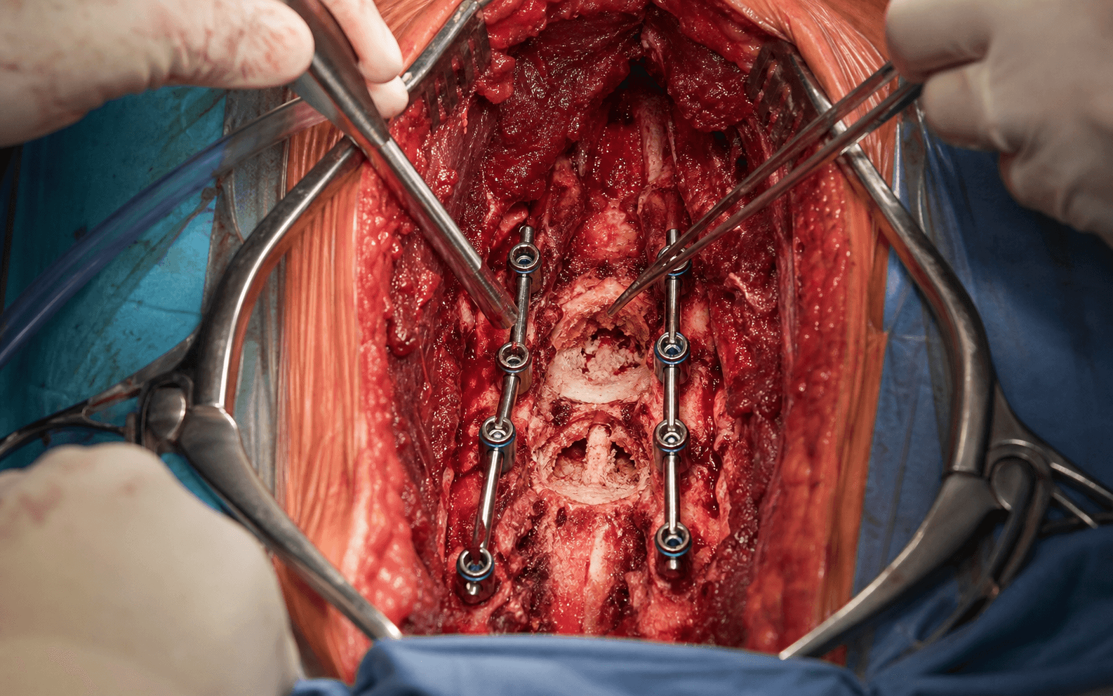
The goal is to decompress the compressed cord or cauda, debulk epidural tumour to create a margin for stereotactic radiosurgery, and stabilise the unstable segment with pedicle-screw instrumentation. The exposure is a standard posterior midline approach, laid out in full below as the first operative steps. Setup and equipment. Prone on a Jackson or Wilson radiolucent frame, arms tucked or abducted 90 degrees, head neutral with the eyes protected. Establish baseline MEPs and SSEPs before positioning (alert if amplitude loss is over 50 percent or latency increase over 10 percent). Set up a cell saver — metastatic tumour surgery can be bloody (a relative contraindication in haematological malignancy). Use fluoroscopy or navigation to confirm levels. Implants: polyaxial pedicle screws (5.5-7.5 mm), titanium or cobalt-chrome rods (5.5 or 6.0 mm), cross-links, PMMA for cement augmentation, and expandable cages for anterior column reconstruction.
Operative sequence
- Prone on a Jackson frame, arms tucked or abducted; head neutral, eyes protected.
- Lateral fluoroscopy to confirm levels — COUNT FROM THE SACRUM superiorly.
- Mark a midline incision spanning 2-3 levels above and below the tumour.
- Establish the neuromonitoring baseline (MEPs and SSEPs).
- Midline incision over the spinous processes.
- Subperiosteal dissection out to the transverse processes bilaterally, exposing 2-3 levels above and below the planned decompression.
- Identify the segmental vessels along the vertebral bodies if lateral extension is needed; the dura is not yet in the field.
- This posterior corridor gives access to the posterior elements, the epidural space, the pedicle-screw fixation points, and transpedicular access to the vertebral body.
- Place pedicle screws BEFORE laminectomy — bone landmarks are preserved and easier to identify, and the construct gives immediate stability if neurological deterioration occurs during decompression.
- Entry point: junction of the transverse process and the lateral border of the superior articular facet.
- Freehand technique or navigation-guided (navigation is especially useful in pathological, distorted bone).
- In osteopenic tumour-bearing bone, use fenestrated screws with cement augmentation.
- Thin the lamina bilaterally with a high-speed burr down to "eggshell" thickness — safer for a compromised dura and gives tactile feedback before dural contact.
- Complete the laminectomy with Kerrison rongeurs.
- Preserve the facet joints where possible (unless tumour-involved).
- Identify and protect the dura throughout.
- For posterior-element tumour: excise tumour-involved lamina en bloc where possible, extending laterally into the pedicle if it is involved.
- For epidural compression (separation surgery): identify the plane between tumour and dura and debulk circumferentially.
- The goal is a 2-3 mm gap between residual tumour and the thecal sac to allow high-dose SBRT — NOT complete resection (palliative intent).
- If the anterior column is involved, curette through the pedicle (transpedicular approach) to remove tumour from the vertebral-body cavity.
- Preserve the anterior and lateral cortical shell if it is intact.
- This avoids the morbidity of a separate anterior approach for contained vertebral-body lesions.
- If the vertebral body has been debulked or is at risk of collapse, inject PMMA under fluoroscopy through a transpedicular cannula.
- Fill the cavity to restore mechanical strength.
- Watch for extravasation into the epidural, foraminal or venous spaces.
- For significant vertebral-body destruction or corpectomy, place an expandable cage through a posterolateral approach, or plan a separate anterior approach with a strut graft or cage.
- This provides load-sharing to protect the posterior instrumentation.
- Pre-contour the rods to match sagittal alignment.
- Insert the rods and secure with set screws; apply compression or distraction as needed.
- Add cross-links for rotational stability (at least one per construct — mandatory in tumour surgery where bone quality is compromised), especially for constructs of 3 or more levels.
- Inspect the decompression site for residual compression.
- Check neuromonitoring — MEPs and SSEPs should be stable or improved.
- Meticulous haemostasis (the tumour bed bleeds significantly); use topical agents such as Floseal or Surgicel and irrigate copiously.
- Place subfascial Jackson-Pratt drains to prevent haematoma, close the deep fascia with a heavy absorbable suture (0-Vicryl), then subcutaneous and skin closure, and apply a dry dressing.
The goal of separation surgery is to create space for radiotherapy, not curative resection. Attempting complete resection increases morbidity without survival benefit in metastatic disease. Leave residual tumour for SBRT.
For osteopenic bone, use fenestrated screws and inject PMMA under fluoroscopy. Avoid cement extravasation into the canal or vessels, and allow polymerization before rod insertion.
Level-counting errors are a never event. Count superiorly from the sacrum and verify with lateral fluoroscopy before incision; mark levels with a spinal needle if uncertain.
Placing pedicle screws before decompression preserves the bony landmarks and gives immediate stability should neurological deterioration occur during the laminectomy.
Cross-links are mandatory for rotational stability in tumour surgery where bone quality is compromised. Place at least one cross-link per construct.
Aftercare & Complications
Immediate postoperative care. Overnight ICU or HDU observation with neurological checks every 1-2 hours. Start DVT prophylaxis (LMWH) once haemostasis is secure, and mobilize when medically stable. Adjuvant therapy. Plan radiotherapy within 2-3 weeks; SBRT for separation-surgery cases; oncology follow-up for systemic therapy. Follow-up and surveillance. Clinical review at 2 weeks, 6 weeks and 3 months, with an MRI at 3 months to assess tumour response. Repeat imaging for any new symptoms, with ongoing oncology and palliative-care coordination. Complications.
- Incidence
- 5-15%
- Prevention
- Intraoperative neuromonitoring (MEPs/SSEPs); careful tumour debulking; avoid cord retraction; staged procedures if extensive
- Management
- Intraoperative MEP/SSEP loss: stop, raise MAP (mean over 85 mmHg), warm irrigation, remove any compressing material, exclude haematoma or malpositioned implant. High-dose methylprednisolone is NOT indicated for tumour-related compression. Postop: urgent MRI, return to theatre if haematoma or residual compression
- Incidence
- 5-10%
- Prevention
- Cement augmentation in osteopenic bone; adequate construct length (2-3 levels above and below); cross-links for rotational stability
- Management
- Revision with a longer construct, cement augmentation, consider anterior column support
- Incidence
- 10-20%
- Prevention
- Avoid RT within 2-3 weeks of surgery; meticulous closure; optimise nutrition; control diabetes
- Management
- Superficial: antibiotics, local wound care. Deep: return to OR for washout, may need muscle-flap coverage
- Incidence
- 3-8%
- Prevention
- Careful technique near dura; identify dural invasion preoperatively; avoid aggressive resection near infiltrated dura
- Management
- Primary repair if possible (5-0 Prolene), dural substitute patch, fibrin glue. CSF drain if needed. Flat bed rest 48-72 hours
- Incidence
- 2-5%
- Prevention
- Meticulous haemostasis; subfascial drain; correct coagulopathy; avoid early anticoagulation
- Management
- Urgent MRI if neuro decline, immediate return to OR for evacuation, ensure haemostasis
- Incidence
- Variable
- Prevention
- Timely adjuvant RT (within 2-4 weeks); systemic therapy coordination; appropriate patient selection
- Management
- Repeat MDT discussion, consider re-irradiation, systemic therapy modification, palliative care involvement
- Incidence
- 3-8%
- Prevention
- Prophylactic antibiotics; minimize OR time; optimise nutrition; delay RT until wound healing
- Management
- Superficial: oral antibiotics. Deep: washout, cultures, prolonged IV antibiotics; may need hardware removal if late or chronic
- Incidence
- 2-5%
- Prevention
- Inject under fluoroscopy; low-viscosity cement; staged injection; watch for extravasation
- Management
- Epidural cement: urgent removal if neuro compromise. Venous embolism: supportive care, anticoagulation if PE
- Incidence
- 5-15%
- Prevention
- Mechanical prophylaxis intraop; LMWH postop when safe; early mobilization
- Management
- DVT: therapeutic anticoagulation. PE: anticoagulation, ICU if massive, consider IVC filter if recurrent on anticoagulation
- Incidence
- 5-10% long-term
- Prevention
- Minimize construct length while ensuring stability; preserve facet joints; extend if progressive
- Management
- Extend construct if symptomatic, address new instability, coordinate with ongoing oncology care
Viva & Exam Focus
BLT-KPBLT WITH A KOSHER PICKLE
- Location and risk
- Within the spinal canal, often compressed by tumour
- Protection
- Careful tumour debulking; avoid cord retraction; neuromonitoring (MEPs/SSEPs) essential
- Location and risk
- Exit through the neural foramina
- Protection
- Identify before decompression; protect during laminectomy; trace if involved in tumour
- Location and risk
- Along the vertebral body at each level; at risk in lateral decompression or corpectomy
- Protection
- May require ligation for exposure; anticipate bleeding
- Location and risk
- Left side at the thoracolumbar junction; injury causes chylothorax
- Protection
- Beware left-sided thoracolumbar approaches
- Location and risk
- Anterior to the vertebral bodies; catastrophic if injured
- Protection
- Avoid anterior breach; confirm screw position
- Location and risk
- Surrounds the cord and nerve roots; at risk from tumour invasion or aggressive decompression
- Protection
- Repair if breached; identify dural invasion preoperatively
Clinical Decision Scenarios
Practise clinical reasoning and management decisions out loud
“A 62-year-old woman with known breast cancer presents with 48 hours of progressive lower limb weakness. She has 3/5 power in the hip flexors and is unable to walk. MRI shows T10 vertebral body collapse with epidural compression. How do you manage her?”
“Explain the SINS score and how you would use it in clinical practice.”
“What is separation surgery and when would you use it?”
SINS score (0-18)
- 0-6: Stable — RT alone
- 7-12: Potentially unstable — consider surgery
- 13-18: Unstable — surgery required
- Components: site, pain, lesion type, alignment, collapse, posterolateral involvement
Tokuhashi score
- 0-8: Conservative or palliative (survival under 6 months)
- 9-11: Palliative surgery (survival 6-12 months)
- 12-15: Excisional surgery (survival over 12 months)
- Factors: tumour type, performance status, metastatic burden, neurology
Key evidence
- Patchell 2005 (Lancet): surgery plus RT is superior to RT alone for MSCC
- Requires reasonable prognosis and a single level of compression
- Surgery within 48 hours preserves ambulatory function
- Separation surgery plus SBRT for radioresistant tumours
Surgical principles
- Place pedicle screws before laminectomy (landmarks preserved)
- Fixate 2-3 levels above and below the tumour
- Cement augmentation for osteopenic bone
- Cross-links for rotational stability
- Separation surgery: 2-3 mm gap for SBRT, not complete resection
Exam tips
- Know SINS score components and thresholds
- Cite the Patchell trial for MSCC management
- Radioresistant tumours: renal, melanoma, sarcoma (separation surgery)
- Radiosensitive tumours: myeloma, lymphoma (RT first-line)
- Common primaries: breast, lung, thyroid, kidney, prostate (BLT with a Kosher Pickle)
Background & Evidence
Epidemiology and pattern of spread. The spine is the most common site of skeletal metastasis. Most metastatic deposits follow a predictable location pattern: the vertebral body in 70-80 percent of cases (haematogenous spread via Batson's plexus), the posterior elements in 20-30 percent (often extending from the body), and the epidural space, where compression arises from vertebral-body extension or a soft-tissue mass. Batson's venous plexus. A valveless venous network connecting the pelvic and thoracic veins to the vertebral venous plexus. It explains the spine's predilection for metastases. The most common primaries are breast, lung, thyroid, kidney and prostate ("BLT with a Kosher Pickle"). Surgical corridors. A posterior approach gives access to the posterior elements and epidural space, the pedicle-screw fixation points, and transpedicular access to the vertebral body. The separation-surgery corridor creates a 2-3 mm gap between tumour and thecal sac, allowing high-dose SBRT without cord myelopathy — essential for radioresistant tumours such as renal cell carcinoma and melanoma. Spinal cord blood supply. The anterior spinal artery supplies the anterior two-thirds of the cord. The artery of Adamkiewicz is the major radicular contributor, typically entering on the LEFT between T9 and L2; it should be preserved during thoracolumbar tumour surgery.
- Scoring (highest to lowest)
- Junctional (occipitocervical, cervicothoracic, thoracolumbar, lumbosacral) = 3; mobile (C3-C6, L2-L4) = 2; semi-rigid (T3-T10) = 1; rigid (S2-S5) = 0
- Scoring (highest to lowest)
- Mechanical (worse with movement) = 3; occasional / non-mechanical = 1; pain-free = 0
- Scoring (highest to lowest)
- Lytic = 2; mixed = 1; blastic = 0
- Scoring (highest to lowest)
- Subluxation / translation = 4; de novo kyphosis or scoliosis = 2; normal = 0
- Scoring (highest to lowest)
- Over 50% = 3; under 50% = 2; no collapse but over 50% body involved = 1; none = 0
- Scoring (highest to lowest)
- Bilateral = 3; unilateral = 1; none = 0
Decision thresholds. SINS 0-6 stable (radiotherapy alone appropriate); 7-12 potentially unstable (surgical opinion, often stabilization); 13-18 unstable (stabilization indicated). The score flags when a patient with a spinal neoplasm should be referred for surgical consultation — it does not dictate the operation.
- Expected survival
- Under 6 months
- Surgical goal
- Conservative / palliative
- Expected survival
- 6 months or more
- Surgical goal
- Palliative surgery or single-level excision
- Expected survival
- 1 year or more
- Surgical goal
- Excisional surgery (curative intent)
- Key recommendation
- Treat suspected MSCC as an emergency; definitive treatment (surgery or radiotherapy) before further neurological deterioration and ideally within 24 hours of diagnosis in a patient with deficit or instability
- Key recommendation
- MDT-led pathway; surgery for instability, high-grade epidural compression, or radioresistant tumour, followed by SBRT
- Key recommendation
- Use the NOMS framework and SINS to standardise decision-making globally
- Key recommendation
- Early decompressive surgery plus RT for good-prognosis patients with cord compression; dexamethasone for symptomatic oedema
Registry and outcome context. Modern targeted and immunotherapy agents (for example in renal cell carcinoma and melanoma) have lengthened survival, so historical prognostic scores increasingly underestimate survival for some histologies — reassess prognosis at the MDT rather than relying on the score alone. Minimally invasive and percutaneous cement-augmentation or stabilization techniques are increasingly used for patients who are poor surgical candidates, to control mechanical pain without open decompression.
References
Direct decompressive surgical resection in spinal cord compression caused by metastatic cancer: a randomised trial
- Multi-institutional RCT, 101 patients with MSCC randomised to decompressive surgery plus radiotherapy versus radiotherapy alone (both arms received 30 Gy in 10 fractions)
- Significantly more patients walked after surgery plus RT than RT alone: 84% (42/50) versus 57% (29/51), odds ratio 6.2 (95% CI 2.0-19.8), p=0.001
- Surgery patients retained ambulation far longer: median 122 days versus 13 days, p=0.003
- Of those non-ambulatory at entry, 62% regained walking with surgery versus 19% with RT alone, p=0.01; steroid and opioid requirements were also reduced
A novel classification system for spinal instability in neoplastic disease: an evidence-based approach and expert consensus from the Spine Oncology Study Group
- Systematic review plus modified Delphi consensus that created the Spinal Instability Neoplastic Score (SINS)
- Six components: global spinal location, presence and mechanical nature of pain, bone lesion quality (lytic/mixed/blastic), spinal alignment, degree of vertebral body collapse, and posterolateral element involvement
- Total 0-18: 0-6 stable, 7-12 potentially unstable (warrants surgical opinion), 13-18 unstable
- Designed to flag when a patient with spinal neoplasm should be referred for surgical consultation, not to dictate the operation
Spinal instability neoplastic score: an analysis of reliability and validity from the Spine Oncology Study Group
- Validation study with 30 spinal tumour cases scored on two occasions by Spine Oncology Study Group members
- Near-perfect reliability of the total score: interobserver ICC 0.846 and intraobserver ICC 0.886
- Sensitivity 95.7% and specificity 79.5% for detecting potentially unstable or unstable lesions against expert consensus
- Predictive validity kappa 0.712
A revised scoring system for preoperative evaluation of metastatic spine tumor prognosis
- Semi-prospective study of 246 patients refining the original Tokuhashi score; six parameters give a total of 0-15 (the primary tumour parameter scores 0-5, the other five score 0-2 each)
- Parameters: general (performance) status, number of extraspinal bone metastases, number of vertebral metastases, metastases to major internal organs, primary tumour type, and palsy severity
- Treatment thresholds: 0-8 conservative/palliative (predicted survival under 6 months), 9-11 palliative or single-level excision (6 months or more), 12-15 excisional surgery (1 year or more)
- Prognosis-actual survival consistency was 86.4% prospectively and 82.5% retrospectively across the three score bands
Local disease control for spinal metastases following 'separation surgery' and adjuvant hypofractionated or high-dose single-fraction stereotactic radiosurgery: outcome analysis in 186 patients
- Retrospective series of 186 patients undergoing separation surgery (epidural decompression and instrumentation) plus postoperative stereotactic radiosurgery
- Overall 1-year cumulative local progression 16.4%, independent of tumour radiosensitivity
- High-dose hypofractionated SRS (24-30 Gy in 3 fractions) gave 1-year local progression under 5% (HR 0.12, p=0.04), superior to low-dose hypofractionated SRS (22.6%)
- Single-fraction SRS (24 Gy) produced 1-year local progression under 10%
Further reading. 1. Bilsky MH, Laufer I, Fourney DR, et al. Reliability analysis of the epidural spinal cord compression scale. J Neurosurg Spine. 2010;13(3):324-328. 2. National Institute for Health and Care Excellence (NICE). Metastatic spinal cord compression in adults: risk assessment, diagnosis and management. CG75. 2008 (updated 2014). 3. Rades D, Fehlauer F, Schulte R, et al. Prognostic factors for local control and survival after radiotherapy of metastatic spinal cord compression. J Clin Oncol. 2006;24(21):3388-3393. 4. Yamada Y, Bilsky MH, Lovelock DM, et al. High-dose, single-fraction image-guided intensity-modulated radiotherapy for metastatic spinal lesions. Int J Radiat Oncol Biol Phys. 2008;71(2):484-490. 5. Klimo P Jr, Thompson CJ, Kestle JR, Schmidt MH. A meta-analysis of surgery versus conventional radiotherapy for the treatment of metastatic spinal epidural disease. Neuro Oncol. 2005;7(1):64-76.